

Cortical processing of human somatic and visceral sensation
- PMID: 10729346
- PMCID: PMC6772246
- DOI: 10.1523/JNEUROSCI.20-07-02657.2000
Cortical processing of human somatic and visceral sensation
Abstract
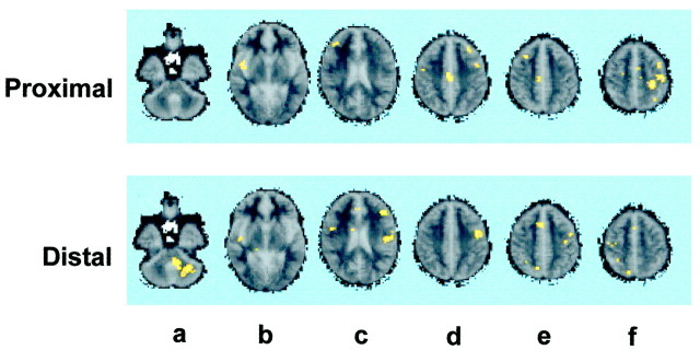
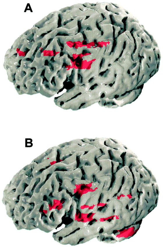
Somatic sensation can be localized precisely, whereas localization of visceral sensation is vague, possibly reflecting differences in the pattern of somatic and visceral input to the cerebral cortex. We used functional magnetic resonance imaging to study the cortical processing of sensation arising from the proximal (somatic) and distal (visceral) esophagus in six healthy male subjects. Esophageal stimulation was performed by phasic distension of a 2 cm balloon at 0.5 Hz. For each esophageal region, five separate 30 sec periods of nonpainful distension were alternated with five periods of similar duration without distension. Gradient echoplanar images depicting bold contrast were acquired using a 1.5 T GE scanner. Distension of the proximal esophagus was localized precisely to the upper chest and was represented in the trunk region of the left primary somatosensory cortex. In contrast, distension of the distal esophagus was perceived diffusely over the lower chest and was represented bilaterally at the junction of the primary and secondary somatosensory cortices. Different activation patterns were also observed in the anterior cingulate gyrus with the proximal esophagus being represented in the right midanterior cingulate cortex (BA 24) and the distal esophagus in the perigenual area (BA32). Differences in the activation of the dorsolateral prefrontal cortex and cerebellum were also observed for the two esophageal regions. These findings suggest that cortical specialization in the sensory-discriminative, affective, and cognitive areas of the cortex accounts for the perceptual differences observed between the two sensory modalities.
Figures
Similar articles
-
A study of the cortical processing of ano-rectal sensation using functional MRI.Brain. 2001 Feb;124(Pt 2):361-8. doi: 10.1093/brain/124.2.361. Brain. 2001. PMID: 11157563 Clinical Trial.
-
Functional neuroimaging of visceral sensation.J Clin Neurophysiol. 2000 Nov;17(6):604-12. doi: 10.1097/00004691-200011000-00006. J Clin Neurophysiol. 2000. PMID: 11151978 Review.
-
Differentiation of visceral and cutaneous pain in the human brain.J Neurophysiol. 2003 Jun;89(6):3294-303. doi: 10.1152/jn.01048.2002. Epub 2003 Feb 12. J Neurophysiol. 2003. PMID: 12611986
-
Somatic and limbic cortex activation in esophageal distention: a functional magnetic resonance imaging study.Ann Neurol. 1998 Nov;44(5):811-5. doi: 10.1002/ana.410440516. Ann Neurol. 1998. PMID: 9818938
-
Pathobiology of visceral pain: molecular mechanisms and therapeutic implications V. Central nervous system processing of somatic and visceral sensory signals.Am J Physiol Gastrointest Liver Physiol. 2000 Jul;279(1):G1-6. doi: 10.1152/ajpgi.2000.279.1.G1. Am J Physiol Gastrointest Liver Physiol. 2000. PMID: 10898740 Review.
Cited by
-
Functional brain imaging and central control of the bladder in health and disease.Front Physiol. 2022 Aug 12;13:914963. doi: 10.3389/fphys.2022.914963. eCollection 2022. Front Physiol. 2022. PMID: 36035497 Free PMC article. Review.
-
Deconstructing Chronic Low Back Pain in the Older Adult: Step by Step Evidence and Expert-Based Recommendations for Evaluation and Treatment: Part IV: Depression.Pain Med. 2015 Nov;16(11):2098-108. doi: 10.1111/pme.12935. Epub 2015 Nov 5. Pain Med. 2015. PMID: 26539754 Free PMC article. Review.
-
Whole-brain functional connectivity identification of functional dyspepsia.PLoS One. 2013 Jun 17;8(6):e65870. doi: 10.1371/journal.pone.0065870. Print 2013. PLoS One. 2013. PMID: 23799056 Free PMC article.
-
Abdominal Pain, the Adolescent and Altered Brain Structure and Function.PLoS One. 2016 May 31;11(5):e0156545. doi: 10.1371/journal.pone.0156545. eCollection 2016. PLoS One. 2016. PMID: 27244227 Free PMC article.
-
Can Interoception Improve the Pragmatic Search for Biomarkers in Psychiatry?Front Psychiatry. 2016 Jul 25;7:121. doi: 10.3389/fpsyt.2016.00121. eCollection 2016. Front Psychiatry. 2016. PMID: 27504098 Free PMC article. Review.
References
-
- Aine CJ. A conceptual overview and critique of functional neuroimaging in humans: I. MRI/fMRI and PET. Crit Rev Neurobiol. 1995;9:229–309. - PubMed
-
- Augustine JR. Circuitry and functional aspects of the insular lobe in primates including humans. Brain Res Rev. 1996;22:229–244. - PubMed
-
- Aziz Q, Andersson JLR, Valind S, Sundin A, Hamdy S, Jones AKP, Foster ER, Langstrom B, Thompson DG. Identification of human brain loci processing esophageal sensation using positron emission tomography. Gastroenterology. 1997;113:50–59. - PubMed
-
- Brammer MJ, Bullmore ET, Simmons A, Williams SCR, Grasby PM, Howard RJ, Woodruff PWR, Rabe-Hesketh S. Generic brain activation mapping in fMRI: a nonparametric approach. Magn Reson Imaging. 1997;15:763–770. - PubMed
Publication types
MeSH terms
Grants and funding
LinkOut - more resources
Full Text Sources