

Multivariate feature analysis of sonographic findings of metastatic cervical lymph nodes: contribution of blood flow features revealed by power Doppler sonography for predicting metastasis
- PMID: 10730652
- PMCID: PMC8174999
Multivariate feature analysis of sonographic findings of metastatic cervical lymph nodes: contribution of blood flow features revealed by power Doppler sonography for predicting metastasis
Abstract
Background and purpose: Sonographic criteria of the lymph node have been found to be good indicators for metastatic lymph nodes. We determined which sonographic features are most predictive of metastasis in cervical lymph nodes among patients with head and neck cancer.
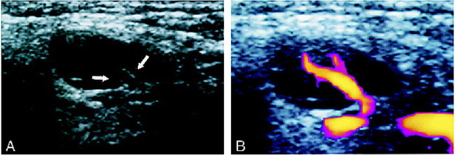
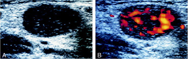
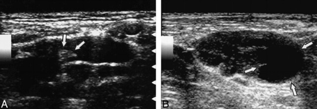
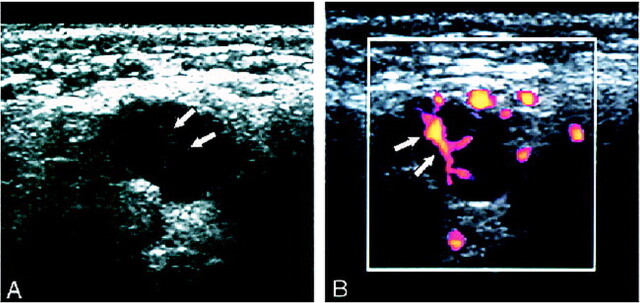
Methods: Gray-scale and power Doppler sonograms were retrospectively analyzed in 133 cervical lymph nodes (57 metastatic and 76 reactive nodes) from 52 patients with head and neck cancer. The gray-scale sonographic features of the presence or absence of hilar echoes, parenchymal echogenicity, and short and long axis lengths as well as the power Doppler features of normal hilar flow and abnormal parenchymal flow were evaluated. Univariate and multivariate logistic regression analyses were conducted to determine the relative value of each sonographic feature.
Results: At univariate analysis, all sonographic features assessed were found to be important. Multivariate analysis, however, suggested that the presence or absence of hilar echoes, increases in short axis length, and the presence of normal hilar flow were the only sonographic features that were predictive of reactive (presence of hilar echoes and hilar flow) and metastatic (increases in short axis length) lymph nodes. Although multivariate analysis did not indicate any significant contribution of the color-flow criteria for predicting metastatic nodes, the color-flow criteria appeared to improve the overall diagnostic accuracy for the less experienced observer.
Conclusion: The sonographic criteria most predictive of metastatic cervical lymph nodes were absent hilar echoes and increases in short axis length, as assessed by logistic regression analysis. Compared with these gray-scale criteria, color-flow criteria had fewer predictive advantages.
Figures
References
-
- Savoury LW, Gluckman JL. Cervical metastasis. In: Paparella MM, Shumrick DA, Gluckman JL, Myerhoff WL, eds. Otolaryngology. 3rd ed. Philadelphia: Saunders; 1991: 2565-2578
-
- Farr HW, Goldfarb PM, Farr CM. Epidermoid carcinoma of the mouth and pharynx at Memorial Sloan-Kettering Cancer Center, 1965 to 1969. Am J Surg 1980;140:563-567 - PubMed
-
- Van den Brekel MWM, Stel HV, Castelijns JA, et al. Cervical lymph node metastasis: assessment of radiologic criteria. Radiology 1990;177:379-384 - PubMed
-
- Som PM. Detection of metastasis in cervical lymph nodes: CT and MR criteria and differential diagnosis. AJR Am J Roentgenol 1992;158:961-969 - PubMed
-
- Vassallo PV, Wernecke K, Roos N, Peters PE. Differentiation of benign from malignant superficial lymphadenopathy: the role of high-resolution US. Radiology 1992;183:215-220 - PubMed
MeSH terms
LinkOut - more resources
Full Text Sources
Other Literature Sources
Medical