

Case Reports
The persistent stapedial artery
Affiliations
- PMID: 10730654
- PMCID: PMC8174972
Item in Clipboard
Case Reports
The persistent stapedial artery
AJNR Am J Neuroradiol.
2000 Mar.
Abstract
The persistent stapedial artery is a rare congenital vascular anomaly that may present as a pulsatile middle ear mass or that may appear as an incidental finding. Five cases of persistent stapedial artery are presented. The CT findings include the absence of the ipsilateral foramen spinosum and a soft-tissue prominence in the region of the tympanic segment of the facial nerve. Three cases were associated with an aberrant internal carotid artery. Imaging identification of this variant may obviate unnecessary surgery and may help in planning surgical or endovascular interventions.
Figures
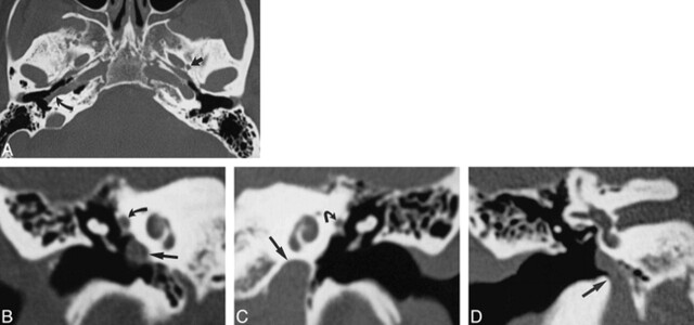
Case 1: 52-year-old woman with buzzing in right ear. A, Axial CT scan of skull base shows a normal left foramen spinosum (straight arrow). The right foramen spinosum is absent. The right ICA is laterally displaced into the middle ear (curved arrow). The left ICA is in a normal position. B, Coronal CT scan through right middle ear shows the ICA in the middle ear cavity (straight arrow). The soft tissue at the tympanic segment of the facial nerve is prominent because of the presence of a stapedial artery (curved arrow). C, Coronal CT scan through left middle ear shows the ICA in a normal position below the cochlea (straight arrow). The tympanic facial nerve is normal in size (curved arrow). D, Coronal CT scan at level of vestibule shows the entrance of the aberrant ICA into the right middle ear (arrow).

Case 2: 14-year-old boy with headache, vomiting, dizziness, and ringing in the ear. A, Axial CT scan shows a normal right foramen spinosum (arrow) and absence of the left foramen spinosum. B, Coronal CT scan shows the aberrant ICA on the left (straight arrow) and the soft-tissue density of a PSA (curved arrow). C, Left carotid arteriogram, lateral view, shows a PSA arising from the aberrant ICA (arrow). D, Left carotid arteriogram, frontal view, shows a PSA arising from the aberrant ICA (arrow).

Case 3: 6-year-old girl with vertigo. A, Axial CT scan shows a normal left foramen spinosum (arrow) and absence of the right foramen spinosum. B, Axial CT scan through middle ear shows prominent soft tissue, representing facial nerve and PSA (arrow). C, Coronal CT scan through middle ear shows prominent soft tissue, representing facial nerve and PSA (arrow).

Case 4: 25-year-old woman with pulsatile tinnitus. A, Axial CT scan shows a normal left foramen spinosum (arrow) and absence of the right foramen spinosum. B, Coronal CT scan through right middle ear shows the soft tissue at the tympanic segment of the facial nerve is prominent because of the presence of a stapedial artery (arrow). C, Coronal CT scan through left middle ear shows the tympanic facial nerve is normal in size (arrow).

Case 5: 5-year-old boy headache for 3 years. Carotid arteriogram, lateral view, shows a PSA (curved arrow) arising from the aberrant ICA, which represents the inferior tympanic branch of the ascending pharyngeal artery assuming the role of the ICA (straight arrow)

Schematic representation of the developmental stages of the stapedial artery. A, The hyoid artery arises from the ICA. The stapedial artery arises from the hyoid artery near its origin. The stapedial artery branches into upper and lower divisions after passing through the stapes. The ventral pharyngeal arteries are the precursors of the definitive ECA. The stapedial artery is the only supply to the upper and lower divisions. B, Anastomosis forms between the ventral pharyngeal artery and the lower division branches. C, The stapedial artery decreases in size. D, Normal adult anatomy with involution of the stapedial artery. E, Anatomic configuration of typical PSA.
References
-
- Congdon ED. Transformation of the aortic-arch system during the development of the human embryo. Contrib Embryol 1922;14:47-110
-
- Padget DH. The development of the cranial arteries in the human embryo. Contrib Embryol 1948;32:205-261
-
- Steffen TN. Vascular anomalies of the middle ear. Laryngoscope 1968;78:171-197 - PubMed
-
- Altman F. Anomalies of the internal carotid artery and its branches; their embryologic and comparative significance: report of a new case of persistent stapedial artery in man. Laryngoscope 1947;57:313-319 - PubMed
-
- Hogg ID, Stephens CB, Arnold GE. Theoretical anomalies of the stapedial artery. Ann Otol Rhinol Laryngol 1972;81:860-870 - PubMed
Publication types
MeSH terms
LinkOut - more resources
Full Text Sources
Medical