

Case Reports
Primary amyloidoma of the cervical spine
Affiliations
- PMID: 10730660
- PMCID: PMC8174966
Item in Clipboard
Case Reports
Primary amyloidoma of the cervical spine
AJNR Am J Neuroradiol.
2000 Mar.
Abstract
Primary solitary amyloidoma of the spine is a disease characterized by localized deposits of amyloid. We describe and illustrate the radiologic appearance of primary solitary amyloidoma of the spine on plain radiographs, CT scans, and MR images. The imaging findings revealed features of a nonspecific soft-tissue mass with calcifications. Epidural extension of the amyloidoma caused spinal cord compression.
Figures
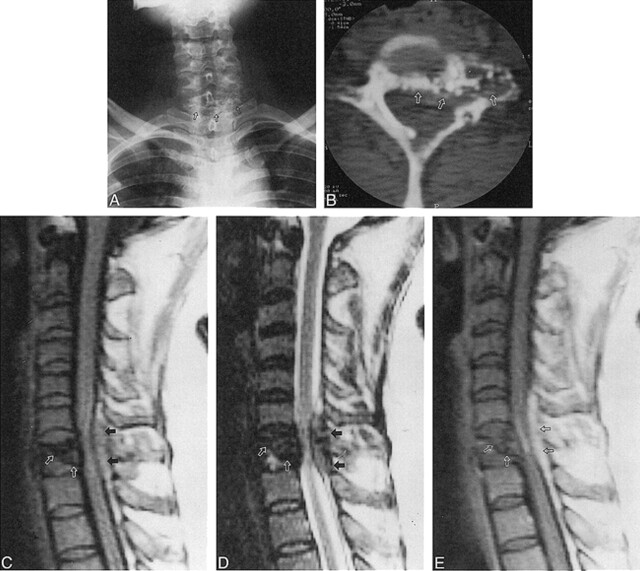
45-year-old woman with paraparesis, urinary incontinence, and posterior neck pain. A, Anteroposterior radiograph of cervical spine shows osteolytic lesion with scattered calcifications (arrows) in the C7 vertebra. B, Axial CT scan reveals osteolytic lesion with scattered calcifications involving the left lateral mass, pedicle, and anterior portion of the lamina of C7 (arrows). C and D, Sagittal T1- (C) and T2-weighted (D) MR images show an inhomogeneous low-signal mass and a partially collapsed C7 body (white arrows), with extension to the posterior extradural space (black arrows). E, Subtle enhancement of amyloid deposits within the partially collapsed vertebral body (white arrows) and extradural mass (black arrows) are noted on sagittal contrast-enhanced T1-weighted MR image.
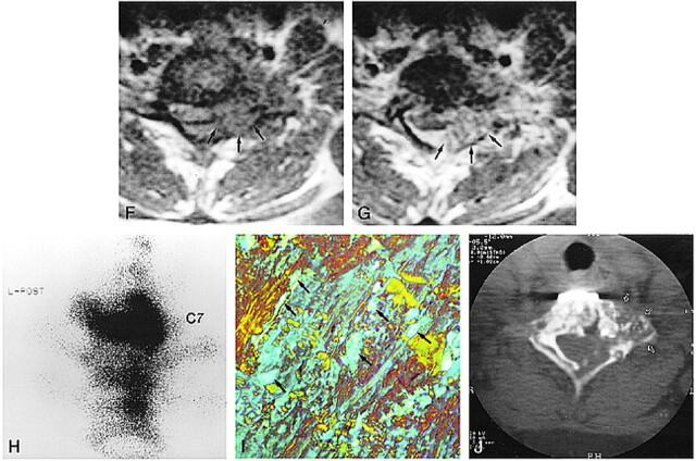
Continued. F and G, Axial T1-weighted MR images show the extradural mass encircling the anterior and lateral aspect of the dural sac (arrows, F). After contrast administration (G), the enhancing mass partially compresses the spinal cord (arrows). H, Posterior pinhole bone scintigram of cervicothoracic junction obtained with 99mTc-MDP shows increased radioactivity in C7. Minimally increased radioactivity is also noted in T1 and T2 vertebral bodies. I, Biopsy specimen viewed with polarized light is positive for Congo red stain, showing a characteristic green or apple-green birefringence (arrows) (original magnification ×80). J, At 3-year follow-up, CT scan shows no growth of the residual mass in C7 vertebra (arrows). The metallic instrument for fixation is noted in the anterior aspect of C7.
References
-
- Bauer WH, Kuzma JF. Solitary “tumors” of atypical amyloid (paramyloid). Am J Clin Pathol 1949;19:1097-1112 - PubMed
-
- Dickmam CA, Sonntag VK, Johnson P, Medina M. Amyloidoma of the cervical spine: a case report. Neurosurgery 1988;22:419-422 - PubMed
-
- Meyers SP, Mullins KJ, Kazee AM. Unifocal primary amyloidoma of the spine causing compression of the cervical spinal cord: MR findings. J Comput Assist Tomogr 1996;20:592-593 - PubMed
-
- Schindel S. Amyloid tumor of the larynx (case report with E.M.). Ann Otolarngol 1972;81:438-444 - PubMed
-
- Pawar S, Kay C, Anderson H. Primary amyloidoma of the spine. J Comput Assist Tomogr 1982;6:1175-1177 - PubMed
Publication types
MeSH terms
LinkOut - more resources
Full Text Sources
Medical