

Involvement of the MAP kinase cascade in resetting of the mammalian circadian clock
Affiliations
- PMID: 10733524
- PMCID: PMC316464
Item in Clipboard
Involvement of the MAP kinase cascade in resetting of the mammalian circadian clock
Genes Dev.
.
Abstract
Although the suprachiasmatic nucleus (SCN) is the major pacemaker in mammals, the peripheral cells or immortalized cells also contain a circadian clock. The SCN and the periphery may use different entraining signals-light and some humoral factors, respectively. We show that induction of the circadian oscillation of gene expression is triggered by TPA treatment of NIH-3T3 fibroblasts, which is inhibited by a MEK inhibitor, and that prolonged activation of the MAPK cascade is sufficient to trigger circadian gene expression. Therefore, such prolonged activation of MAPK by entraining cues may be involved in the resetting of the circadian clock.
Figures
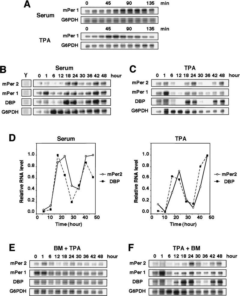
TPA-triggered induction of circadian gene expression in NIH-3T3 fibroblasts. (A) Cells were shifted to medium containing 50% serum or 50 nm TPA (t = 0). The levels of mPer1 mRNA were determined by RNase protection assays. G6PDH is a loading control. Cells were shifted to medium containing 50% serum (B) or 50 nm TPA (C) (t = 0), and incubated for 2 hr, after which the medium was replaced with serum-free medium. Whole-cell RNA was prepared at the indicated times, and the relative levels of the mRNAs were determined by RNase protection assays. Yeast RNA (Y) was used as negative control. Three independent experiments gave similar results. (D) The signals obtained the RNase protection assays in B and C for mPer2(○) and DBP(█) mRNAs were quantified and normalized to signals obtained for G6PDH mRNA. The maximum value was set to 1.0. (E,F) Bisindolylmaleimide I (BM, 5 μm final concentration), a PKC inhibitor, was added 30 min before the addition of TPA (E) or 8 hr after the addition of TPA (F).
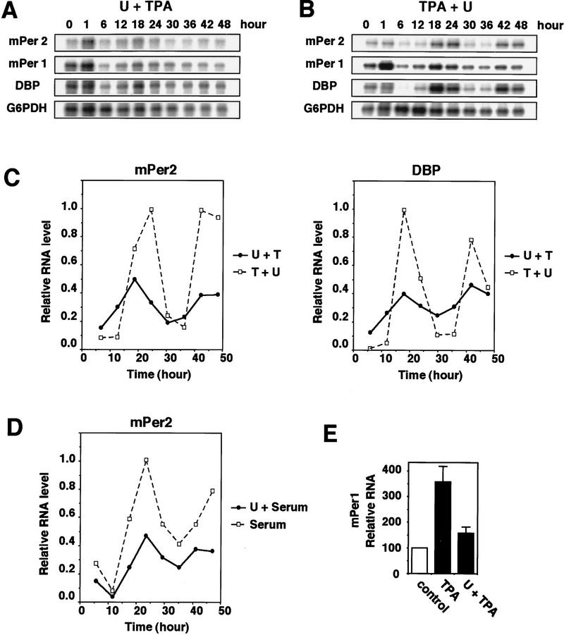
Inhibition of TPA-triggered induction of circadian gene expression by pretreatment with MEK inhibitor U0126. (A) 10 μm U0126 was added 30 min before the addition of 50 nm TPA (A) or 8 hr after the addition of TPA (B). Three independent experiments gave similar results. (C) The signals obtained in the RNase protection assays shown in A (●, U + TPA) and B (□, TPA + U) for mPer2 and DBP mRNAs were quantified and normalized as in Fig. 1D. (D) Inhibition of serum shock-triggered induction of circadian gene expression by pretreatment with U0126. The signals obtained in the RNase protection assays for mPer2 mRNA were quantified and normalized. (●); 20 μm U0126 before 50% serum treatment; (□) 50% serum treatment. (E) Relative levels of mPer1 mRNA induction at t = 70 min (means ± s.d. ; n = 3).
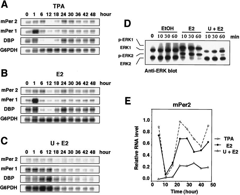
Induction of circadian gene expression triggered by activation of the Raf/MEK/ERK cascade in ΔB-Raf:ER NIH-3T3 cells. TPA (A, 50 nm ) or estradiol-17β (E2) (B, 1 μm ) was added to the medium (t = 0) and incubated for 2 hr (TPA) or 1 hr (E2), as described. Thirty minutes before the addition of 1 μm E2 (C) 10 μm U0126 was added. The relative levels of the mRNAs were determined by RNase protection assays. Three independent experiments gave similar results. (D) Cells were pretreated with or without 10 μm U0126 for 30 min prior to E2 treatment, and the protein extracts prepared at the indicated times were subjected to immunoblotting with anti-ERK/MAPK antibody. The electrophoretically retarded bands represent active forms. (E) Signals obtained in the RNase protection assays shown in A (□), B (●), and C (▵) for mPer2 mRNAs were quantified and normalized.
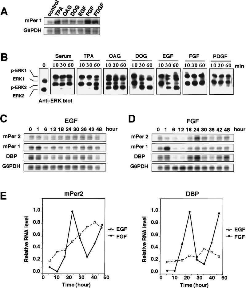
Prolonged activation of ERK MAPK is able to trigger the induction of robust circadian gene expression. NIH-3T3 cells were treated with 50% serum, 50 nm TPA, 200 μm OAG, 200 μm DOG, 30 nm EGF, 25 ng/ml FGF, or 30 ng/ml PDGF. (A) Whole-cell RNA was prepared at 70 min after treatment, and the relative levels of the mRNAs were determined by RNase protection assays. (B) Protein extracts prepared at the indicated times were subjected to immunoblotting with anti-ERK/MAPK antibody. EGF (C, 30 nm ) or FGF (D, 25 ng/ml) was added to the medium (t = 0) and incubated for 2 hr as described. Relative levels of the mRNAs were determined by RNase protection assays. Three independent experiments gave similar results. (E) Signals obtained in the RNase protection assays shown in C and D (□, EGF; ●, FGF) for mPer2 and DBP mRNAs were quantified and normalized.
References
-
- Akiyama M, Kouzu Y, Takahashi S, Wakamatsu H, Moriya T, Maetani M, Watanabe S, Tei H, Sakaki Y, Shibata S. Inhibition of light- or glutamate-induced mPer1 expression represses the phase shifts into the mouse circadian locomotor and suprachiasmatic firing rhythms. J Neurosci. 1999;19:1115–1121. - PMC - PubMed
-
- Albrecht U, Sun ZS, Eichele G, Lee CC. A differential response of two putative mammalian circadian regulators, mper1 and mper2, to light. Cell. 1997;91:1055–1064. - PubMed
-
- Balsalobre A, Damiola F, Schibler U. A serum shock induces circadian gene expression in mammalian tissue culture cells. Cell. 1998;93:929–937. - PubMed
-
- Bina KG, Rusak B. Nerve growth factor phase shifts circadian activity rhythms in Syrian hamsters. Neurosci Lett. 1996;206:97–100. - PubMed
Publication types
MeSH terms
Substances
LinkOut - more resources
Full Text Sources