. 2000 Mar 15;14(6):655-65.

The KEN box: an APC recognition signal distinct from the D box targeted by Cdh1

Affiliations

The KEN box: an APC recognition signal distinct from the D box targeted by Cdh1

C M Pfleger et al. Genes Dev. .

Abstract

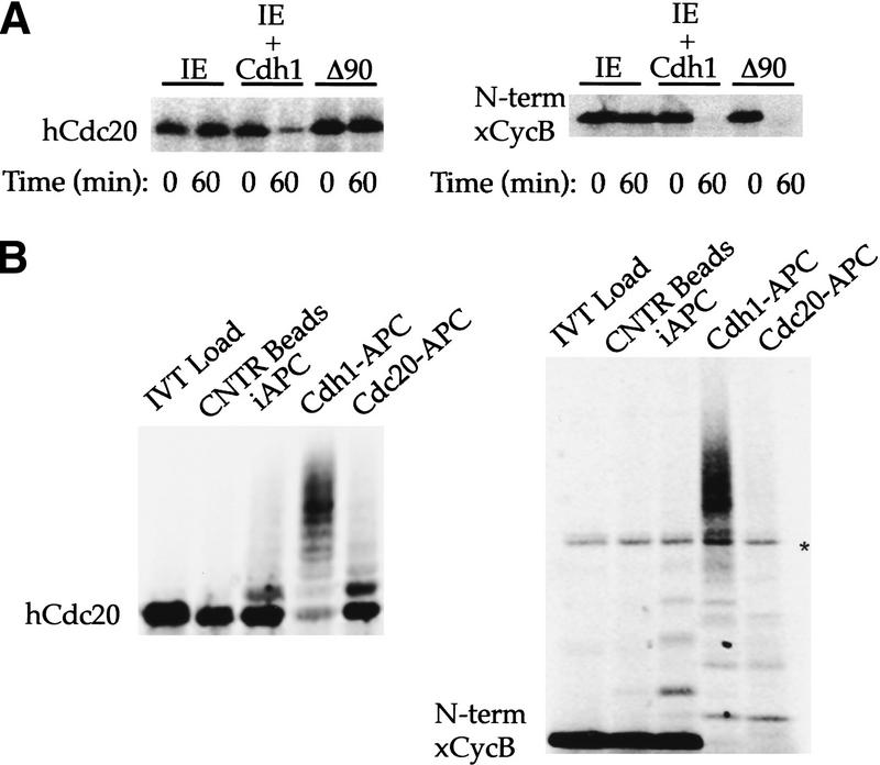
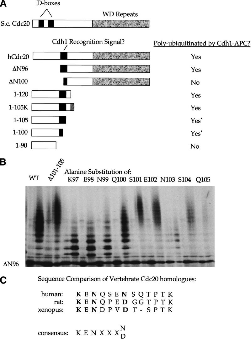
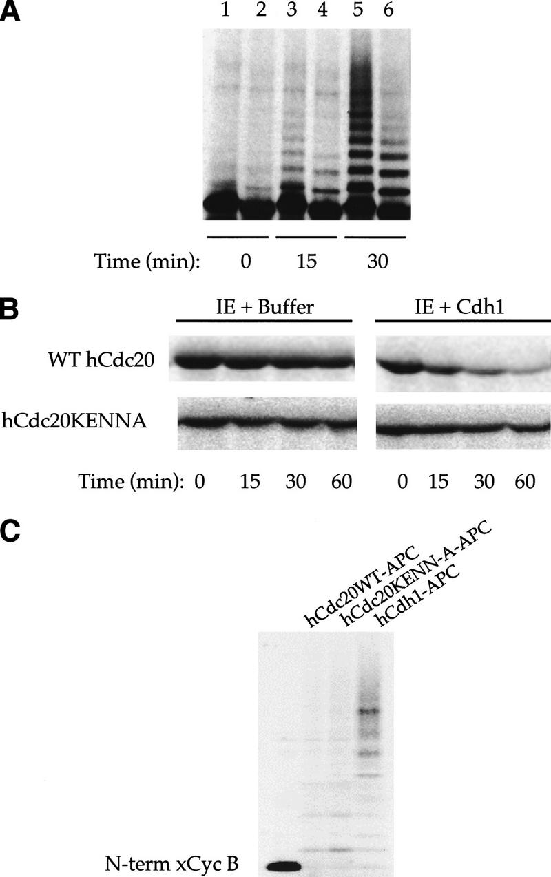
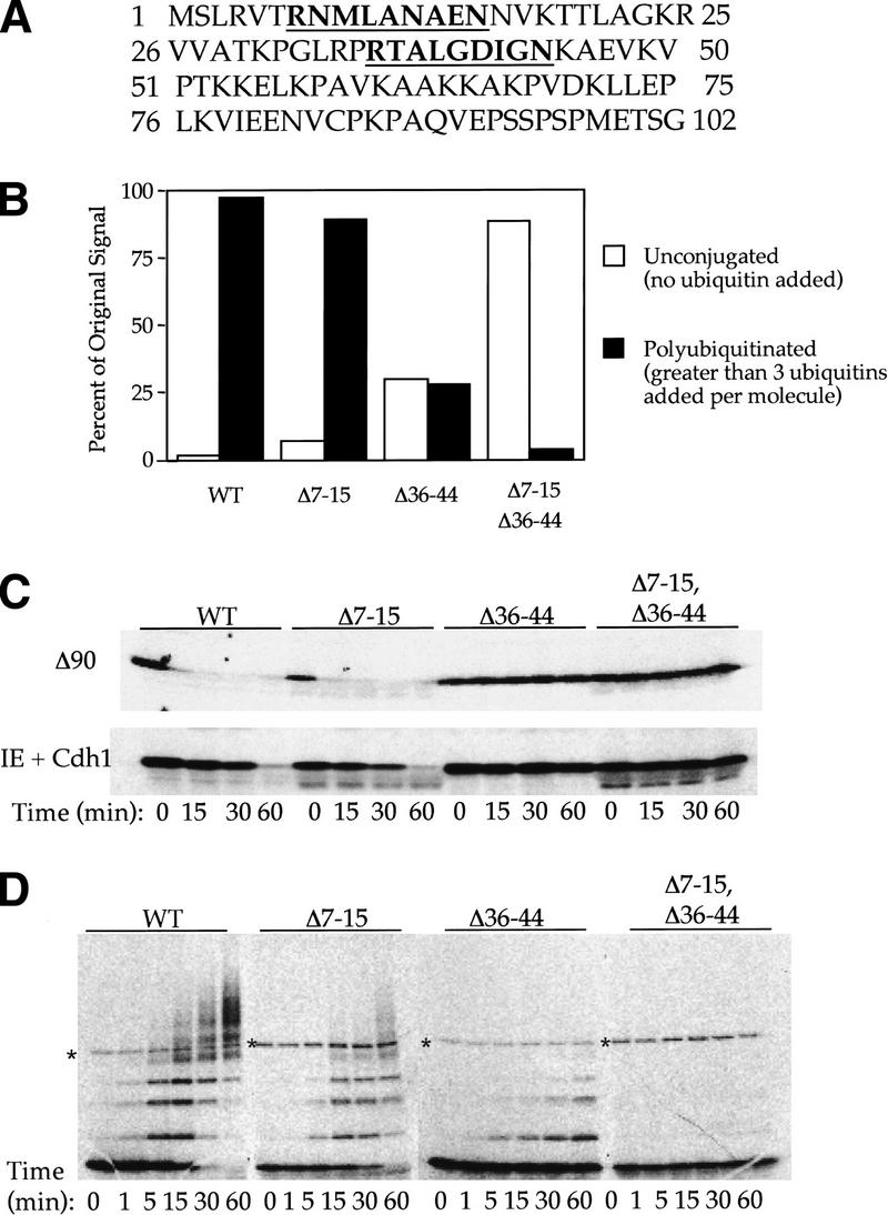
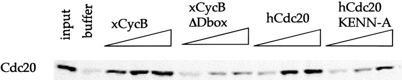
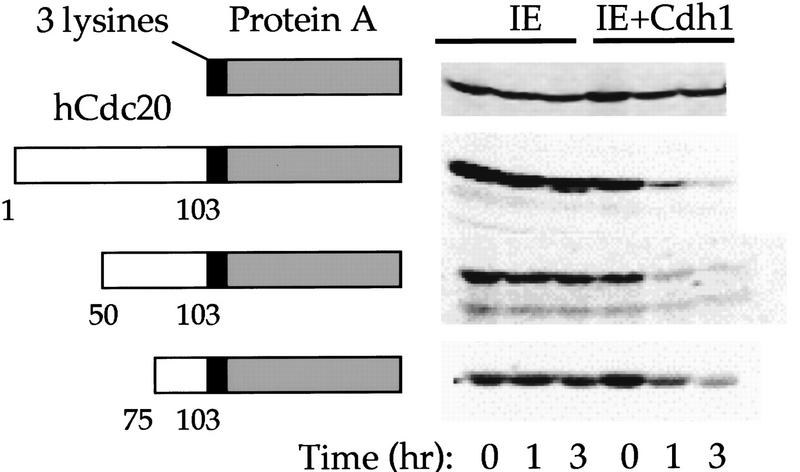
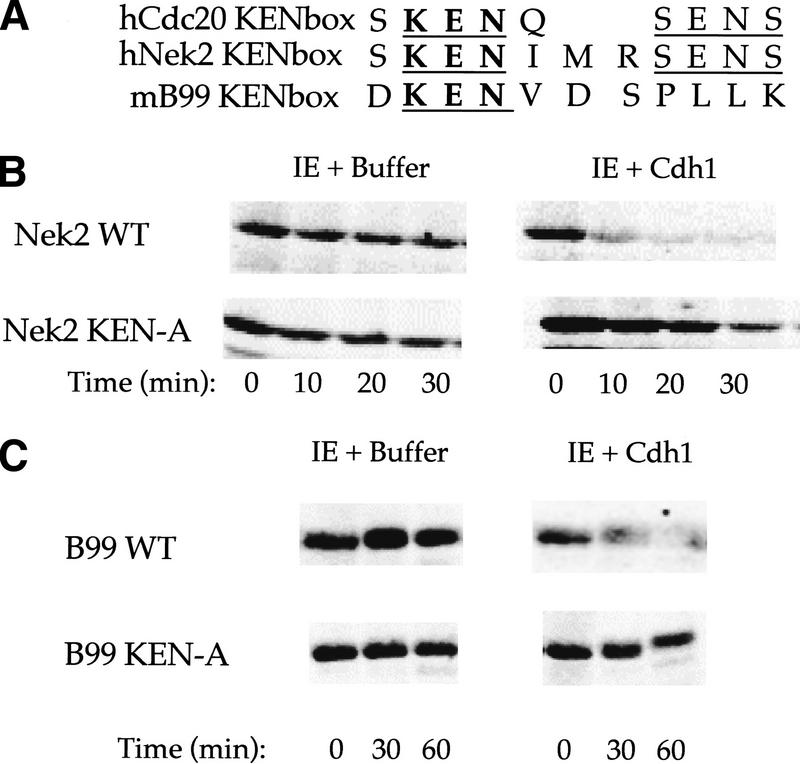
The ordered progression through the cell cycle depends on regulating the abundance of several proteins through ubiquitin-mediated proteolysis. Degradation is precisely timed and specific. One key component of the degradation system, the anaphase promoting complex (APC), is a ubiquitin protein ligase. It is activated both during mitosis and late in mitosis/G(1), by the WD repeat proteins Cdc20 and Cdh1, respectively. These activators target distinct sets of substrates. Cdc20-APC requires a well-defined destruction box (D box), whereas Cdh1-APC confers a different and as yet unidentified specificity. We have determined the sequence specificity for Cdh1-APC using two assays, ubiquitination in a completely defined and purified system and degradation promoted by Cdh1-APC in Xenopus extracts. Cdc20 is itself a Cdh1-APC substrate. Vertebrate Cdc20 lacks a D box and therefore is recognized by Cdh1-APC through a different sequence. By analysis of Cdc20 as a substrate, we have identified a new recognition signal. This signal, composed of K-E-N, serves as a general targeting signal for Cdh1-APC. Like the D box, it is transposable to other proteins. Using the KEN box as a template, we have identified cell cycle genes Nek2 and B99 as additional Cdh1-APC substrates. Mutation in the KEN box stabilizes all three proteins against ubiquitination and degradation.


Figures

Figure 1
Figure 1
Recognition of Cdc20 as a substrate by Cdh1–APC but not Cdc20–APC. (A) Incubation of 35S-labeled in vitro-translated hCdc20 and the amino terminus of Xenopus cyclin B (xCycB) in interphase extract supplemented with buffer (IE), interphase extract supplemented with Cdh1 protein (IE + Cdh1), or interphase extract pushed into mitosis by sea urchin cyclin B Δ90 (Δ90). Both hCdc20 and xCycB are stable in IE, but degrade rapidly in IE + Cdh1. xCycB, and not hCdc20, disappears in the mitotic extract. (B) Incubation of hCdc20 and amino-terminal xCycB in a purified system ubiquitination assay using control beads (Cntr Beads), immunopurified interphase APC on beads (iAPC), or iAPC on beads activated by Cdh1 (Cdh1–APC) or Cdc20 (Cdc20–APC). Neither substrate forms high molecular weight conjugates in the presence of control beads or iAPC, but both form significant higher molecular weight conjugates in the presence of Cdh1–APC. Only xCycB forms higher molecular weight conjugates in the presence of Cdc20–APC. Polyubiquitinated species of xCyc B spread out more in the presence of Cdc20–APC than in the presence of Cdh1–APC, creating a much lighter smearing not always easily visualized for the 35S-labeled cyclin fragment as with iodinated substrate in previous reports. A darker exposure (not shown) makes visualization of the conjugates more obvious. An asterisk indicates the presence of a background band present in the in vitro translation products.
Figure 2
Figure 2
Isolation of the region in hCdc20 recognized by Cdh1–APC. (A) Schematic representation comparing yeast and human Cdc20. D boxes are indicated by black boxes. The WD repeat region is indicated by a textured gray box. Amino- and carboxy-terminal deletion mutants of hCdc20 were incubated in the purified system ubiquitination assay in the presence of Cdh1–APC, and the results are summarized at right. Deletion mutants narrowed the region recognized by Cdh1–APC, shown as a solid gray box. (B) Ubiquitination assays of hCdc20 point mutants using Cdh1–APC. Each pair represents an IVT load and the Cdh1–APC ubiquitination reaction. (WT) An internal deletion of amino acids 101–105, individual alanine substitution mutants of amino acids 97–105 within the Δ96 context are shown. All alanine mutants produced a ubiquitination profile showing high molecular weight conjugates except for K97A, E98A, N99A, and N103A. These four substitution mutants formed only lower molecular weight conjugates representing monoubiquitinated species. (C) Comparison of various vertebrate Cdc20 sequences of this region. Residues shown to play a role in recognition by the alanine scanning mutagenesis are shown in bold. Below the sequence comparisons, a putative consensus for Cdh1 recognition on the basis of sequence comparison and experimental results is suggested. The Cdc2 site is highly conserved among species (even in clam, not shown) even though it is not required for recognition by Cdh1.
Figure 3
Figure 3
Cdh1–APC does not recognize hCdc20 with mutant KEN-box recognition signal. (A) Time series of ubiquitination of wild-type hCdc20 (lanes 1,3,5) and mutant hCdc20KENN-A (key recognition residues changed to alanine—97AAAQSEA103, lanes 2,4,6). The side-by-side comparison shows reduced and slower ubiquitination of the mutant by Cdh1–APC. After 30 min, significant polyubiquitinated species have formed for wild-type hCdc20, whereas only monoubiquitinated species exist for the mutant. (B) Degradation assays using interphase extracts supplemented with buffer or Cdh1 show rapid degradation of wild-type but not mutant hCdc20. (C) Ubiquitination assay of Xenopus amino-terminal cyclin B using similar amounts of cold in vitro translated wild-type hCdc20–APC, hCdc20KENN-A–APC, and hCdh1–APC to activate iAPC on beads. Wild-type and mutant Cdc20 show similar ability to recognize amino-terminal xcyclin B.
Figure 4
Figure 4
Cdh1 recognizes the R-L-N destruction box motif. (A) Sequence of Xenopus cyclin B amino terminus. Both R-L-N motifs are underlined and shown in bold. The first R-L-N motif, at position 7–15 is not conserved between species, whereas the second, at position 36–44, is well conserved and well characterized as the B-type cyclin D box. (B) Quantitation of ubiquitination assays in the purified system using Cdh1–APC in the presence of 35S-labeled in vitro translated substrate. Bar graph depicting unconjugated signal remaining after a 1-hr incubation with Cdh1–APC (open bar) and polyubiquitinated (greater than three ubiquitins added per molecule) species formed for each mutant (solid bar). Polyubiquitinated species form for wild-type Xenopus amino-terminal cyclin B as well as the Δ7–15 and Δ36–44 mutants. Only monoubiquitination occurs for the double mutant, and most of the signal remains unconjugated. The unconjugated and polyubiquitinated percentages do not add up to 100%; the remaining signal reflects monoubiquitinated species not shown in the graph. (C) Degradation assays of cyclin and D-box mutants in the extract system Cdh1-supplemented interphase extracts and Δ90 extracts. Both mutants that lack the 36–44 D box are stable in the Δ90 and Cdh1-supplemented extracts. (D) Time series of Cdh1–APC ubiquitination assay of xCycB and D-box deletion mutants using 20% of ubiquitin in the other assays. By limiting the addition of ubiquitin, the specificity of the extract system from C is restored in the purified system. The Δ36–44 mutant no longer forms significant polyubiquitin chains. An asterisk indicates a background band from the in vitro translation products.
Figure 5
Figure 5
Peptides containing, but not lacking, the D box or KEN box inhibit degradation of Cdc20 in Cdh1-supplemented extracts. Degradation of full-length hCdc20 in Cdh1-supplemented interphase extracts. The left-most lane shows the initial input of substrate for each condition. Subsequent lanes indicate 1-hr incubations of buffer, followed by incubation with 1, 5, and 10 μm amounts of amino-terminal xcyclin B, Δbox amino-terminal xcyclin B, amino-terminal hCdc20, and amino-terminal hCdc20KENN-A.
Figure 6
Figure 6
The Cdc20 KEN box acts as a transposable ubiquitination and degradation signal. (Open box) Schematic representation of fusion proteins containing the hCdc20 KEN box; (shaded box) and the immunoglobulin-binding region of protein A. Three lysines have been inserted at the hinge region (small black box). The schematic is not scaled to size. Degradation assays using in vitro translated proteins in IE or IE + Cdh1 are shown at right.
Figure 7
Figure 7
Cdh1–APC targets cell cycle proteins Nek2 and B99 and recognizes the KEN box as a general targeting signal. (A) Sequence comparison of the Cdc20 KEN box with the KEN-containing regions of human Nek2 and mouse B99. KEN residues are in bold and conserved residues are underlined. Incubation of Nek2 (B) and B99 (C) and corresponding alanine substitution KEN mutants IE and IE + Cdh1. Both Nek2 and B99 form polyubiquitin chains in the presence of Cdh1–APC in the purified system and degrade rapidly in the presence of Cdh1 in the extracts. Alanine substitution of the KEN box stabilizes these mutants in the extract.

References

    1. Bischoff J, Plowman GD. The Auror/Ip1p kinase family: Regulators of chromosome segregation and cytokinesis. Trends Cell Biol. 1999;9:454–459. - PubMed
    1. Ciechanover A. The ubiquitin-proteasome proteolytic pathway. Cell. 1994;79:13–21. - PubMed
    1. Cohen-Fix O, Peters J-M, Kirschner MW, Koshland D. Anaphase initiation in Saccharomyces cerevisiae is controlled by the APC-dependent degradation of the anaphase inhibitor Pds1p. Genes & Dev. 1996;10:3081–3093. - PubMed
    1. Coux O, Tanaka K, Goldberg AL. Structure and functions of the 20S and 26S proteasomes. Annu Rev Biochem. 1996;65:801–847. - PubMed
    1. Fang G, Yu H, Kirschner MW. Direct binding of CDC20 protein family members activates the anaphase-promoting complex in Mitosis and G1. Mol Cell. 1998;2:163–171. - PubMed

Publication types

LinkOut - more resources