. 2000 Mar 15;14(6):704-18.

PERP, an apoptosis-associated target of p53, is a novel member of the PMP-22/gas3 family

Affiliations

PERP, an apoptosis-associated target of p53, is a novel member of the PMP-22/gas3 family

L D Attardi et al. Genes Dev. .

Erratum in

  • Genes Dev 2000 Jul 15;14(14):1835

Abstract

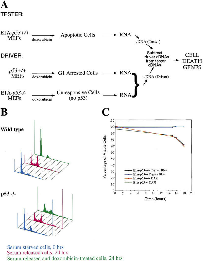
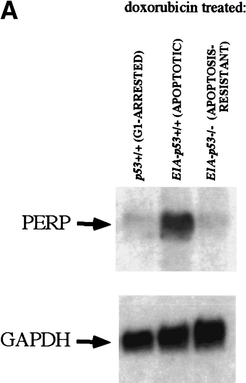
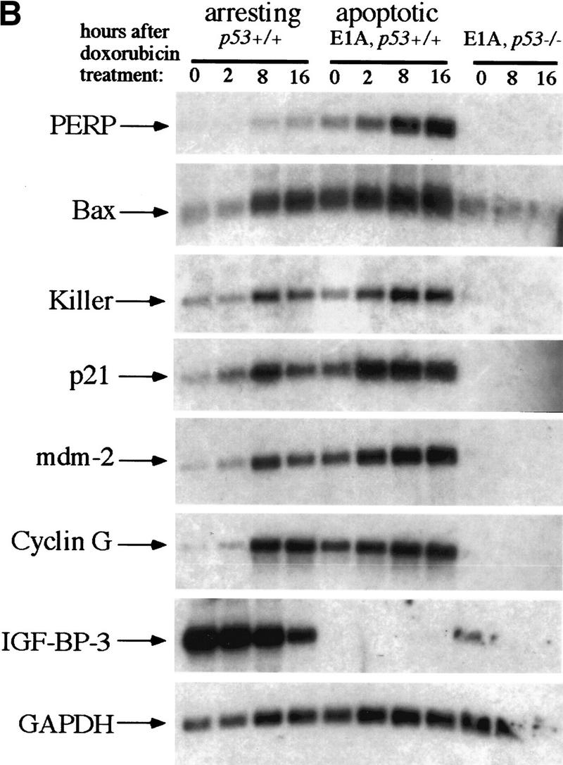
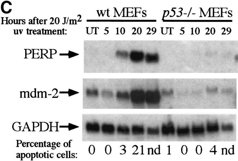
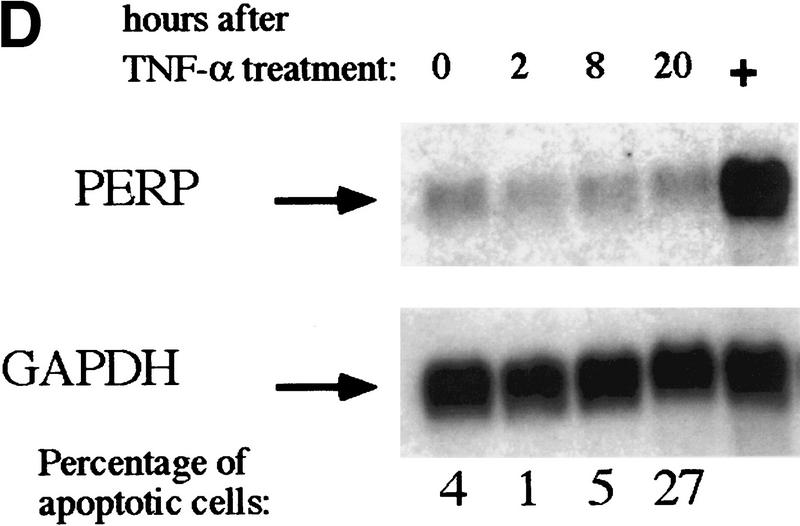
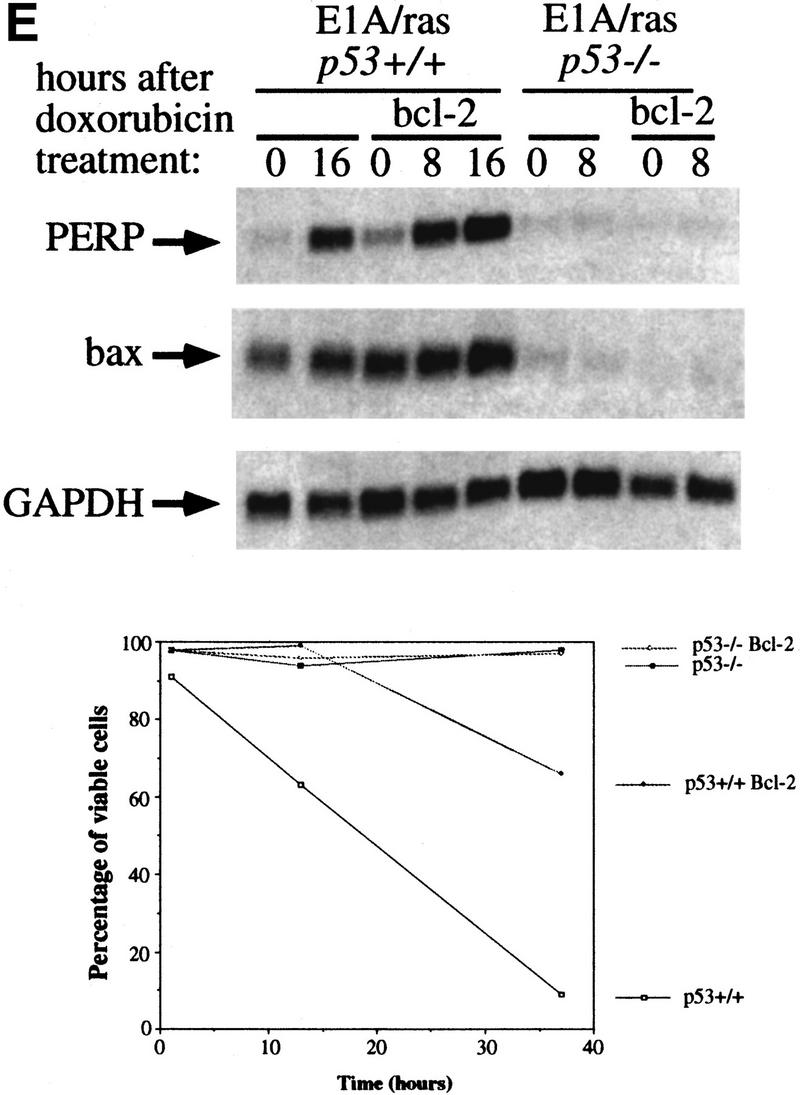
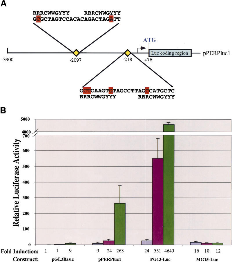
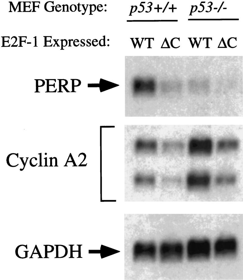
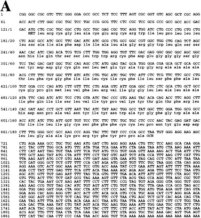
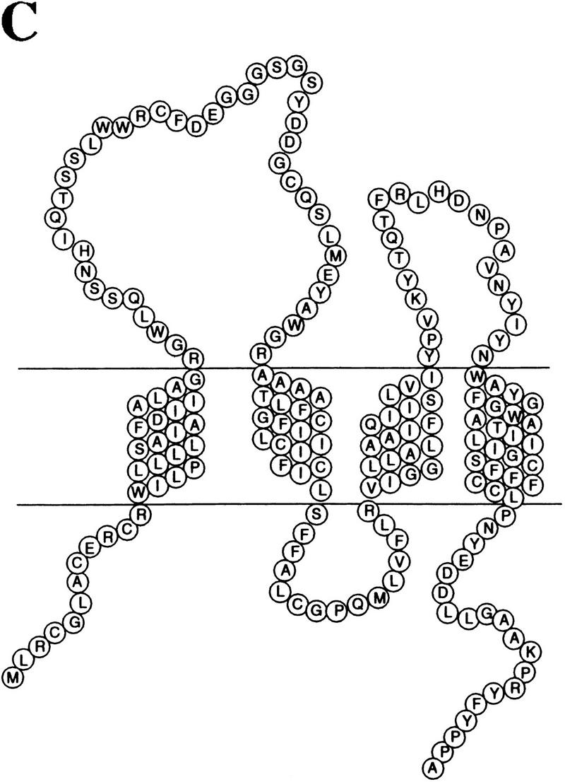
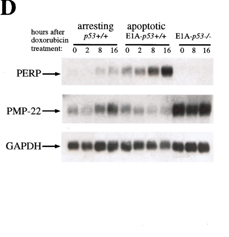
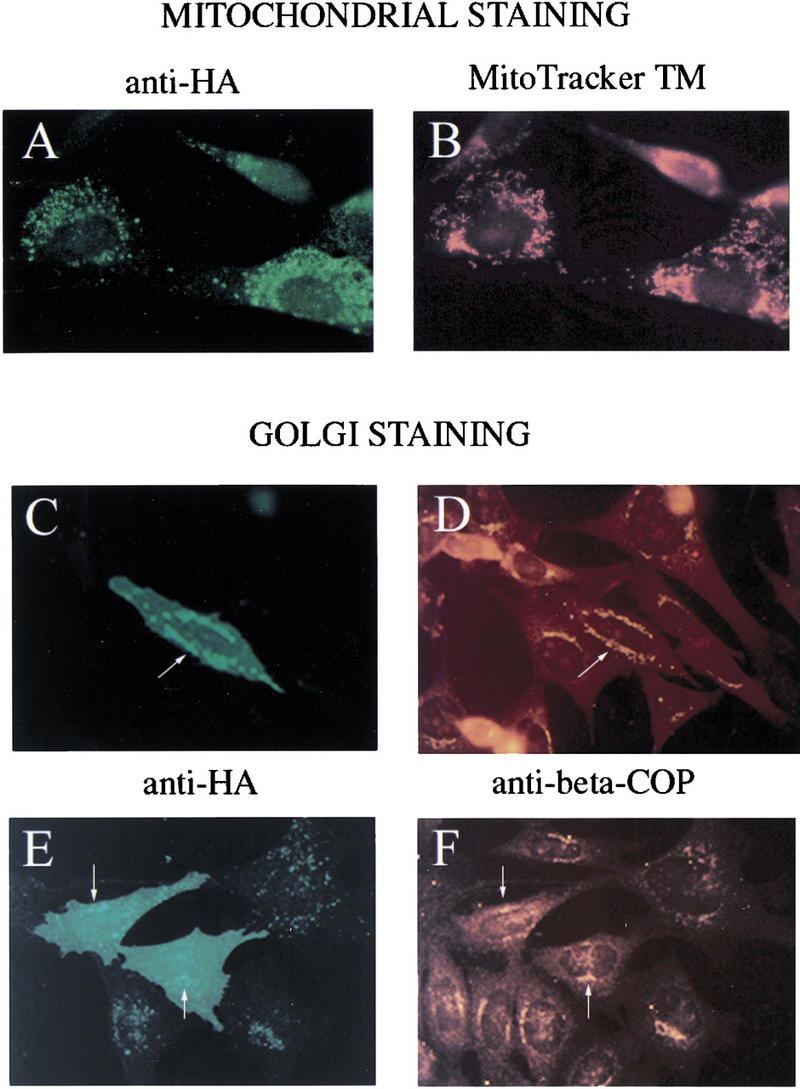
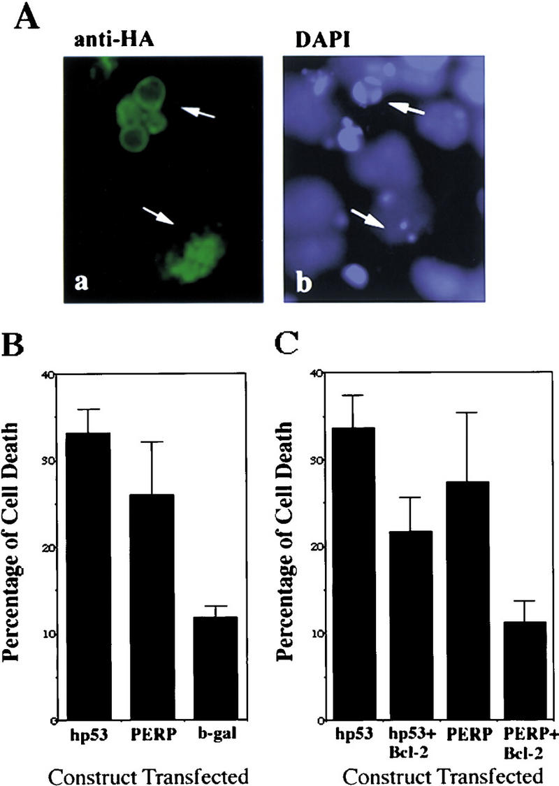
The p53 tumor suppressor activates either cell cycle arrest or apoptosis in response to cellular stress. Mouse embryo fibroblasts (MEFs) provide a powerful primary cell system to study both p53-dependent pathways. Specifically, in response to DNA damage, MEFs undergo p53-dependent G(1) arrest, whereas MEFs expressing the adenovirus E1A oncoprotein undergo p53-dependent apoptosis. As the p53-dependent apoptosis pathway is not well understood, we sought to identify apoptosis-specific p53 target genes using a subtractive cloning strategy. Here, we describe the characterization of a gene identified in this screen, PERP, which is expressed in a p53-dependent manner and at high levels in apoptotic cells compared with G(1)-arrested cells. PERP induction is linked to p53-dependent apoptosis, including in response to E2F-1-driven hyperproliferation. Furthermore, analysis of the PERP promoter suggests that PERP is directly activated by p53. PERP shows sequence similarity to the PMP-22/gas3 tetraspan membrane protein implicated in hereditary human neuropathies such as Charcot-Marie-Tooth. Like PMP-22/gas3, PERP is a plasma membrane protein, and importantly, its expression causes cell death in fibroblasts. Taken together, these data suggest that PERP is a novel effector of p53-dependent apoptosis.


Figures

Figure 1
Figure 1
Subtractive hybridization strategy. (A) Scheme for differential screen. cDNA from G1-arrested MEFs (doxorubicin-treated MEFs) and doxorubicin-treated E1A–p53−/− MEFs was pooled and subtracted from cDNA from apoptotic MEFs (doxorubicin-treated E1A–p53+/+ MEFs). (B) Doxorubicin induces a p53-dependent G1 arrest. For the G1-arrested population in the screen, wild-type MEFs were synchronized by serum starvation, released into the cell cycle, and then treated with doxorubicin to induce G1 arrest. FACS profiles of both wild-type and p53-null MEFs are shown and include synchronized cells, cells stimulated to re-enter the cell cycle for 24 hr, and cells G1-arrested with doxorubicin. (C) Doxorubicin induces a p53-dependent apoptotic response in E1A MEFs. Shown here are data from one experiment of two performed to prepare RNA for the subtraction experiment. The percentage of viable E1A–p53+/+ and E1A–p53−/− cells after doxorubicin treatment is plotted as a function of time. Cell death was measured both by trypan blue exclusion, to measure loss of membrane integrity, and DAPI staining, to examine nuclear morphology, and the techniques gave equivalent results.
Figure 2
Figure 2
PERP induction is correlated with activation of the p53-dependent apoptotic pathway. (A) Northern blot analysis shows a 1.9-kb message, PERP, that is up-regulated in apoptotic E1A MEFs (middle lane) compared with G1-arrested MEFs (left lane) and E1A–p53−/− MEFs (right lane). The blot was reprobed with GAPDH as a loading control. (B) Time course analysis of PERP and p53 target gene message levels in MEFs undergoing G1 arrest, E1A MEFs undergoing apoptosis, and doxorubicin-treated E1A–p53−/− MEFs. PERP message accumulates to significantly higher levels in apoptotic cells than in G1-arrested cells. The blot was probed with GAPDH as a loading control. (C) PERP is induced in another context of p53-dependent apoptosis. Northern blot analysis shows that PERP is induced as wild-type MEFs undergo apoptosis in response to UV light. It is not induced in the p53−/− MEFs, which are not undergoing apoptosis. The percentages of cells undergoing apoptosis are indicated (bottom). The blot was probed with GAPDH as a loading control. (D) PERP is not induced during p53-independent apoptosis. Northern blot analysis shows RNA derived from TNF-α-treated E1A/rasp53−/− (transformed) fibroblasts. The percentages of cells undergoing apoptosis are indicated (bottom). For comparison, lane 5 includes RNA from E1A MEFs undergoing apoptosis, in which PERP mRNA levels are much higher than in TNF-α-treated samples. The blot was probed with GAPDH as a loading control. (E) PERP Induction is not inhibited by Bcl-2. Northern blot analysis shows that PERP is still induced in response to doxorubicin in cells that express Bcl-2. Bax levels, in contrast, remain stable in response to doxorubicin. The blot was probed with GAPDH as a loading control. The graph (bottom) shows the cell death profiles in cells of various genotypes, with only p53+/+, non-Bcl-2-expressing cells undergoing apoptosis.
Figure 2
Figure 2
PERP induction is correlated with activation of the p53-dependent apoptotic pathway. (A) Northern blot analysis shows a 1.9-kb message, PERP, that is up-regulated in apoptotic E1A MEFs (middle lane) compared with G1-arrested MEFs (left lane) and E1A–p53−/− MEFs (right lane). The blot was reprobed with GAPDH as a loading control. (B) Time course analysis of PERP and p53 target gene message levels in MEFs undergoing G1 arrest, E1A MEFs undergoing apoptosis, and doxorubicin-treated E1A–p53−/− MEFs. PERP message accumulates to significantly higher levels in apoptotic cells than in G1-arrested cells. The blot was probed with GAPDH as a loading control. (C) PERP is induced in another context of p53-dependent apoptosis. Northern blot analysis shows that PERP is induced as wild-type MEFs undergo apoptosis in response to UV light. It is not induced in the p53−/− MEFs, which are not undergoing apoptosis. The percentages of cells undergoing apoptosis are indicated (bottom). The blot was probed with GAPDH as a loading control. (D) PERP is not induced during p53-independent apoptosis. Northern blot analysis shows RNA derived from TNF-α-treated E1A/rasp53−/− (transformed) fibroblasts. The percentages of cells undergoing apoptosis are indicated (bottom). For comparison, lane 5 includes RNA from E1A MEFs undergoing apoptosis, in which PERP mRNA levels are much higher than in TNF-α-treated samples. The blot was probed with GAPDH as a loading control. (E) PERP Induction is not inhibited by Bcl-2. Northern blot analysis shows that PERP is still induced in response to doxorubicin in cells that express Bcl-2. Bax levels, in contrast, remain stable in response to doxorubicin. The blot was probed with GAPDH as a loading control. The graph (bottom) shows the cell death profiles in cells of various genotypes, with only p53+/+, non-Bcl-2-expressing cells undergoing apoptosis.
Figure 3
Figure 3
The PERP promoter is p53 responsive. (A) Schematic of the pPERPluc1 luciferase reporter containing the PERP promoter region. The arrow represents the putative start site of transcription. The position of two potential p53 binding sites (at −2097 and −218) with significant homology to the published consensus sequence (18 out of 20 and 16 out of 20 matches, respectively) are indicated by yellow diamonds and their sequences are shown. Mismatches are indicated in red. (B) The 4-kb region containing the PERP promoter is activated by p53. The pPERPluc1 reporter was transfected into p53−/− MEFs (lavender bars), wild-type MEFs (fuchsia bars), or p53−/− MEFs cotransfected with human p53 (green bars). Values shown are given relative to the pGL3Basic backbone plasmid in p53−/− MEFs and represent the average of three separate experiments. Reporters include pPERPluc1 (diagramed above); pGL3Basic luciferase reporter with no promoter/enhancer (Promega); PG13–Luc1 (13 copies of a p53 binding site placed upstream of the polyoma promoter and luciferase); and MG15–Luc (15 copies of a mutant p53 binding site upstream of the polyoma promoter and luciferase).
Figure 4
Figure 4
PERP is up-regulated during E2F-1-induced cell death. Wild-type and p53-null MEFs were infected with retroviruses carrying either a full-length or a mutant form of E2F-1 lacking the transactivation domain (E2F-1ΔC, comprising residues 1–409 of E2F-1). Forty-eight hours after infection, PERP expression was observed only in the wild-type cells infected with full-length E2F-1, correlating with a visible induction of apoptosis. The E2F-1 target gene cyclin A, in contrast, is induced in full-length E2F-1-infected wild-type and p53−/− MEFs, relative to background endogenous cyclin A levels (seen in E2F-1ΔC-infected cells). The blot was probed with GAPDH as a loading control.
Figure 5
Figure 5
PERP is a new member of the PMP-22/gas3 family. (A) cDNA and amino acid sequence of PERP. (B) Alignment of PERP and PMP-22/gas3 from several species (human, mouse, rat) is shown. Identical amino acids are highlighted in red, and similar amino acids are highlighted in yellow. (C) Molecular model of PERP in the plasma membrane, as determined by transmembrane prediction programs. (D) Northern blot analysis shows that PMP-22/gas3 is expressed in a pattern distinct from PERP during p53-dependent G1 arrest in MEFs and apoptosis in E1A MEFs. The blot was probed with GAPDH as a loading control.
Figure 5
Figure 5
PERP is a new member of the PMP-22/gas3 family. (A) cDNA and amino acid sequence of PERP. (B) Alignment of PERP and PMP-22/gas3 from several species (human, mouse, rat) is shown. Identical amino acids are highlighted in red, and similar amino acids are highlighted in yellow. (C) Molecular model of PERP in the plasma membrane, as determined by transmembrane prediction programs. (D) Northern blot analysis shows that PMP-22/gas3 is expressed in a pattern distinct from PERP during p53-dependent G1 arrest in MEFs and apoptosis in E1A MEFs. The blot was probed with GAPDH as a loading control.
Figure 6
Figure 6
PERP localizes to the Golgi apparatus and the plasma membrane. (A,B) Fibroblasts were transfected with PERP–HA and immunostained with anti-HA antibodies (A) and MitoTracker (B). Many regions of staining are nonoverlapping, suggesting that PERP is not exclusively localized to the mitochondria. (CF) E1A–p53−/− MEFs were transfected with PERP–HA and immunostained with anti-HA (C,E) and anti-β-COP (D,F). Regions of vesicular HA staining coincide with β-COP staining, suggesting that PERP is localizing to the Golgi apparatus (arrows). In addition, some cells show plasma membrane staining, seen as uniform cell staining (E).
Figure 7
Figure 7
PERP is sufficient to induce cell death. (A) Expression of PERP induces apoptotic morphology. Cells transfected with PERP–HA and immunostained with anti-HA and DAPI show apoptotic morphology (shrunken cell bodies and blebbing nuclei) when examined by immunofluorescence. (B) Expression of PERP induces cell death. PERP induces cell death in E1A–p53−/− MEFs at a level intermediate between p53 and the background level of the negative control (β-galactosidase). Data represent the average of four experiments. (C) Bcl-2 expression inhibits PERP-induced cell death. Coexpression of Bcl-2 lowers the percentage of cells undergoing apoptosis in both p53- and PERP-expressing samples. Data represent the average of three experiments.

References

    1. Attardi LD, Lowe SW, Brugarolas J, Jacks T. Transcriptional activation by p53, but not induction of the p21 gene, is essential for oncogene-mediated apoptosis. EMBO J. 1996;15:3693–3701. - PMC - PubMed
    1. Attardi LD, Jacks T. The role of p53 in tumour suppression lessons from mouse models. Cell Mol Life Sci. 1999;55:48–63. - PMC - PubMed
    1. Bates S, Phillips AC, Clark PA, Stott F, Peters G, Ludwig RL, Vousden KH. p14ARF links the tumour suppressors RB and p53. Nature. 1998;395:124–125. - PubMed
    1. Brancolini C, Marzinotto S, Edomi P, Agostoni E, Fiorentini C, Muller HW, Schneider C. Rho-dependent regulation of cell spreading by the tetraspan membrane protein Gas 3/PMP22. Mol Biol Cell. 1999;10:2441–2459. - PMC - PubMed
    1. Brugarolas J, Chandrasekaran C, Gordon JI, Beach D, Jacks T, Hannon GH. Radiation-induced cell cycle arrest compromised by p21 deficiency. Nature. 1995;377:552–557. - PubMed

Publication types

MeSH terms