. 2000 Apr;20(8):2818-26.
doi: 10.1128/MCB.20.8.2818-2826.2000.

The DNA binding protein BTEB mediates acetaldehyde-induced, jun N-terminal kinase-dependent alphaI(I) collagen gene expression in rat hepatic stellate cells

Affiliations

The DNA binding protein BTEB mediates acetaldehyde-induced, jun N-terminal kinase-dependent alphaI(I) collagen gene expression in rat hepatic stellate cells

A Chen et al. Mol Cell Biol. 2000 Apr.

Abstract

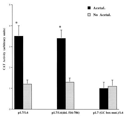
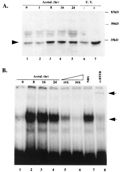
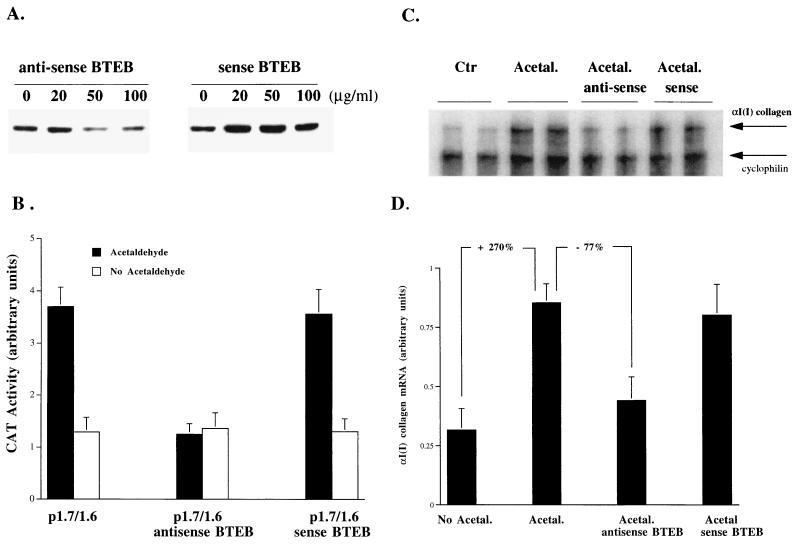
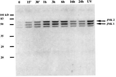
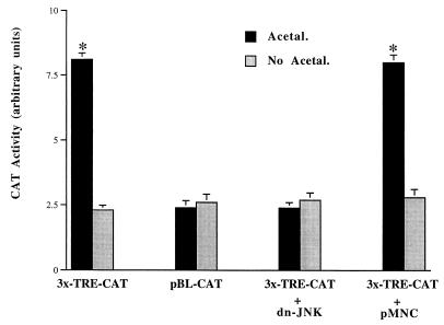
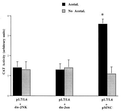
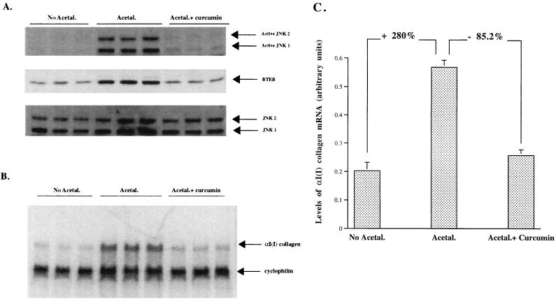
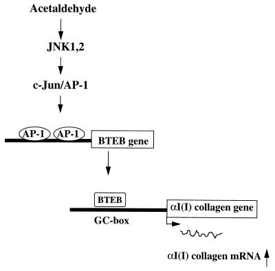
Alcohol-induced cirrhosis results partially from the excessive production of collagen matrix proteins, which, predominantly alphaI(I) collagen, are produced and secreted by activated hepatic stellate cells (HSC). The accumulation of alphaI(I) collagen in HSC during cirrhosis is largely due to an increase in alphaI(I) collagen gene expression. Acetaldehyde, the major active metabolite of alcohol, is known to stimulate alphaI(I) collagen production in HSC. However, the mechanisms responsible for it remain unknown. The aim of this study was to elucidate the mechanisms by which alphaI(I) collagen gene expression is induced by acetaldehyde in rat HSC. In the present study, the acetaldehyde response element was located in a distal GC box, previously described as the UV response element, in the promoter of the alphaI(I) collagen gene (-1484 to -1476). The GC box was predominantly bound by the DNA binding transcription factor BTEB (basic transcription element binding protein), expression of which was acetaldehyde and UV inducible. Blocking BTEB protein expression significantly reduced the steady-state levels of the acetaldehyde-induced alphaI(I) collagen mRNA, suggesting that BTEB is required for this gene expression. Further studies found that acetaldehyde activated Jun N-terminal kinase (JNK) 1 and 2 and activator protein 1 (AP-1) transactivating activity. Inhibition of JNK activation resulted in the reduction of the acetaldehyde-induced BTEB protein abundance and alphaI(I) collagen mRNA levels, indicating that the expression of both genes is JNK dependent in HSC. Taken together, these studies demonstrate that BTEB mediates acetaldehyde-induced, JNK-dependent alphaI(I) collagen gene expression in HSC.


Figures

FIG. 1
FIG. 1
The distal GC box is required for acetaldehyde-induced αI(I) collagen gene transcription. Serum-starved HSC were transfected with 2 μg of one of the collagen CAT reporter plasmids [wild-type plasmid p1.7/1.6, p1.7/1.6(del. 516-786), which contains a deletion of a fragment containing a putative AP-1 binding motif in the first intron, or p1.7 (GC box mut.)/1.6, which contains a site-directed mutation in the distal GC box] as detailed in Materials and Methods. After transfection and recovery, cells were left untreated or treated with acetaldehyde for an additional 36 hr. CAT assays were performed as described in Materials and Methods. The transfection efficiency was normalized by β-galactosidase activity as described in Materials and Methods. Values presented here reflect the means ± standard deviations (n = 6). ∗, P < 0.05 compared with the control (No Acetal.).
FIG. 2
FIG. 2
Acetaldehyde induces BTEB protein abundance and enhances BTEB DNA binding activity. Sixty-eight-percent confluent HSC were preincubated in DMEM with 0.4% serum for 48 h before the acetaldehyde treatment (100 μM) for the indicated times (hours). Nuclear protein extracts were prepared as described in Materials and Methods. A whole-cell extract from HSC treated with UV irradiation (10 J/m2) as previously described (8) was used to study whether BTEB is UV inducible. (A) Twenty micrograms of nuclear extract proteins or 30 μg of whole-cell extracts of each sample were analyzed by Western blotting using a polyclonal anti-BTEB serum. A representative Western blot assay is shown here (repeated three times with similar results). (B) Ten micrograms of nuclear extract proteins from HSC exposed to acetaldehyde for the indicated times were analyzed by EMSA. [32P]-labeled double-stranded oligonucleotides containing a GC box identical to the sequence in the 5′ promoter of the αI(I) collagen gene were used as a probe (see Materials and Methods). The lower arrow indicates the oligonucleotide-BTEB complex. Ten- to 50-fold excesses of the unlabeled double-stranded oligonucleotides were used in the competition assays. Two microliters of the anti-BTEB serum (α-BTEB) or NRS was used in the supershift assays. Incubation with the anti-BTEB serum caused a supershift band, as indicated by the upper arrow, and a significant reduction in the oligo-BTEB complex band. A representative gel is shown here.
FIG. 3
FIG. 3
Inhibition of BTEB by antisense BTEB oligonucleotides significantly reduces acetaldehyde-induced αI(I) collagen mRNA levels. Serum-starved HSC were treated with or without acetaldehyde (100 μM) plus sense or antisense BTEB oligonucleotides at the indicated concentrations for 48 h. The medium was replaced once with fresh DMEM containing acetaldehyde and antisense or sense BTEB oligonucleotides. (A) To determine the optimal concentration of antisense BTEB oligonucleotides, whole-cell protein extracts (30 μg) were analyzed by Western blotting using a polyclonal anti-BTEB serum. (B) Compared to the BTEB sense oligonucleotides, the effectiveness of the BTEB antisense oligonucleotides at 50 μg/ml in blocking acetaldehyde-induced αI(I) collagen gene expression was confirmed in HSC transfected with the αI(I) collagen reporter P1.7/1.6 by CAT assays. Values presented here reflect the means ± standard deviations (n = 6). (C) A representative αI(I) collagen RPA gel is shown. Ten micrograms of total RNA from HSC treated with or without acetaldehyde (100 μM) plus sense or antisense BTEB oligonucleotides at 50 μg/ml were used. Upper arrow, αI(I) collagen mRNA; lower arrow, cyclophilin mRNA, as a control. (D) Quantitation of αI(I) collagen mRNA in an RPA (Fig. 3B) by computer-aided phosphorimaging densitometry. Loading variation was normalized by cyclophilin mRNA. Representative gels are shown.
FIG. 4
FIG. 4
Acetaldehyde induces rapid JNK activation. Whole-cell extracts were prepared from serum-starved HSC exposed to acetaldehyde (100 μM) for the indicated times. A whole-cell extract from HSC treated with UV irradiation (10 J/m2) was used as a positive control for active forms of JNK as previously described (8). Twenty micrograms of proteins of each sample was used for Western blot analysis. Active forms of JNK were probed with a polyclonal antibody specific to the dual phosphorylated and activated JNK1 and JNK2 (Promega), as indicated by the arrows on the right of the gel. A representative gel is shown here.
FIG. 5
FIG. 5
Acetaldehyde induces AP-1 activation via a JNK-dependent pathway. Sixty- to eighty-percent confluent HSC were transfected with either an AP-1 reporter plasmid, 3x-TRE-CAT, or an empty control plasmid, pBL-CAT. The AP-1 reporter plasmid 3x-TRE-CAT contains three AP-1 binding sites upstream of a CAT reporter gene. In cotransfection experiments, HSC were cotransfected with 3x-TRE-CAT and a dominant-negative JNK expression plasmid (dn-JNK) or an empty control plasmid, pMNC. After recovery, the transfected cells were treated with or without acetaldehyde (100 μM) for an additional 36 h in DMEM containing 0.4% fetal bovine serum. Transfection efficiency was normalized by measurement of β-galactosidase activity (see Materials and Methods). Values are expressed as means ± standard deviations (n = 6). ∗, P < 0.05 compared with the control (No Acetal).
FIG. 6
FIG. 6
Acetaldehyde induces αI(I) collagen gene promoter activation by a JNK-dependent mechanism. Serum-starved HSC were cotransfected with the wild-type collagen CAT reporter plasmid p1.7/1.6 and either a dominant-negative JNK expression plasmid (dn-JNK), a dominant-negative c-Jun expression plasmid (dn-Jun), or an empty control plasmid, pMNC. After transfection and recovery, cells were treated with or without acetaldehyde for an additional 36 h as described for Fig. 1 and in Materials and Methods. Cotransfected β-galactosidase activity was used for normalization of transfection efficiency (n ± 6). ∗, P < 0.05 compared with the control (No Acetal.).
FIG. 7
FIG. 7
Inhibition of JNK by curcumin reduced BTEB protein abundance and decreased the endogenous αI(I) collagen mRNA levels. Serum-starved HSC were pretreated with (Acetal.+ curcumin) or without (Acetal.) curcumin at 15 μM for 3 h before being treated with acetaldehyde for 24 h. Cells treated with 0.2% ethyl alcohol (EtOH) only were used as a control (No Acetal.), as curcumin was dissolved in EtOH. Each treatment was performed in triplicate. (A) Nuclear proteins (20 μg/sample) were used for Western blot analyses and detected by anti-ACTIVE JNK pAb, a polyclonal antibody specific to the dual phosphorylated and activated JNK1,2, by anti-BTEB, or by anti-JNK1,2 total proteins as a control for the normalization of loading. (B) Total RNA from the cells (10 μg/lane) was analyzed for the endogenous αI(I) collagen mRNA by RPA. Cyclophilin was used as an internal control to normalize the loading of the total RNA in each lane. (C) The radioactivity in each band in panel B was quantified and normalized by computer-aided phosphorimaging densitometry. Representative gels are shown.
FIG. 8
FIG. 8
Schema of acetaldehyde-induced αI(I) collagen gene expression in HSC. Exposure of HSC to acetaldehyde rapidly activates JNK1,2, though it remains unclear how JNKs are activated. Activated JNKs phosphorylate and activate c-Jun/AP-1, which, in turn, up-regulates BTEB gene expression by binding to the putative AP-1 binding sites in the promoter of BTEB gene. The acetaldehyde-induced BTEB acts as a mediator, binding to the distal GC box in the promoter and stimulating αI(I) collagen gene expression in HSC.

References

    1. Bornstein P, McKay J. The first intron of the alpha 1(I) collagen gene contains several transcriptional regulatory elements. J Biol Chem. 1988;263:1603–1606. - PubMed
    1. Brenner D A, Chojkier M. Acetaldehyde increases collagen gene transcription in cultured human fibroblasts. J Biol Chem. 1987;262:17690–17695. - PubMed
    1. Brenner D A, Rippe R A, Veloz L. Analysis of the collagen alpha 1(I) promoter. Nucleic Acids Res. 1989;17:6055–6064. - PMC - PubMed
    1. Casini A, Cunningham M, Rojkind M, Lieber C S. Acetaldehyde increases procollagen type I and fibronectin gene transcription in cultured rat fat-storing cells through a protein synthesis-dependent mechanism. Hepatology. 1991;13:758–765. - PubMed
    1. Casini A, Galli G, Salzano R, Ceni E, Franceschelli F, Rotella C M, Surrenti C. Acetaldehyde induces c-fos and c-jun proto-oncogenes in fat-storing cell cultures through protein kinase C activation. Alcohol Alcohol. 1994;29:303–314. - PubMed

Publication types

MeSH terms