. 2000 Apr;20(8):2880-9.
doi: 10.1128/MCB.20.8.2880-2889.2000.

Atypical protein kinases Clambda and -zeta associate with the GTP-binding protein Cdc42 and mediate stress fiber loss

Affiliations

Atypical protein kinases Clambda and -zeta associate with the GTP-binding protein Cdc42 and mediate stress fiber loss

M P Coghlan et al. Mol Cell Biol. 2000 Apr.

Abstract

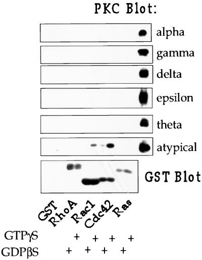
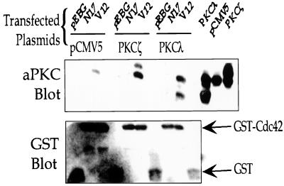
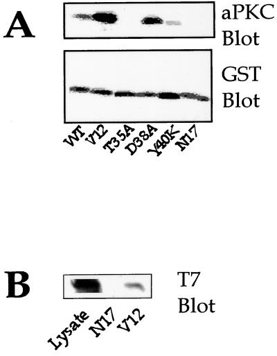
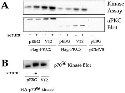
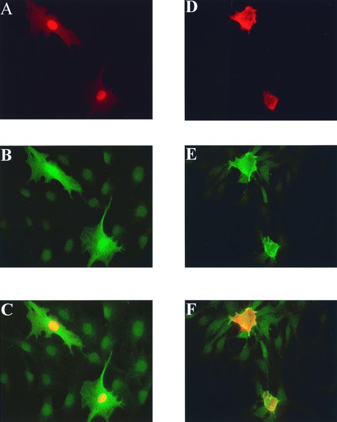
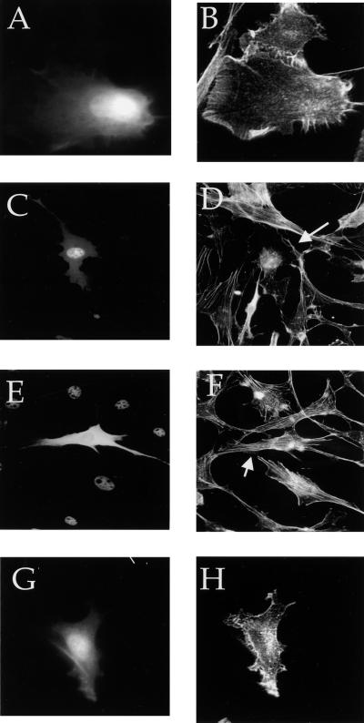
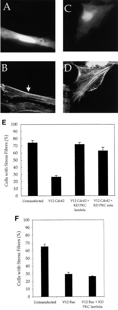
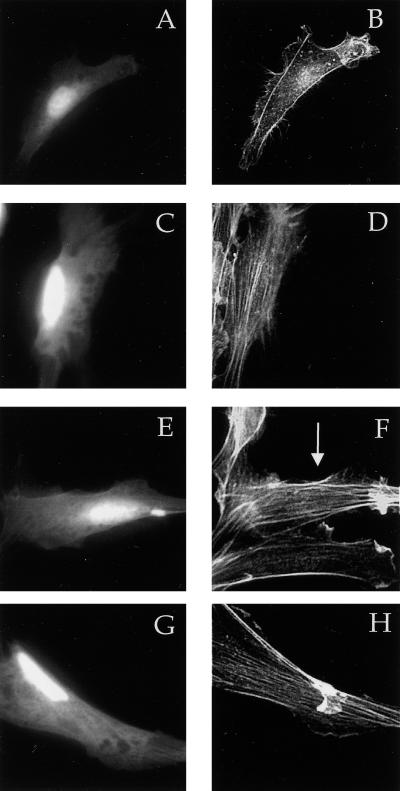
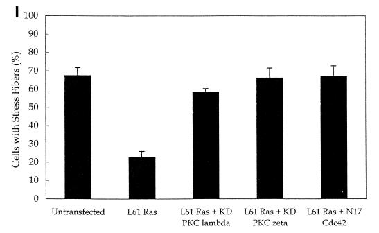
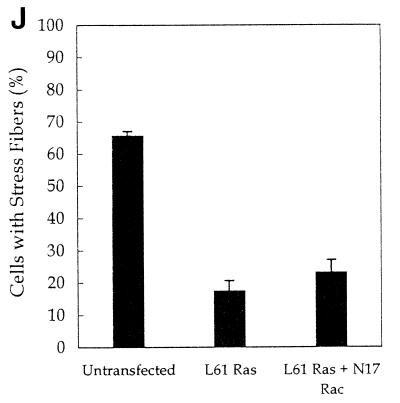
Both the Rho family of low-molecular-weight GTP-binding proteins and protein kinases C (PKCs) mediate responses to a variety of extracellular and intracellular signals. They share many downstream targets, including remodeling of the actin cytoskeleton, activation of p70(S6) kinase and c-jun N-terminal kinase (JNK), and regulation of transcription and cell proliferation. We therefore investigated whether Rho family GTP-binding proteins bind to PKCs. We found that Cdc42 associates with atypical PKCs (aPKCs) PKCzeta and -lambda in a GTP-dependent manner. The regulatory domain of the aPKCs mediates the interaction. Expression of activated Cdc42 results in the translocation of PKClambda from the nucleus into the cytosol, and Cdc42 and PKClambda colocalize at the plasma membrane and in the cytoplasm. Expression of activated Cdc42 leads to a loss of stress fibers, as does overexpression of either the wild type or an activated form of PKClambda. Kinase-dead PKClambda and -zeta constructs acted as dominant negatives and restored stress fibers in cells expressing the activated V12 Cdc42 mutant, indicating that Cdc42-dependent loss of stress fibers requires aPKCs. Kinase-dead PKClambda and -zeta and dominant-negative N17 Cdc42 also blocked Ras-induced loss of stress fibers, suggesting that this pathway may also be important for Ras-dependent cytoskeletal changes. N17 Rac did not block Ras-induced loss of stress fibers, nor did kinase-dead PKClambda block V12 Rac-stimulated loss of stress fibers. These results indicate that Cdc42 and Rac use different pathways to regulate stress fibers.


Figures

FIG. 1
FIG. 1
Cdc42 associates with aPKCs in rat brain cytosol. GST alone or GST fusion proteins of RhoA, Rac1, Cdc42, or Ras, bound to glutathione-Sepharose beads, were loaded with GTPγS or GDPβS and then incubated with rat brain cytosol as described in Materials and Methods. G protein-associated PKCs were detected by Western blotting with PKC isoform-specific antibodies. These antibodies were also used to immunoprecipitate PKC from rat brain cytosol, and these immunoprecipitates are included as positive controls on the right side of each blot. The GST fusion proteins were visualized by Western blotting with a GST antibody.
FIG. 2
FIG. 2
V12 Cdc42 associates with PKCλ and -ζ in vivo. NIH 3T3 cells were transfected with GST-tagged V12 or N17 Cdc42 and either vector alone (pCMV5), PKCζ, or PKCλ, as described in Materials and Methods. Forty-eight hours after transfection, the cells were lysed, and Cdc42 was isolated by incubating the cell lysates with glutathione-Sepharose beads. The beads were washed, and the associated proteins were detected by Western blotting with the pan-aPKC antibody (upper panel). Lysates were blotted to determine PKC expression (three right-hand lanes in the upper panel). The amount of GST or GST-Cdc42 associated with the beads was determined by Western blotting with a GST antibody (lower panel).
FIG. 3
FIG. 3
Characterization of the domains of Cdc42 and aPKC involved in their association. (A) COS cells were transiently transfected with the indicated GST-tagged Cdc42 mutants. Forty-eight hours after transfection, the cells were lysed, and Cdc42 was isolated by incubating the lysates with glutathione-Sepharose beads. Associated endogenous aPKC was detected by Western blotting with the pan-aPKC antibody (upper panel), and Cdc42 bound to the beads was detected by Western blotting with GST antibody (lower panel). (B) 293 cells were transfected with V12 or N17 GST-Cdc42 and the regulatory domain of PKCζ with a T7 epitope tag. Forty-eight hours after transfection, Cdc42 was isolated as described above. Association of the regulatory domain of PKCζ with V12 or N17 GST-Cdc42 was determined by Western blotting with a T7-specific monoclonal antibody. Expression of the regulatory domain was determined by Western blotting of the lysate with T7 antibody.
FIG. 4
FIG. 4
Cdc42 does not activate aPKC. (A) 293 cells were transfected with Flag epitope-tagged PKCζ or -λ and V12 Cdc42 or vector alone (pEBG). Cells were serum starved for 24 h and then stimulated with or without 10% fetal calf serum for 10 min. Lysates were immunoprecipitated with the Flag antibody. Half of the immunoprecipitate was used to assay PKC activity with myelin basic protein as the substrate (upper panel), and the other half was used for Western blotting with the aPKC antibody (lower panel). (B) 293 cells were transfected with hemagglutinin (HA)-p70S6 kinase and V12 Cdc42 or pEBG. Cells were stimulated with serum as described above. p70S6 kinase was immunoprecipitated with HA antibody and visualized by Western blotting with HA antibody.
FIG. 5
FIG. 5
Cdc42 induces translocation of aPKCλ from the nucleus to the cytosol in NIH 3T3 cells. NIH 3T3 cells were transfected with Flag epitope-tagged PKCλ (A to F) and either pEBG vector (A to C) or V12 Cdc42 (panels D to F) as described in Materials and Methods. Fixed cells were stained for PKCλ by using Flag primary antibody and a rhodamine-conjugated secondary antibody (A and D). GST and GST-V12 Cdc42 were stained with GST primary antibody and Cy2-conjugated secondary antibody (B and E). Panels A and B and D and E were merged to give the images shown in panels C and F, respectively.
FIG. 6
FIG. 6
Cdc42 and PKCλ expression in NIH 3T3 fibroblasts reduces the stress fiber content. NIH 3T3 cells were transfected with hemagglutinin (HA)-tagged V12 Cdc42 alone (A and B) or Flag epitope-tagged PKCλ (C and D), T7 epitope-tagged PKCζ (E and F), or Flag epitope-tagged constitutively active PKCλ (G and H) as described in Materials and Methods. The cells were serum starved overnight and then fixed. Actin was stained with rhodamine-conjugated phalloidin (B, D, F, and H). The epitope-tagged aPKCs were stained with the appropriate epitope-directed antibodies followed by fluorescein isothiocyanate-conjugated secondary antibody (C and E), or cells were cotransfected with pEGFP, and the fluorescence of GFP was used to identify the transfected cells (A and G). The arrows indicate the transfected cells.
FIG. 7
FIG. 7
Kinase-dead PKCλ and -ζ block Cdc42-induced loss of stress fibers. NIH 3T3 cells were transfected with 0.25 μg of pEGFP, 0.5 μg of hemagglutinin (HA)-tagged V12 Cdc42 (A to E) or 0.5 μg of myc V12 Rac (F) and 2.5 μg of kinase-dead PKCλ (A, B, and F) or kinase-dead PKCζ (C and D). Twenty-four hours after transfection, the cells were serum starved. Forty-eight hours after transfection, the cells were fixed and stained with rhodamine-phalloidin (B and D). Transfected cells were identified by GFP fluorescence (A and C). The arrows indicate the transfected cells. (E) The numbers of cells with stress fibers were counted in the experiments described above. (F) The numbers of cells with stress fibers were counted in untransfected cells, V12 Rac-expressing cells, and cells expressing V12 Rac and kinase-dead (KD) PKCλ. Data are the means of three to four experiments in which at least 100 cells were counted per experiment. The error bars represent the standard error of the mean.
FIG. 8
FIG. 8
Cdc42, but not Rac, is necessary for Ras-induced loss of stress fibers. NIH 3T3 cells were transfected by using Superfect with 0.25 μg of pEGFP, 0.5 μg of L61 Ras, and either vector only (A and B), 2.5 μg of kinase-dead (KD) PKCλ (C and D), kinase-dead PKCζ (E and F), N17 Cdc42 (G and H), or N17 Rac (J). Twenty-four hours after transfection, the cells were serum starved. Forty-eight hours after transfection, the cells were fixed and stained with rhodamine-phalloidin (B, D, F, and H). Transfected cells were identified by GFP fluorescence (A, C, E, and G). The arrows indicate the transfected cells (I and J). The number of cells with stress fibers was counted in the experiments described above. Data are the means of three to four experiments in which at least 100 cells were counted per experiment. The error bars represent the standard error of the mean.
FIG. 8
FIG. 8
Cdc42, but not Rac, is necessary for Ras-induced loss of stress fibers. NIH 3T3 cells were transfected by using Superfect with 0.25 μg of pEGFP, 0.5 μg of L61 Ras, and either vector only (A and B), 2.5 μg of kinase-dead (KD) PKCλ (C and D), kinase-dead PKCζ (E and F), N17 Cdc42 (G and H), or N17 Rac (J). Twenty-four hours after transfection, the cells were serum starved. Forty-eight hours after transfection, the cells were fixed and stained with rhodamine-phalloidin (B, D, F, and H). Transfected cells were identified by GFP fluorescence (A, C, E, and G). The arrows indicate the transfected cells (I and J). The number of cells with stress fibers was counted in the experiments described above. Data are the means of three to four experiments in which at least 100 cells were counted per experiment. The error bars represent the standard error of the mean.

References

    1. Akimoto K, Nakaya M, Yamanaka T, Tanaka J, Matsuda S, Weng Q P, Avruch J, Ohno S. Atypical protein kinase Clambda binds and regulates p70 S6 kinase. Biochem J. 1998;335:417–424. - PMC - PubMed
    1. Akimoto K, Takahashi R, Moriya S, Nishioka N, Takayanagi J, Kimura K, Fukui Y, Osada S, Mizuno K, Hirai S, Kazlauskas A, Ohno S. EGF or PDGF receptors activate atypical PKClambda through phosphatidylinositol 3-kinase. EMBO J. 1996;15:788–798. - PMC - PubMed
    1. Arber S, Barbayannis F A, Hanser H, Schneider C, Stanyon C A, Bernard O, Caroni P. Regulation of actin dynamics through phosphorylation of cofilin by LIM-kinase. Nature. 1998;393:805–809. - PubMed
    1. Behre G, Whitmarsh A J, Coghlan M P, Hoang T, Carpenter C L, Zhang D E, Davis R J, Tenen D G. c-Jun is a JNK-independent coactivator of the PU.1 transcription factor. J Biol Chem. 1999;274:4939–4946. - PubMed
    1. Bjorkoy G, Perander M, Overvatn A, Johansen T. Reversion of Ras- and phosphatidylcholine-hydrolyzing phospholipase C-mediated transformation of NIH 3T3 cells by a dominant interfering mutant of protein kinase C lambda is accompanied by the loss of constitutive nuclear mitogen-activated protein kinase/extracellular signal-regulated kinase activity. J Biol Chem. 1997;272:11557–11565. - PubMed

Publication types