. 2000 Apr;20(8):2902-6.
doi: 10.1128/MCB.20.8.2902-2906.2000.

Mesothelin is not required for normal mouse development or reproduction

Affiliations

Mesothelin is not required for normal mouse development or reproduction

T K Bera et al. Mol Cell Biol. 2000 Apr.

Abstract

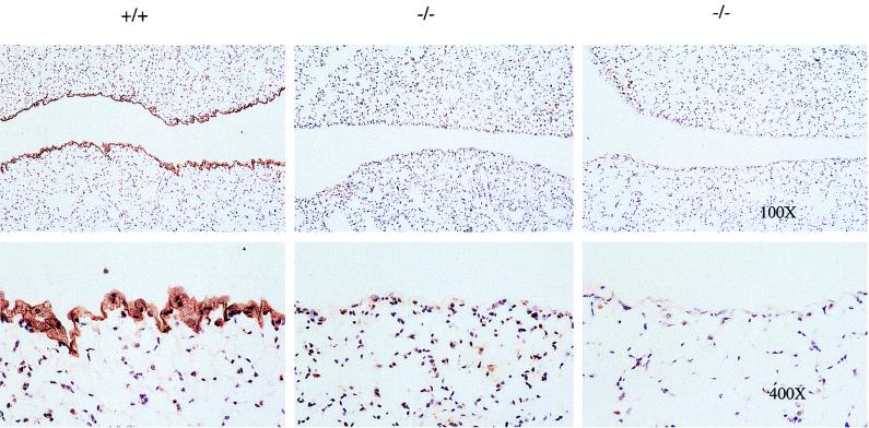
Mesothelin is a glycosylphosphatidylinositol-linked glycoprotein highly expressed in mesothelial cells, mesotheliomas, and ovarian cancer, but the biological function(s) of the protein is not known. We have analyzed the expression of the mouse mesothelin gene in different developmental stages and in various adult tissues by Northern hybridization. The 2.5-kb mesothelin transcript was detected in the mRNA of E 7.0, E 15.0, and E 17.0 stages of mouse development. In adult tissues the mesothelin gene was expressed in lung, heart, spleen, liver, kidney, and testis. To directly assess the function of the mesothelin in vivo, we generated mutant mice in which the mesothelin gene was inactivated by replacing it with the neomycin resistance gene. In homozygous mutant mice neither mesothelin mRNA nor the protein product was detected. Null mutant mice were obtained in accordance with Mendelian laws, and both males and females produced offspring normally. No anatomical or histological abnormalities were detected in any tissues where mesothelin was reportedly expressed in wild-type mice. Our results demonstrate that mesothelin function is not essential for growth or reproduction in mice.


Figures

FIG. 1
FIG. 1
Tissue- and development-specific expression of mesothelin transcripts in mice. Shown are the results of Northern blot analysis of mesothelin expression in developmental stages (A) and eight adult tissues (B). The filters were obtained from Clontech and contained 2 μg of poly(A)+ RNA in each lane. Beta actin, blots hybridized with the β-actin probe; Sk muscle, skeletal muscle.
FIG. 2
FIG. 2
Targeted disruption of the mesothelin gene. (A) Strategy used for mesothelin gene targeting. Construction of the targeting vector, organization of the m-mesothelin gene, and the structure of the targeted genome are shown. Restriction sites are indicated as follows: Bm, BamHI; E, EcoRI; Ev, EcoRV; H, HindIII, N, NotI; S, SalI; Xb, XbaI; Xh, XhoI. The DNA fragments used as 5′ and 3′ probes are indicated. WT, wild type; TK, thymidine kinase. (B) Southern blot analysis of BamHI-digested genomic DNA from representative pups. +/+, wild type; +/−, heterozygote; −/−, homozygote.
FIG. 2
FIG. 2
Targeted disruption of the mesothelin gene. (A) Strategy used for mesothelin gene targeting. Construction of the targeting vector, organization of the m-mesothelin gene, and the structure of the targeted genome are shown. Restriction sites are indicated as follows: Bm, BamHI; E, EcoRI; Ev, EcoRV; H, HindIII, N, NotI; S, SalI; Xb, XbaI; Xh, XhoI. The DNA fragments used as 5′ and 3′ probes are indicated. WT, wild type; TK, thymidine kinase. (B) Southern blot analysis of BamHI-digested genomic DNA from representative pups. +/+, wild type; +/−, heterozygote; −/−, homozygote.
FIG. 3
FIG. 3
Expression of mesothelin transcript in mutant null mice. (A) Northern blot analysis of the mesothelin transcript. Total RNA was prepared from lung tissues of wild-type (+/+) and mutant (−/−) mice. RNA (20 μg from each) was separated on a 1.2% agarose formaldehyde gel and hybridized with a mouse cDNA mesothelin probe covering most of the mesothelin coding sequence (top). Bottom, methylene blue staining of total RNA transferred onto the nylon membrane shown at the top prior to hybridization. (B) RT-PCR analysis of the mesothelin transcript. RNAs from wild type and mutant mice were reverse transcribed, amplified by PCR using the primer pair T75 and T76 (see Materials and Methods), and analyzed in 2% agarose gel.
FIG. 3
FIG. 3
Expression of mesothelin transcript in mutant null mice. (A) Northern blot analysis of the mesothelin transcript. Total RNA was prepared from lung tissues of wild-type (+/+) and mutant (−/−) mice. RNA (20 μg from each) was separated on a 1.2% agarose formaldehyde gel and hybridized with a mouse cDNA mesothelin probe covering most of the mesothelin coding sequence (top). Bottom, methylene blue staining of total RNA transferred onto the nylon membrane shown at the top prior to hybridization. (B) RT-PCR analysis of the mesothelin transcript. RNAs from wild type and mutant mice were reverse transcribed, amplified by PCR using the primer pair T75 and T76 (see Materials and Methods), and analyzed in 2% agarose gel.
FIG. 4
FIG. 4
Immunohistochemical detection of mesothelin in lung tissue sections from wild-type (+/+) and mutant (−/−) adult mice with antimesothelin antibodies. Tissues were fixed and embedded in paraffin, and serial sections 5 to 6 μm in width were cut. The sections were stained with rabbit polyclonal antibodies to m-mesothelin. Unlike what was observed in mesothelial cells in wild-type lung tissue, no staining was observed in mutant tissue. Magnifications are indicated.
FIG. 5
FIG. 5
Histological analysis of selected tissues from wild-type and mutant mice. Tissues were fixed in 4% paraformaldehyde, sectioned, and stained with hematoxylin and eosin. Magnification, ×100.

References

    1. Brinkmann U, Brinkmann E, Bera T K, Wellmann A, Pastan I. Tissue specific alternatively spliced variants of hCSE1/CAS may be regulators of nuclear transporter of tissue specific proteins. Genomics. 1999;58:41–49. - PubMed
    1. Chang K, Pai L H, Pass H, Pogrebniak H W, Tsao M S, Pastan I, Willingham M C. Monoclonal antibody K1 reacts with epithelial mesothelioma but not with lung adenocarcinoma. Am J Surg Pathol. 1992;16:259–268. - PubMed
    1. Chang K, Pastan I. Molecular cloning of mesothelin, a differentiation antigen present on mesothelium, mesotheliomas, and ovarian cancers. Proc Natl Acad Sci USA. 1996;93:136–140. - PMC - PubMed
    1. Dustin M L, Selvara P, Mattaliano R J, Springer T A. Anchoring mechanisms for lfa-3 cell-adhesion glycoprotein at membrane-surface. Nature. 1987;329:846–848. - PubMed
    1. Kojima T, Oheda M, Hattori K, Taniguchi Y, Tamura M, Ochi N, Yamaguchi N. Molecular-cloning and expression of megakaryocyte potentiating factor cDNA. J Biol Chem. 1995;270:21984–21990. - PubMed