. 2000 Apr;20(8):2926-32.
doi: 10.1128/MCB.20.8.2926-2932.2000.

Functionally significant secondary structure of the simian virus 40 late polyadenylation signal

Affiliations

Functionally significant secondary structure of the simian virus 40 late polyadenylation signal

H Hans et al. Mol Cell Biol. 2000 Apr.

Abstract

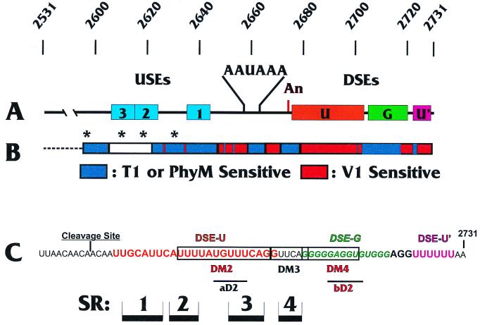
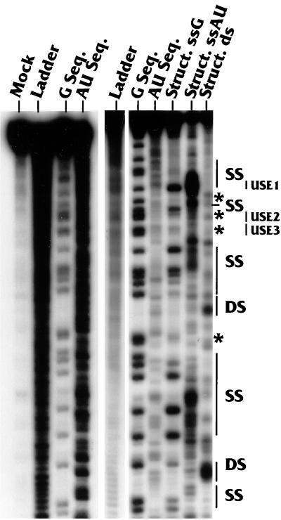
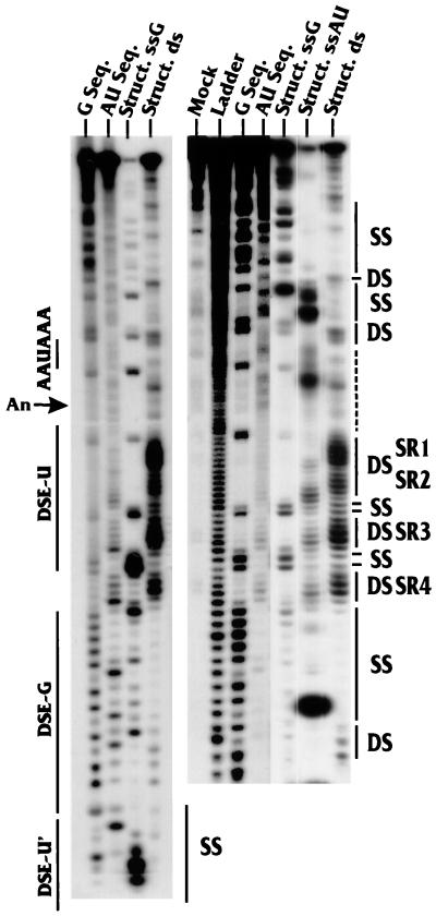
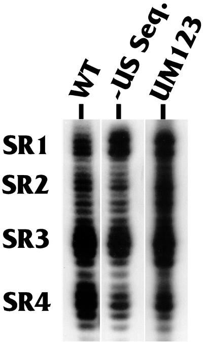
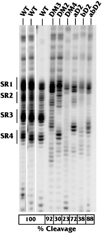
The structure of the highly efficient simian virus 40 late polyadenylation signal (LPA signal) is more complex than those of most known mammalian polyadenylation signals. It contains efficiency elements both upstream and downstream of the AAUAAA region, and the downstream region contains three defined elements (two U-rich elements and one G-rich element) instead of the single U- or GU-rich element found in most polyadenylation signals. Since many reports have indicated that the secondary structure in RNA may play a significant role in RNA processing, we have used nuclease structure analysis techniques to determine the secondary structure of the LPA signal. We find that the LPA signal has a functionally significant secondary structure. Much of the region upstream of AAUAAA is sensitive to single-strand-specific nucleases. The region downstream of AAUAAA has both double- and single-stranded characteristics. Both U-rich elements are predominately sensitive to the double-strand-specific nuclease RNase V(1), while the G-rich element is primarily single stranded. The U-rich element closest to AAUAAA contains four distinct RNase V(1)-sensitive regions, which we have designated structural region 1 (SR1), SR2, SR3, and SR4. Linker scanning mutants in the downstream region were analyzed both for structure and for function by in vitro cleavage analyses. These data show that the ability of the downstream region, particularly SR3, to form double-stranded structures correlates with efficient in vitro cleavage. We discuss the possibility that secondary structure downstream of the AAUAAA may be important for the functions of polyadenylation signals in general.


Figures

FIG. 1
FIG. 1
SV40 LPA signal. (A) Features of the SV40 LPA signal. AAUAAA and the cleavage site (An) are shown along with the three USEs (USE1 to -3; blue boxes) and the three DSEs (DSE-U [orange box], a predominantly U-rich element just downstream of the cleavage site; DSE-G [green box], a G-rich region downstream of DSE-U; and DSE-U′ [purple box], a second U-rich element further downstream). The numbering above the diagram indicates the SV40 nucleotide numbering. (B) Summary of nuclease structure analyses of the LPA signal. Regions sensitive to the double-strand-specific RNase V1 are indicated in red, and regions sensitive to the single-strand-specific RNase T1 or PhyM are indicated in blue. The white box and asterisks indicate regions which appeared to be inaccessible to the nucleases in the structural analyses (see the text). Note that the diagram does not continue as far upstream as does the structural analysis shown in Fig. 2. (C) Sequence of the region downstream of the cleavage site. The approximate regions of the three DSEs are shown; DSE-U is in orange letters, DSE-G is in green italic letters, and DSE-U′ is in purple letters. Regions of the double-stranded structures SR1 to SR4 identified in this study are shown as black bars. The locations of the various linker substitution mutations used in these studies are indicated (see the text).
FIG. 2
FIG. 2
RNase sequencing and structure analyses of the region upstream of AAUAAA. Sequencing and structure analysis reactions were carried out with a substrate in which the wild-type LPA signal was labeled at its 5′ end with 32P as described in Materials and Methods. The labeling of the lanes indicates the nucleotide(s) (G or AU) being analyzed and whether the analysis is for sequence (Seq.) or structure (Struct.). The lane marked Struct. ds reflects results of the structural analysis using RNase V1, which is specific for double-stranded RNA with no nucleotide preference. The lane marked Ladder contains the oligonucleotide ladder generated by alkaline hydrolysis of the substrate RNA. The panel on the left shows the mock-digested sample (lane Mock), an additional hydrolysis ladder, and the results of repeated sequencing analyses, which provided for better analysis of the structural data. The positions of single-stranded (SS) and double-stranded (DS) regions are indicated on the right as well as the positions of the three USEs, USE1, USE2, and USE3. The asterisks indicate G nucleotides which were not well cleaved by either RNase T1 or V1 in the structure analyses (see the text).
FIG. 3
FIG. 3
RNase sequencing and structure analyses of the region downstream of AAUAAA. Sequencing and structure analysis reactions were carried out with an RNA substrate in which the wild-type LPA signal was labeled at its 3′ end with 32P as described in Materials and Methods. The lanes indicate the nucleotide(s) (G or AU) being analyzed and whether the analysis is for sequence (Seq.) or structure (Struct.). The lane marked Struct. ds shows the results of the structural analysis using RNase V1, which is specific for double-stranded RNA with no nucleotide preference. The lane marked Ladder contains the oligonucleotide ladder generated by alkaline hydrolysis of the substrate RNA. The lane marked Mock contains the mock-digested sample. The panel on the left provides additional sequence and structural analyses to show reproducibility. The locations of AAUAAA, the cleavage site (An), DSE-U, DSE-G, and part of DSE-U′ are indicated at the left of the panels. The positions of single-stranded (SS) and double-stranded (DS) regions as well as the positions of the four prominent double-stranded regions, SR1 to SR4 (see the text), are indicated at the right of the panels. It should be noted that the sample used in the Struct. ssAU lane was overly digested with RNase PhyM, which resulted in low levels of cleavage at A's and U's in the double-stranded regions SR1 to SR4.
FIG. 4
FIG. 4
Integrity of SR1 to SR4 during mutation of the upstream region. The RNase V1 sensitivity of the prominent downstream double-stranded regions SR1 to SR4 was analyzed using substrates in which the wild-type LPA signal RNA was labeled at its 3′ end with 32P (lane WT) and two mutants in which (i) the entire region upstream of AAUAAA was deleted (lane −US Seq.) or (ii) the three USEs were mutated by specific linker substitution mutagenesis (lane UM123).
FIG. 5
FIG. 5
Correlation of downstream structure with in vitro cleavage efficiency. The RNase V1 sensitivity of the downstream region, particularly SR1 to SR4, was analyzed using 32P-3′-end-labeled substrates representing the wild-type LPA signal (WT) and the LPA signal containing linker substitution mutants in the downstream region (DM2, DM3, DM4, aD2, bD2, and abD2). The locations of these mutations in the LPA signal are shown in Fig. 1C. The results of three WT structure analyses are provided to demonstrate reproducibility. The boxes at the bottom show the percentage of cleavage by each of the substrates as measured in an in vitro cleavage reaction by using a HeLa cell extract (see Materials and Methods). The wild-type cleavage activity is set at 100%, and the standard error of the analyses is ±5%.

Similar articles

Cited by

References

    1. Ahmed Y F, Gilmartin G M, Hanly S M, Nevins J R, Greene W C. The HTLV-I rex response element mediates a novel form of mRNA polyadenylation. Cell. 1991;64:727–737. - PubMed
    1. Bagga P S, Arhin G K, Wilusz J. DSEF-1 is a member of the hnRNP H family of RNA-binding proteins and stimulates pre-mRNA cleavage and polyadenylation in vitro. Nucleic Acids Res. 1998;26:5343–5350. - PMC - PubMed
    1. Bagga P S, Ford L P, Chen F, Wilusz J. The G-rich auxiliary downstream element has distinct sequence and position requirements and mediates efficient 3′ end pre-mRNA processing through a trans-factor. Nucleic Acids Res. 1995;23:1625–1631. - PMC - PubMed
    1. Bar-Shira A, Panet A, Honigman A. An RNA secondary structure juxtaposes two remote genetic signals for human T-cell leukemia virus type I RNA 3′-end processing. J Virol. 1991;65:5165–5173. - PMC - PubMed
    1. Bhat B M, Wold W S M. ATTAAA as well as downstream sequences are required for RNA 3′-end formation in the E3 complex transcription unit of adenovirus. Mol Cell Biol. 1985;5:3183–3193. - PMC - PubMed

Publication types

MeSH terms

LinkOut - more resources