. 2000 Apr;20(8):2933-40.
doi: 10.1128/MCB.20.8.2933-2940.2000.

Protein kinase C-theta participates in NF-kappaB activation induced by CD3-CD28 costimulation through selective activation of IkappaB kinase beta

Affiliations

Protein kinase C-theta participates in NF-kappaB activation induced by CD3-CD28 costimulation through selective activation of IkappaB kinase beta

X Lin et al. Mol Cell Biol. 2000 Apr.

Abstract

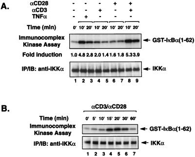
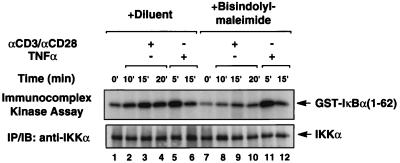
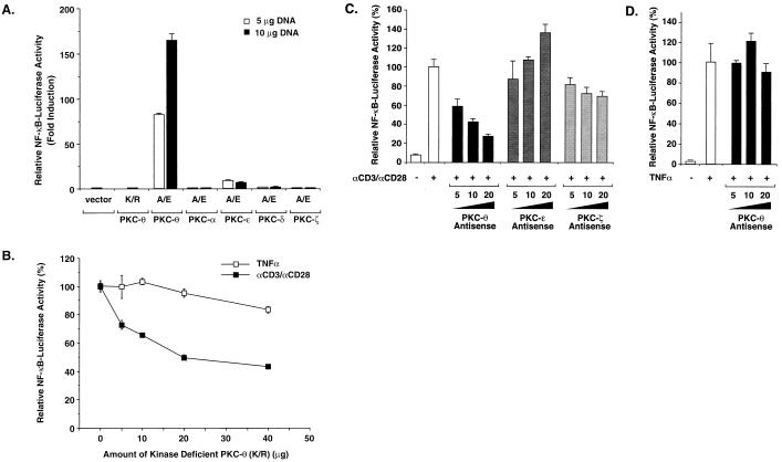
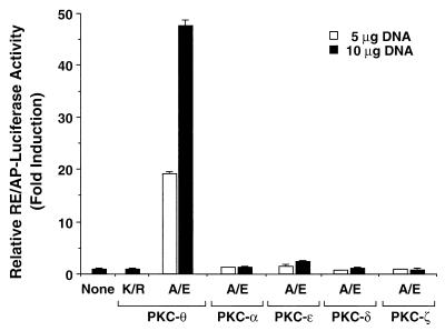
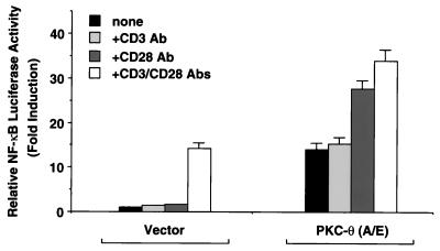
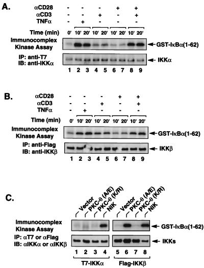
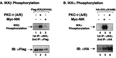
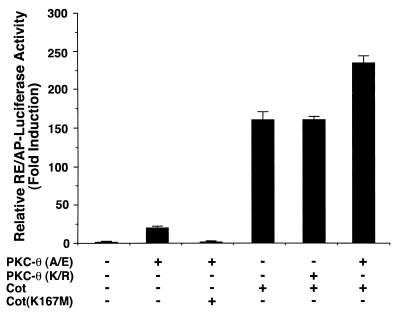
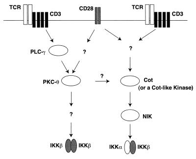
The NF-kappaB/Rel family of eukaryotic transcription factors plays an essential role in the regulation of inflammatory, antiapoptotic, and immune responses. NF-kappaB is activated by many stimuli including costimulation of T cells with ligands specific for the T-cell receptor (TCR)-CD3 complex and CD28 receptors. However, the signaling intermediates that transduce these costimulatory signals from the TCR-CD3 and CD28 surface receptors leading to nuclear NF-kappaB expression are not well defined. We now show that protein kinase C-theta (PKC-theta), a novel PKC isoform, plays a central role in a signaling pathway induced by CD3-CD28 costimulation leading to activation of NF-kappaB in Jurkat T cells. We find that expression of a constitutively active mutant of PKC-theta potently induces NF-kappaB activation and stimulates the RE/AP composite enhancer from the interleukin-2 gene. Conversely, expression of a kinase-deficient mutant or antisense PKC-theta selectively inhibits CD3-CD28 costimulation, but not tumor necrosis factor alpha-induced activation of NF-kappaB in Jurkat T cells. The induction of NF-kappaB by PKC-theta is mediated through the activation of IkappaB kinase beta (IKKbeta) in the absence of detectable IKKalpha stimulation. PKC-theta acts directly or indirectly to stimulate phosphorylation of IKKbeta, leading to activation of this enzyme. Together, these results implicate PKC-theta in one pathway of CD3-CD28 costimulation leading to NF-kappaB activation that is apparently distinct from that involving Cot and NF-kappaB-inducing kinase (NIK). PKC-theta activation of NF-kappaB is mediated through the selective induction of IKKbeta, while the Cot- and NIK-dependent pathway involves induction of both IKKalpha and IKKbeta.


Figures

FIG. 1
FIG. 1
CD3-CD28 transiently activates endogenous IKK signalsome complexes. (A) Jurkat T cells (∼2 × 10⁶) were stimulated with antibodies specific for CD3 (2 μg/ml) or CD28 (2 μg/ml) alone or in combination as indicated. Endogenous IKK complexes were immunoprecipitated (IP) using anti-IKKα antibodies, and then in vitro kinase reactions were performed with the immunoprecipitates using GST–IκBα(1-62) as an exogenously added substrate. The kinase reaction products were analyzed by SDS-PAGE, transferred to a nitrocellulose membrane, and subjected to autoradiography. The level of IKKα present in each immunoprecipitate is shown in the lower blot. (B) IKK enzymatic activity was assessed as described for panel A over a longer time course. IB, immunoblot.
FIG. 2
FIG. 2
A PKC inhibitor blocks IKK activation induced by CD3-CD28 costimulation. Jurkat T cells (∼2 × 10⁶) were incubated in medium with or without bisindolylmaleimide (12 μM) for 30 min and then stimulated with a combination of antibodies specific for CD3 (2 μg/ml) and CD28 (2 μg/ml) or TNF-α (10 ng/ml). The enzymatic activity of the endogenous IKK complexes was assessed using phosphorylated GST–IκBα(1-62) as described for Fig. 1. The kinase reactions are shown in the upper panel. The lower panel shows the level of immunoprecipitated endogenous IKKα in each kinase reaction mixture.
FIG. 3
FIG. 3
PKC-θ plays a role in NF-κB induction following CD3-CD28 costimulation. (A) Jurkat T cells (∼2 × 10⁷) were transfected with 5 or 10 μg of plasmids encoding either a constitutively active form (A/E) of PKC-θ, -α, -ɛ, -δ, and -ζ or a kinase-deficient form (K/R) of PKC-θ together with 10 μg of 5× κB luciferase and 5 μg of β-actin–β-galactosidase reporter plasmids. Cell lysates were prepared from the cultures 20 h after transfection, and luciferase activities were determined. (B to D) Jurkat T cells were transfected with 10 μg of 5× κB luciferase and 5 μg of β-actin–β-galactosidase reporter plasmids together with 0, 5, 10, 20, or 40 μg of PKC-θ (K/R) (B) or antisense expression vectors of PKC-θ, -ɛ, or -ζ (C and D). The total DNA concentration was held constant by supplementation with the parental vector DNA. Twenty hours after the transfection, the cells were stimulated with a combination of antibodies specific for CD3 (2 μg/ml) and CD28 (2 μg/ml) or TNF-α (10 ng/ml) for 5 h. Cell lysates were prepared from the cultures, and luciferase activities were determined. The β-galactosidase activities in these lysates were determined and used to normalize for differences in transfection efficiency. The standard deviations were derived from independent transfections performed in triplicate.
FIG. 4
FIG. 4
PKC-θ activates the CD28 response element of the IL-2 promoter. Jurkat T cells (∼2 × 10⁷) were transfected with 5 μg of plasmids encoding a constitutively active form (A/E) or a kinase-deficient form (K/R) of PKC-θ, -α, -ɛ, -δ, and -ζ together with 10 μg of a luciferase reporter plasmid containing a composite enhancer motif from the human IL-2 gene corresponding to the CD28RE and AP-1 sites (RE/AP-Luciferase) and 5 μg of β-actin–β-galactosidase reporter plasmids. Cell lysates were prepared 20 h after transfection and assayed for luciferase activity. The β-galactosidase activities in these lysates were determined and used to normalize for differences between transfection efficiencies in the various cultures. The standard deviations were derived from independent triplicate transfections.
FIG. 5
FIG. 5
CD28 stimulation enhances PKC-θ-induced NF-κB activation. Jurkat T cells (∼2 × 10⁷) were transfected with 2.5 μg of plasmids encoding a constitutively active form (A/E) of PKC-θ together with 10 μg of 5 × κB luciferase and 5 μg of β-actin–β-galactosidase reporter plasmids or with the luciferase and reporter plasmids alone. About 15 h after the transfection, the cells were stimulated for 5 h with antibodies specific for CD3 (2 μg/ml) and CD28 (2 μg/ml), either alone or in combination as indicated. Cell lysates were then prepared from the cultures, and luciferase activities were determined. The β-galactosidase activities in these lysates were assayed and used to normalize for differences in transfection efficiency. The standard deviations were derived from independent transfections performed in triplicate.
FIG. 6
FIG. 6
PKC-θ activates IKKβ but not IKKα. Jurkat T cells (∼2 × 10⁷) were transfected with 5 μg of IKKα-T7 (A) or 2 μg of IKKβ-Flag (B). Twenty hours after transfection, the cultures were stimulated with antibodies specific for CD3 (2 μg/ml) and CD28 (2 μg/ml), either alone or in combination as indicated. The transfected IKKα-T7 and IKKβ-Flag were then immunoprecipitated (IP) with anti-T7 and anti-Flag antibody-coupled agarose, respectively. The immunoprecipitates were then subjected to an in vitro kinase reaction using GST–IκBα(1-62) as an exogenously added substrate. The kinase reaction products were then analyzed by SDS-PAGE and transferred to a nitrocellulose membrane, and the resultant autoradiographs are shown in the upper portion of each panel. The phosphorylated GST–IκBα(1-62) is indicated on the right. The lower portion of each panel shows the levels of immunoprecipitated IKKα-T7 or IKKβ-Flag present in each kinase reaction mixture. (C) Jurkat T cells (∼2 × 10⁷) were transfected with 5 μg of IKKα-T7 or 2 μg of IKKβ-Flag plasmids in the presence or absence of plasmids encoding PKC-θ (A/E), PKC-θ (K/R), or NIK. Twenty hours after transfection, the transfected IKKα-T7 and IKKβ-Flag proteins were immunoprecipitated using anti-T7 and anti-Flag antibody-conjugated agarose, respectively, and then subjected to an in vitro kinase reaction as described for panels A and B. The resultant autoradiographs are shown in the upper portion of the panel. The phosphorylated GST–IκB(1-62) is indicated on the right. The lower portion of the panel shows the level of immunoprecipitated IKKα-T7 or IKKβ-Flag present in each kinase reaction mixture. IB, immunoblot.
FIG. 7
FIG. 7
PKC-θ selectively induced IKKβ phosphorylation but not IKKα phosphorylation. Jurkat T cells (∼2 × 10⁷) were transfected with kinase-deficient mutants of IKKα(K44M)-HA or IKKβ(K44A)-Flag in the presence or absence of a constitutively active form (A/E) of PKC-θ or NIK. After 20 h, cell lysates were prepared from the cultures. Physiological signalsome complexes containing these IKKs were immunoprecipitated (IP) using anti-IKKγ antibodies. The immunoprecipitates were subjected to an in vitro kinase assay. The IKK complexes in the kinase reaction mixtures were then dissociated in a buffer containing a high concentration of SDS (10%) by boiling for 15 min. IKKα(K44M)-HA and IKKβ(K44A)-Flag in the heat-dissociated samples were then reimmunoprecipitated after dilution of the SDS using anti-HA and anti-Flag antibody-conjugated agarose, respectively. The immunoprecipitates were then analyzed by SDS-PAGE and transferred to a nitrocellulose membrane, and the autoradiographs are shown in the upper portions of the panels. The lower portions show the level of immunoprecipitated IKKα(K44M)-HA or IKKβ(K44A)-Flag proteins present in each kinase reaction mixture. IB, immunoblot.
FIG. 8
FIG. 8
PKC-θ functions upstream or in a parallel pathway to Cot. Jurkat T cells (∼2 × 10⁷) were transfected with 5 μg of plasmids encoding a constitutively active form (A/E) of PKC-θ or Cot with 10 μg of a luciferase reporter plasmid containing a composite enhancer motif from the human IL-2 gene corresponding to the CD28RE and AP-1 sites (RE/AP-Luciferase) and 5 μg of β-actin–β-galactosidase reporter plasmids. In some samples, 10 μg of plasmids encoding the kinase-deficient form (K/R) of PKC-θ or the kinase-deficient form (K167M) of Cot was added. Cell lysates were prepared 20 h after transfection and assayed for luciferase activity. The β-galactosidase activities in these lysates were determined and used to normalize for differences in transfection efficiency in the various cultures. The standard deviations were derived from independent triplicate transfections.
FIG. 9
FIG. 9
A working model for IKK activation induced by CD3-CD28 costimulation. CD3-CD28 costimulation induces at least two parallel pathways leading to activation of NF-κB. One pathway involves the activation of signalsomes containing IKKα-IKKβ heterodimers through NIK and Cot (or a Cot-like kinase). The other pathway leads to activation of IKKβ-IKKβ homodimers through the induction of PKC-θ. PLC-γ, phospholipase C-γ.

References

    1. Baeuerle P A, Baltimore D. NF-κB: ten years after. Cell. 1996;87:13–20. - PubMed
    1. Baier G, Telford D, Giampa L, Coggeshall K M, Baier-Bitterlich G, Isakov N, Altman A. Molecular cloning and characterization of PKC-θ, a novel member of the protein kinase C (PKC) gene family expressed predominantly in hematopoietic cells. J Biol Chem. 1993;268:4997–5004. - PubMed
    1. Baier-Bitterlich G, Überall F, Bauer B, Fresser F, Wachter H, Grunicke H, Utermann G, Altman A, Baier G. Protein kinase C-θ isoenzyme selective stimulation of the transcription factor complex AP-1 in T lymphocytes. Mol Cell Biol. 1996;16:1842–1850. - PMC - PubMed
    1. Baldwin A J. The NF-κB and IκB proteins: new discoveries and insights. Annu Rev Immunol. 1996;14:649–683. - PubMed
    1. Brockman J A, Scherer D C, McKinsey T A, Hall S M, Qi X, Lee W Y, Ballard D W. Coupling of a signal response domain in IκBα to multiple pathways for NF-κB activation. Mol Cell Biol. 1995;15:2809–2818. - PMC - PubMed

Publication types

LinkOut - more resources