

Mass spectrometry of single-stranded restriction fragments captured by an undigested complementary sequence
- PMID: 10734208
- PMCID: PMC102835
- DOI: 10.1093/nar/28.8.e31
Mass spectrometry of single-stranded restriction fragments captured by an undigested complementary sequence
Abstract
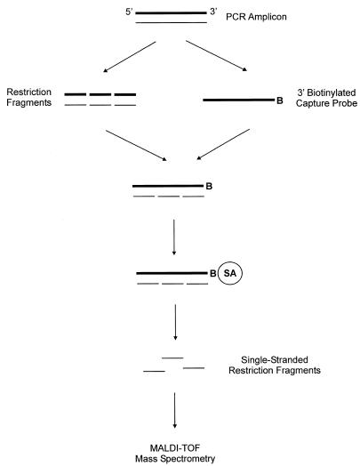
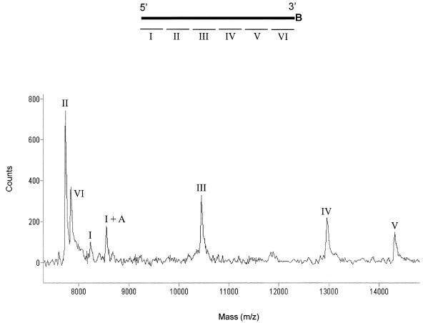
In this report, we describe a simple and accurate method to analyze restriction fragments using matrix-assisted laser desorption/ionization time-of-flight mass spectrometry. The two complementary strands of restriction fragments are separated through hybridization to a capture probe, which is a single-stranded undigested fragment. Using the biotin-streptavidin linkage, the hybrid is immobilized on streptavidin-coated magnetic beads. After conditioning the captured restriction fragments, they are eluted from the probe and their molecular weights are determined. The proposed method greatly improves the quality, and reduces the complexity of the mass spectrum by analyzing only one of the complementary strands of restriction fragments.
Figures
References
-
- Crain P.F. and McCloskey,J.A. (1998) Curr. Opin. Biotechnol., 9, 25–34. - PubMed
-
- Graber J.H., O’Donnell,M.J., Smith,C.L. and Cantor,C.R. (1998) Curr. Opin. Biotechnol., 9, 14–18. - PubMed
-
- Nordhoff E., Kirpekar,F. and Roepstorff, P. (1996) Mass Spectrom. Rev., 15, 67–138. - PubMed
-
- Koster H., Tang,K., Fu,D.J., Braun,A., Boom,D.V.D., Smith,C.L., Cotter,R.J. and Cantor,C.R. (1996) Nat. Biotechnol., 14, 1123–1128. - PubMed
-
- Ross P., Hall,L., Smirnov,I., and Haff,L. (1998) Nat. Biotechnol., 16, 1347–1351. - PubMed
MeSH terms
Substances
LinkOut - more resources
Full Text Sources
Other Literature Sources
