. 2000 Mar 28;97(7):3022-7.
doi: 10.1073/pnas.97.7.3022.

Crystal structure of the functional domain of the splicing factor Prp18

Affiliations

Crystal structure of the functional domain of the splicing factor Prp18

J Jiang et al. Proc Natl Acad Sci U S A. .

Abstract

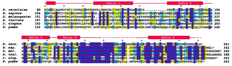
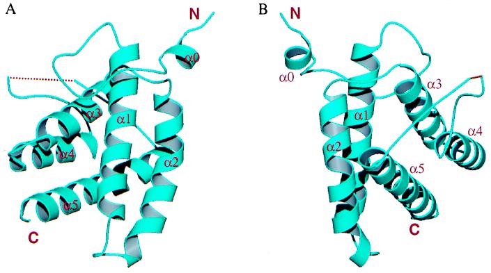
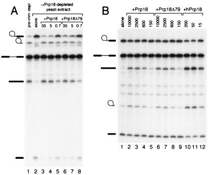
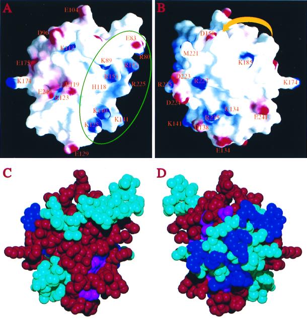
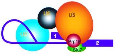
The splicing factor Prp18 is required for the second step of pre-mRNA splicing. We have isolated and determined the crystal structure of a large fragment of the Saccharomyces cerevisiae Prp18 that lacks the N-terminal 79 amino acids. This fragment, called Prp18Delta79, is fully active in yeast splicing in vitro and includes the sequences of Prp18 that have been evolutionarily conserved. The core structure of Prp18Delta79 is compact and globular, consisting of five alpha-helices that adopt a novel fold that we have designated the five-helix X-bundle. The structure suggests that one face of Prp18 interacts with the splicing factor Slu7, whereas the more evolutionarily conserved amino acids in Prp18 form the opposite face. The most highly conserved region of Prp18, a nearly invariant stretch of 19 aa, forms part of a loop between two alpha-helices and may interact with the U5 small nuclear ribonucleoprotein particles. The structure is consistent with a model in which Prp18 forms a bridge between Slu7 and the U5 small nuclear ribonucleoprotein particles.


Figures

Figure 1
Figure 1
Alignment of sequences of five Prp18 proteins. S. cerevisiae Prp18Δ79 was aligned with the Prp18s of H. sapiens, D. melanogaster, A. thaliana, C. elegans, and S. pombe. Positions at which four or more sequences are identical are shown red on blue; positions at which three sequences are identical are shown red on cyan; and positions at which three or more sequences are similar are shown green on yellow. Similarities used are D≈E, K≈R, N≈Q, A≈G, C≈S≈T, V≈L≈I≈M, and F≈Y≈W. The positions of α-helices in the S. cerevisiae structure are indicated above the sequence. Loops 1 through 5, which precede helices 1 through 5, are indicated by red lines; broken lines identify regions where the protein was disordered in the crystal. The A. thaliana Prp18 extends 32 aa beyond the end shown here.
Figure 2
Figure 2
Overall structure of Prp18Δ79. (A) Front view of the Prp18Δ79 structure shown in a ribbon representation. The protein core has a five-helix X bundle structure. The N and C termini and all helices are labeled. The dotted line represents the disordered segment of loop-5. (B) Another view of the Prp18Δ79 structure (rotated 210° with respect to a vertical axis) showing that helices α3, α4, and α5 are antiparallel and located on the same side of the α1–α2 plane. Loop-5 connecting helices α4 and α5 are exposed. The figure is produced with molscript (40) and raster3d (41).
Figure 3
Figure 3
Activity of Prp18Δ79. (A) Prp18 or Prp18Δ79 was added to Prp18-depleted yeast extract, and splicing of actin pre-mRNA was assayed. Lane 1 shows splicing in extract depleted with preimmune serum. Lane 2 shows splicing in extract depleted by anti-Prp18 with no added Prp18. For the splicing reactions displayed in lanes 3–8, the amount of Prp18 or Prp18Δ79 added is shown at the top of each lane in femtomol per microliter of yeast extract. The positions of lariat intermediate, lariat intron, pre-mRNA, mRNA, and exon1 are shown at the left of both A and B. (B) Prp18, Prp18Δ79, or hPrp18 was added to hPrp18-depleted HeLa-cell nuclear extract, and splicing of β-globin pre-mRNA was assayed. Lane 1 shows splicing in hPrp18-depleted extract alone. For lanes 2–12, the amount of Prp18, Prp18Δ79, or hPrp18 added is shown at the top in femtomol per microliter of nuclear extract.
Figure 4
Figure 4
Surface characteristics of the Prp18Δ79 structure. (A) Electrostatic potential on the protein surface, viewed from a similar direction as in Fig. 2A. Positive charges are shown in blue, negative charges in red. Charged residues are labeled by their standard single-letter code followed by the residue number in the sequence. The green circle identifies the proposed Slu7-interacting area. His-118 on α2 is also labeled, although histidines are not included in calculating the electric potential on the surface (charges were assigned only to Lys and Arg, Asp and Glu). The surface was calculated by using a probe radius of 1.4 Å, and the potential is displayed at a −15 kBT to + 15 kBT scale, where kB is the Boltzmann constant. The region on the right side concentrated with positively charged residues is the putative Slu7-binding region. (B) Rear view (the protein is rotated 180° with respect to a vertical axis). The yellow arrowheaded ribbon indicates schematically the location of the disordered segment in loop-5. The direction of the arrowhead indicates the amino to carboxyl direction of the segment. The figures were generated with the program grasp (44). (C) Corey–Pauling–Koltun representation of the Prp18Δ79 structure viewed from the same direction as in A. Residues implicated in interacting with Slu7 by the two-hybrid method are colored red; conserved residues (S. cerevisiae residues shared by three or more other species as shown in Fig. 1) are colored blue; conserved residues that lie within the Slu7 interacting region are colored magenta, and all others are colored cyan. (D) Rear view. Most invariant residues are located on this side of the surface (the disordered segment of loop-5 is not shown, but it is also expected to be located on this side of the protein). These residues are not involved in interacting with Slu7.
Figure 5
Figure 5
A schematic drawing showing the proposed model of the Prp18 function in the second step of splicing. Pre-mRNA is shown in purple; exons 1 and 2 are indicated. Prp18 is shown in red and in contact with both Slu7 (green) and U5 snRNP (orange); the latter two contact the 3′ splice site directly. U2 (cyan) and U6 (black) snRNPs are shown to indicate their involvement in the second step splicing reaction.

References

    1. Krämer A. Annu Rev Biochem. 1996;65:367–409. - PubMed
    1. Staley J P, Guthrie C. Cell. 1998;92:315–326. - PubMed
    1. Burge C B, Tuschl T H, Sharp P A. In: RNA World II. Gesteland R F, Cech T R, Atkins J F, editors. Plainview, NY: Cold Spring Harbor Lab. Press; 1999. pp. 525–560.
    1. Horowitz D S, Abelson J. Genes Dev. 1993;7:320–329. - PubMed
    1. Vijayraghavan U, Abelson J. Mol Cell Biol. 1990;10:324–332. - PMC - PubMed

Publication types

Associated data