Comparative Study
. 2000 Apr;66(4):1211-20.
doi: 10.1086/302857. Epub 2000 Mar 17.

Similar splice-site mutations of the ATP7A gene lead to different phenotypes: classical Menkes disease or occipital horn syndrome

Affiliations
Comparative Study

Similar splice-site mutations of the ATP7A gene lead to different phenotypes: classical Menkes disease or occipital horn syndrome

L B Møller et al. Am J Hum Genet. 2000 Apr.

Abstract

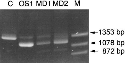
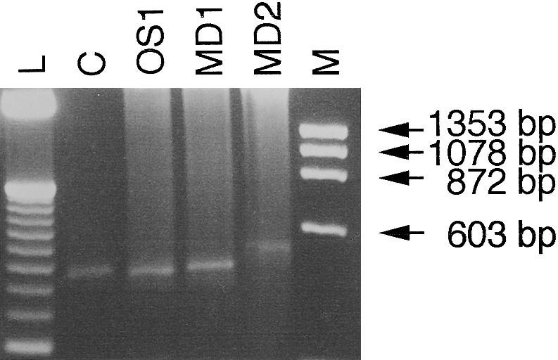
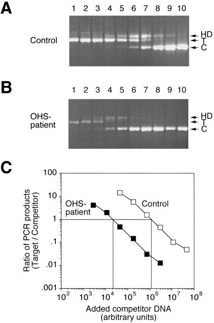
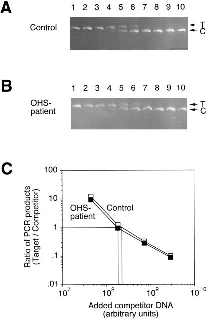
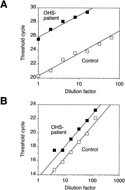
More than 150 point mutations have now been identified in the ATP7A gene. Most of these mutations lead to the classic form of Menkes disease (MD), and a few lead to the milder occipital horn syndrome (OHS). To get a better understanding of molecular changes leading to classic MD and OHS, we took advantage of the unique finding of three patients with similar mutations but different phenotypes. Although all three patients had mutations located in the splice-donor site of intron 6, only two of the patients had the MD phenotype; the third had the OHS phenotype. Fibroblast cultures from the three patients were analyzed by reverse transcriptase (RT)-PCR to try to find an explanation of the different phenotypes. In all three patients, exon 6 was deleted in the majority of the ATP7A transcripts. However, by RT-PCR amplification with an exon 6-specific primer, we were able to amplify exon 6-containing mRNA products from all three patients, even though they were in low abundance. Sequencing of these products indicated that only the patient with OHS had correctly spliced exon 6-containing transcripts. We used two different methods of quantitative RT-PCR analysis and found that the level of correctly spliced mRNA in this patient was 2%-5% of the level found in unaffected individuals. These findings indicate that the presence of barely detectable amounts of correctly spliced ATP7A transcript is sufficient to permit the development of the milder OHS phenotype, as opposed to classic MD.


Figures

Figure  1
Figure 1
Analyses of exon skipping in patients OS1, MD1, and MD2. The region from exon 4 to exon 9 in the ATP7A cDNA sequence was PCR-amplified from cDNA obtained from the three patients (see Materials and Methods). cDNA from an unaffected person was used as a control (C). The PCR products were separated on a 3% Metaphor gel and visualized by ethidium bromide staining. Lane M, φX174DNA-HaeIII digest.
Figure  2
Figure 2
Amplification of exon 6–containing transcripts from patients OS1, MD1, and MD2. Exon 6–containing cDNAs from three patients were amplified by the use of primers located in exons 6 and 9 (see Materials and Methods). cDNA obtained from an unaffected person was used as a control (C). The PCR products were separated on a 3% Metaphor gel and visualized by ethidium bromide staining. Lane M, φX174DNA-HaeIII digest; lane L, 50-bp DNA ladder (Gibco BRL).
Figure  3
Figure 3
Determination of the amount of ATP7A transcript in patient OS1 by competitive RT-PCR. Total RNA isolated from patient OS1 and control fibroblasts, respectively, was used for the synthesis of cDNA. By use of primers MNK-1 and MNK-2, equal amounts of cDNA were amplified in the presence of increasing amounts of homologous ATP7A competitor DNA, pMNKΔ (from left to right). Quantitation of the reaction products was accomplished by labeling with α-[32P]-dCTP included in the PCR reactions. A, Ethidium bromide–stained agarose gel with separated ATP7A RT-PCR products from control fibroblast. “T” and “C” indicate the positions of the target and competitor PCR products, respectively, and HD represents a heteroduplex formed between these. B, Ethidium bromide–stained agarose gel with separated ATP7A RT-PCR products from patient OS1. C, Ratio of PCR products generated from target and competitor DNA, respectively, as a function of the amount of competitor plasmid DNA added to the PCR reactions. The amount of the two different products in the heteroduplex band was taken into account in the calculation of the product ratios. The ratio of target/competitor product has been plotted only for lanes in which both signals are above background—that is, lanes 2–7 in the OHS experiment and lanes 4–9 in the control experiment.
Figure  4
Figure 4
Determination of the amount of 18S rRNA by competitive RT-PCR. Total RNA isolated from OS1 and control fibroblasts, respectively, was used for the synthesis of cDNA. By use of primers 18S-1 and 18S-2, equal amounts of cDNA were amplified in the presence of increasing amounts of homologous 18S competitor DNA, p18SΔ (from left to right). Reaction products were quantitated as in figure 3. A, Ethidium bromide–stained agarose gel with separated 18S RT-PCR products from control fibroblast. T and C indicate the positions of target and competitor PCR products, respectively. B, Ethidium bromide–stained agarose gel with separated 18S RT-PCR products from patient OS1. C, Ratio of PCR products generated from target and competitor DNA, respectively, as a function of the amount of competitor plasmid DNA added to the PCR reactions. The ratio of target/competitor product has been plotted only for lanes in which both signals are above background—that is, lanes 4–7 in either experiment.
Figure  5
Figure 5
Quantitation of ATP7A mRNA by real-time monitoring of the formation of fluorescent RT-PCR products. cDNA was reverse transcribed from mRNA isolated from patient OS1's cells or from control cells, respectively. The amount of exon 6–containing ATP7A transcripts or GAPDH mRNA was estimated from twofold serial dilutions of the cDNAs. A, Plot of the threshold cycle number for the ATP7A amplification as a function of the dilution of the input cDNA sample. B, Plot of the threshold cycle number for the GAPDH amplification as a function of the dilution of the input cDNA sample.

References

Electronic-Database Information

    1. Online Mendelian Inheritance in Man (OMIM), http://www.ncbi.nlm.nih.gov/Omim (for MD, MIM 309400) - PubMed

References

    1. Bouaboula M, Legoux P, Pességué B, Delpech B, Dumont X, Piechaczyk M, Casellas P, et al (1992) Standardization of mRNA titration using a polymerase chain reaction method involving co-amplification with a multispecific internal control. J Biol Chem 267:21830–21838 - PubMed
    1. Das S, Levinson B, Whitney S, Vulpe C, Packman S, Gitschier J (1994) Diverse mutations in patients with Menkes disease often lead to exon skipping. Am J Hum Genet 55:883–889 - PMC - PubMed
    1. Das S, Levinson B, Vulpe C, Whitney S, Gitschier J, Packman S (1995) Similar splicing mutations of the Menkes/mottled copper-transporting ATPase gene in occipital horn syndrome and the blotchy mouse. Am J Hum Genet 56:570–576 - PMC - PubMed
    1. DiDonato M, Sarkar B (1997) Copper transport and its alterations in Menkes and Wilson disease. Biochim Biophys Acta 1360:3–16 - PubMed
    1. Dierick HA, Adam AN, Escara-Wilke JF, Glover TW (1997) Immunocytochemical localization of the Menkes copper transport protein (ATP7A) to the trans-Golgi network. Hum Mol Genet 6:409–416 - PMC - PubMed

Publication types

MeSH terms