. 2000 Mar 31;287(5462):2479-82.
doi: 10.1126/science.287.5462.2479.

Necessity for afferent activity to maintain eye-specific segregation in ferret lateral geniculate nucleus

Affiliations

Necessity for afferent activity to maintain eye-specific segregation in ferret lateral geniculate nucleus

B Chapman. Science. .

Abstract

In the adult mammal, retinal ganglion cell axon arbors are restricted to eye-specific layers in the lateral geniculate nucleus. Blocking neuronal activity early in development prevents this segregation from occurring. To test whether activity is also required to maintain eye-specific segregation, ganglion cell activity was blocked after segregation was established. This caused desegregation, so that both eyes' axons became concentrated in lamina A, normally occupied only by contralateral afferents. These results show that an activity-dependent process is necessary for maintaining eye-specific segregation and suggest that activity-independent cues may favor lamina A as the target for arborization of afferents from both eyes.


Figures
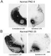
Fig. 1
Fig. 1
Normal development of the LGN. Retinogeniculate afferents from the right eye were labeled by anterograde transport of WGA-HRP. (A) Horizontal sections from PND 9 ferret LGN show good segregation of afferents from the two eyes into lamina A (contralateral) and lamina A1 (ipsilateral). (B) PND 25 ferret afferents show both eye-specific segregation into lamina A and lamina A1 and ON/OFF segregation into inner (Ai and A1i) and outer (Ao and A1o) leaflets. C laminae are also seen. Bar = 100 μm.
Fig. 2
Fig. 2
Physiological effects of APB treatment and controls for non-specific effects of eye injections and possible systemic APB effects. (A) APB (3.5 mM) abolishes both ON and OFF LGN responses. Poststimulus time histograms of multiunit LGN activity in response to flashing light and dark circles (white, 1.5 s; black, 1.5 s). (Top left) Normal PND 30 multiunit ON response from lamina A. (Bottom left) Recording from the same location 20 min after contralateral eye APB injection. (Top right) Normal PND 30 multiunit OFF response from lamina A1. (Bottom right) Recording from the same location 20 min after ipsilateral eye APB injection. y axis, 700 spikes per second; x axis, 3 s. APB was found to block all ON and OFF activity in the PND 30 LGN for 24 to 28 hours in three ferrets. (B) APB (3.5 mM) blocks spontaneous LGN activity in PND 30 ferrets. Ten-second raster plots of spontaneous LGN activity. (Top) Normal spontaneous activity in layer A outer. (Middle) Activity at same location 10 min after contralateral eye APB injection. (Bottom) Thirty min after injection. (C) Effects of 1 μM APB on spontaneous retinal activity in PND 7 ferret. In vitro calcium imaging shows complete blockade of retinal ganglion cell activity. y axis, nM Ca2+; x axis, 40 min. APB was applied at 19 min and washed out at 29 min. Data graciously provided by Wong and Wong (16). (D) APB does not disrupt normal development of gross morphology in the retina. Thionin-stained sections from ferret retina treated with 3.5 mM APB from PND 9 to 25 (right) and from a normal PND 25 animal (left). Bar = 50 μm. (E) Eye injections of saline or low concentration APB or systemic injections of high-concentration APB do not affect retinogeniculate afferent segregation. Binocular WGA-HRP injections reveal the normal PND 25 pattern of eye-specific layers and ON/OFF sublaminae in ferret LGN. Daily eye injections of 0.9% NaCl or 700 μM APB PND 9 to 25 do not alter the projection pattern. Note that 700 μM APB is sufficient to block ON-center cells in PND 30 ferrets for approximately 24 hours, suggesting that OFF-center activity alone may be sufficient to drive axonal segregation in the LGN. Systemic daily (PND 9 to 25) injections of APB directly into the cisterna magna in three ferrets had no effect. The concentration of APB used was the same as that used for eye injections, but the volume was doubled so the total amount of APB administered in cisternal injections was equal to that administered in binocular eye injections. Bar = 100 μm.
Fig. 3
Fig. 3
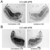
Effects of APB-induced retinal activity blockade on eye-specific segregation of afferents in the LGN. Retinogeniculate afferents were labeled by anterograde transport of WGA-HRP. (A) Horizontal sections from the LGN contralateral (left) and ipsilateral (right) to the injected eye show the same pattern of afferent projection. Cross-sectional areas of the ipsilateral and contralateral projections to lamina A measured in 10 horizontal sections through the middle of LGNs from four APB-treated animals were statistically indistinguishable (two-tailed t test; P < 0.001) from each other and from the area occupied by the contralateral projection to lamina A in similar sections from four normal ferrets [APB contralateral, 0.56 ± 0.06 mm2; APB ipsilateral, 0.49 ± 0.08 mm2; normal contralateral, 0.52 ± 0.07; normal ipsilateral (lamina A1), 0.18 ± 0.04]. Faint segregation of afferents into inner (Ai) and outer (Ao) leaflets can be seen in both eyes' projections. Staining was always darker contralateral to the injected eye, suggesting that the contralateral projection remains stronger than the ipsilateral projection. (B) Sections from the ventral third of the LGN of another ferret again show identical projection patterns contralateral (left) and ipsilateral (right) to the injected eye. This abnormal lack of segregation was found in all six animals studied throughout the depth of the LGN. Bar = 100 μm.
Fig. 4
Fig. 4
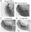
APB-induced retinal activity blockade disrupts development of the interlaminar space in ferret LGN. (A) Faint cell-sparse space between lamina A and lamina A1 can be seen in normal PND 9 ferret LGN (arrows). (B) By PND 25, the interlaminar space is clear. (C) APB-treated ferrets show no interlaminar space on PND25. Nissl-stained section with arrows marking the apparent A/A1 border, as shown by the pattern of the afferent projection seen in an adjacent WGAHRP section from the same LGN (D). Bar = 100 μm.

References

    1. Rakic P. Nature. 1976;261:467. - PubMed
    2. Shatz CJ. J. Neurosci. 1983;3:482. - PMC - PubMed
    3. Sretevan DW, Shatz CJ. J. Neurosci. 1986;6:234. - PMC - PubMed
    1. Linden DC, Guillery RW, Cucchiaro J. J. Comp. Neurol. 1981;203:189. - PubMed
    1. Shatz CJ, Stryker MP. Science. 1988;242:87. - PubMed
    2. Sretevan DW, Shatz CJ, Stryker MP. Nature. 1988;336:468. - PubMed
    1. Penn AA, Riquelme PA, Feller MB, Shatz CJ. Science. 1998;279:2108. - PubMed
    1. Cook PM, Prusky G, Ramoa AS. Vis. Neurosci. 1999;16:491. - PubMed

Publication types

MeSH terms

Substances