. 2000 Apr 7;275(14):10315-22.
doi: 10.1074/jbc.275.14.10315.

Isolation of a novel family of C(2)H(2) zinc finger proteins implicated in transcriptional repression mediated by chicken ovalbumin upstream promoter transcription factor (COUP-TF) orphan nuclear receptors

Affiliations

Isolation of a novel family of C(2)H(2) zinc finger proteins implicated in transcriptional repression mediated by chicken ovalbumin upstream promoter transcription factor (COUP-TF) orphan nuclear receptors

D Avram et al. J Biol Chem. .

Abstract

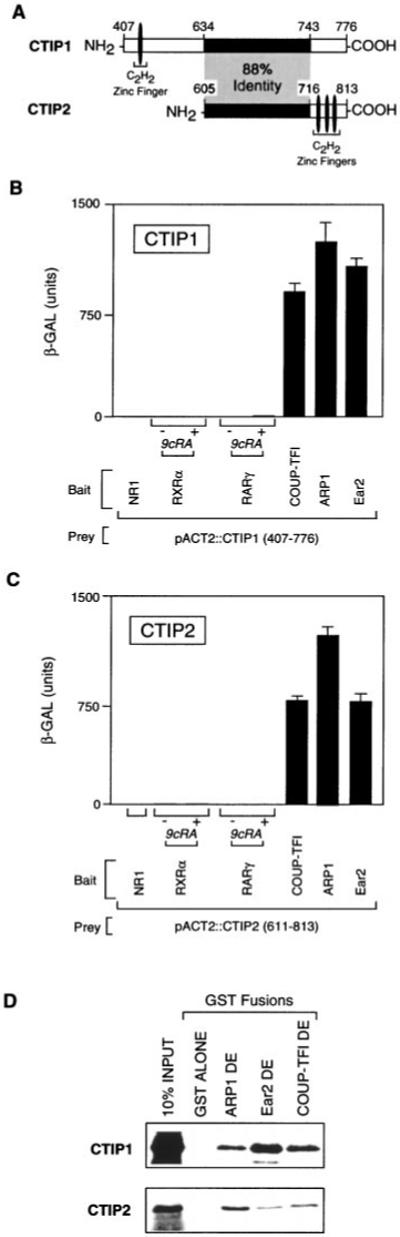
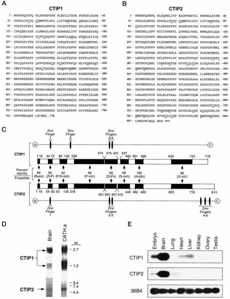
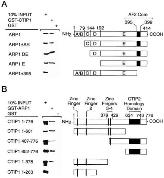
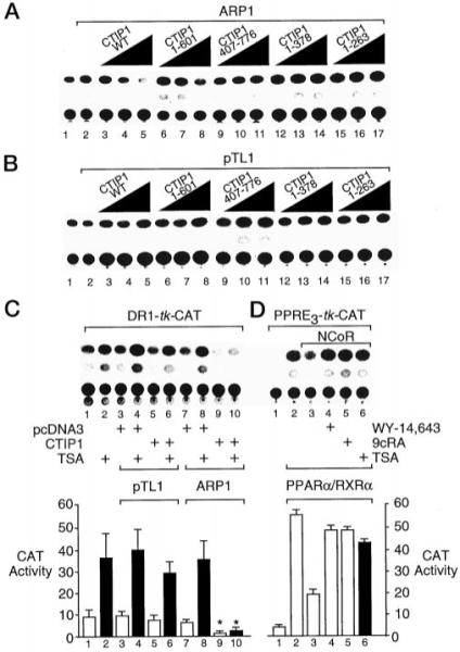
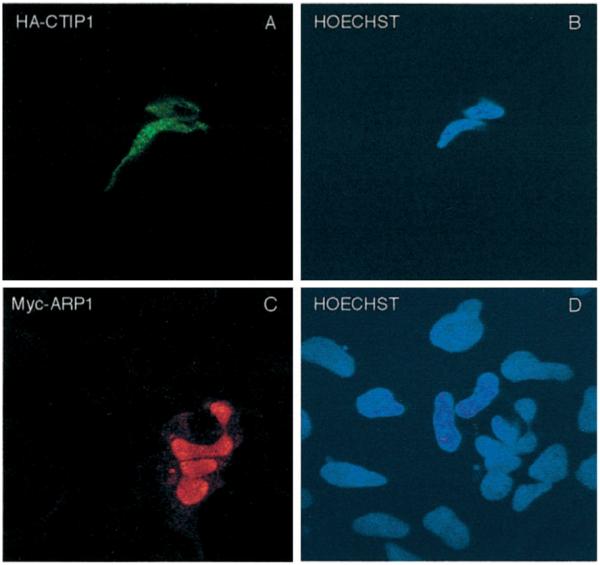
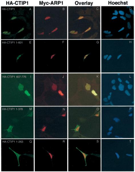
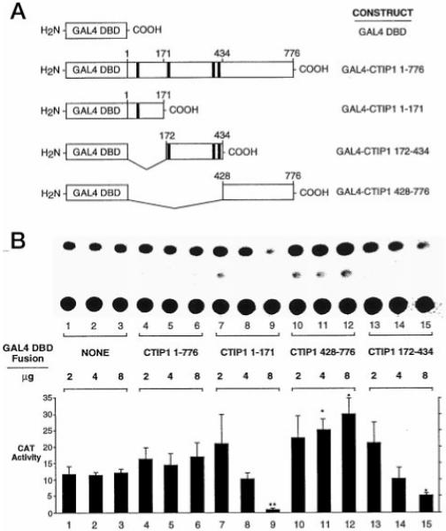
Two novel and related C(2)H(2) zinc finger proteins that are highly expressed in the brain, CTIP1 and CTIP2 (COUP TF-interacting proteins 1 and 2, respectively), were isolated and shown to interact with all members of the chicken ovalbumin upstream promoter transcription factor (COUP-TF) subfamily of orphan nuclear receptors. The interaction of CTIP1 with ARP1 was studied in detail, and CTIP1 was found to harbor two independent ARP1 interaction domains, ID1 and ID2, whereas the putative AF-2 of ARP1 was required for interaction with CTIP1. CTIP1, which exhibited a punctate staining pattern within the nucleus of transfected cells, recruited cotransfected ARP1 to these foci and potentiated ARP1-mediated transcriptional repression of a reporter construct. However, transcriptional repression mediated by ARP1 acting through CTIP1 did not appear to involve recruitment of a trichostatin A-sensitive histone deacetylase(s) to the template, suggesting that this repression pathway may be distinct from that utilized by several other nuclear receptors.


Figures

Fig. 1
Fig. 1. Interaction of COUP-TFs with CTIP proteins in yeast and in vitro
A, diagram of CTIP1 and CTIP2 clones isolated in the yeast two-hybrid screen. The shaded region represents a domain exhibiting 88% identity between the two proteins. B and C, interaction of CTIP1 and CTIP2, respectively, with COUP-TFs, RXRα, RARγ, and the NR1 subunit of the N-methyl-d-aspartate receptor (36). All baits were cloned in pBTM116 as described previously (21) and expressed in S. cerevisiae L40 together with GAL4 activation domain fusions of CTIP1-(407–776) (B) or CTIP2-(611–813) (C). Ligand-dependent interactions were examined in the presence of 1 μm 9-cis-retinoic acid (9cRA). The results shown represent the means ± S.D. of three independent experiments. D, in vitro interaction between COUP-TFs and CTIP1-(602–776) and CTIP2-(611–813). GST/COUP-TFs were bound to glutathione-Sepharose and used as affinity matrices to examine the interaction with the in vitro translated [35S]methionine-labeled CTIPs as described previously (21).
Fig. 2
Fig. 2. Amino acid sequence, diagram of amino acid sequence alignment, and expression of CTIP1 and CTIP2 in several mouse tissues
A and B, amino acid sequence of CTIP1 and CTIP2, respectively. The conserved cysteine and histidine amino acids contributing the C2H2 and C2HC zinc finger motifs of both proteins are underlined. C, schematic diagram of CTIP1 and CTIP2 amino acid alignment. The homologous regions are represented by black boxes and the percentage of identity between each region of CTIP1 and CTIP2 is indicated. The alignment was performed using Clustal X (version 1.63b). The overall characteristics of each domain are indicated. D, Northern blot analysis of CTIP1 and CTIP2 expression in brain and CATH.a cells. Approximately 5 μg of brain and 10 μg of CATH.a poly(A)+ RNA were loaded per lane, and the blots were hybridized with probes derived from the 3′-end of both CTIP1 and CTIP2. E, Southern blot of reverse transcription-polymerase chain reaction analysis of RNA from various tissues of adult mouse and embryo (10–12.5 days postcoitum. Note that partial EST clones of both CTIP1 and CTIP2 have been reported (mi17e04.r1 and mv64 h01.r1, respectively, from the Washington University-HHMI Mouse EST Project).
Fig. 3
Fig. 3. Definition of the in vitro interaction domains of both ARP1 (A) and CTIP1 (B) by deletion mutagenesis
A, CTIP1 interaction domains of ARP1. The ARP1 deletion mutants indicated in the schematic diagram were cloned in pCDNA3 and translated in vitro using the TNT Coupled Reticulocyte Lysate System (Promega). CTIP1-(1–176) was cloned in pGEX-2T. GST/CTIP1 was bound to glutathione-Sepharose and used as affinity matrix to examine the interaction with the in vitro translated [35S]methionine-labeled ARP1 mutants as described previously (21). B, ARP1 interaction interfaces of CTIP1. ARP1-(1–414) was cloned in pGEX-2T, and GST/ARP1 was bound to glutathione-Sepharose and used as affinity matrix to examine the interaction with the in vitro translated [35S]methionine-labeled CTIP1 mutants. CTIP1 deletion mutants indicated in the schematic diagram were cloned in pCDNA3 and in vitro translated using the TNT Coupled Reticulocyte Lysate System (Promega).
Fig. 4
Fig. 4. CTIP1 potentiates the ARP1-mediated repression in a TSA-insensitive manner
A and B, HEK 293 cells were cotransfected with 5 μg of the DR1-TK-CAT reporter, 5 μg of Myc-ARP1 expression vector (A) or empty vector (pTL1; B) and increasing amounts (0.2, 0.66, and 2.22 μg) of expression vectors encoding full-length HA-CTIP1 or HA-CTIP1 deletion mutants as indicated. CAT reporter activity in cell extracts was determined as described previously (21). C, HEK 293 cells were cotransfected with 5 μg of DR1-TK-CAT reporter, 5 μg of Myc-ARP1 expression vector or empty vector (pTL1), and 2.2 μg of HA-CTIP1 or empty vector (pcDNA3). Cells were treated with TSA (100 ng/ml; solid bars) as indicated for 24 h prior to harvesting and CAT assays. D, cotransfection of HEK 293 cells with 2 μg of PPRE-TK-CAT reporter and expression vectors for PPARα/RXRα (0.5 μg each) and NCoR (2 μg) as indicated. Cells were treated with either vehicle (0.1% Me2SO; lane 3); the RXR agonist, 9-cis-retinoic acid (9cRA; 1 μm); or the PPARα agonist, WY-14,643 (10 μm), or TSA as noted for 24 h prior to harvesting and determination of CAT activity. The quantifications shown below B and C represent mean CAT activities ± S.D. derived from three independent experiments. The CAT activity values in lanes 9 and 10 are statistically different from those shown in lanes 7 and 8, respectively, as determined by Student’s t test (p < 0.05, indicated by asterisks).
Fig. 5
Fig. 5. Localization of HA-CTIP1 (A) and Myc-ARP1 (C) in HEK293 cell nuclei
HEK293 cells growing on coverslips were transiently transfected with expression vectors encoding HA-CTIP1 (A) and Myc-ARP1 (C). Forty-eight hours after transfection, the cells were fixed and incubated with anti-HA (A) or anti-Myc (C) antibodies and stained with appropriate fluorescein isothiocyanate- or tetramethylrhodamine isothiocyanate-conjugated secondary antibodies detecting HA-CTIP1 and Myc-ARP1 immune complexes, respectively. B and D, Hoechst-counterstained cells shown in A and C, respectively. HA-CTIP1 exhibited the punctate staining pattern depicted in A in 80% of transfected cells examined, with the balance displaying a combination of focal and diffuse staining (see Table I). In contrast, Myc-ARP1 exhibited the diffuse staining pattern in 100% of 120 transfected cells examined by a naive observer. Images were obtained on a Leica inverted confocal microscope model TCS4D using a × 100 objective. The images shown are derived from a representative experiment that was replicated several times.
Fig. 6
Fig. 6. ARP1 is redistributed in the nucleus when cotransfected with CTIP1
HEK293 cells were transiently cotransfected with expression vectors encoding Myc-ARP1 and either HA-CTIP1 or HA-CTIP1 mutants as indicated. The corresponding proteins were localized 48 h after transfection by indirect immunofluorescence confocal microscopy. Cells were stained for HA-CTIP1 (first column) and Myc-ARP1 (second column) as described in the legend of Fig. 5. An overlay of the images presented in the first two columns is shown in the third column, and the fourth column represents counterstaining of the cells with Hoechst as indicated. Shown are representative experiments that were replicated 3–7 times. Each micrograph was prepared using a × 100 objective on a Leica inverted confocal microscope model TCS4D, and overlays were prepared using Photoshop 5.0.
Fig. 7
Fig. 7. Autonomous transcriptional modulatory activity of CTIP1
A, diagram of GAL4 DBD-CTIP1 fusion constructs used in the transfection experiments illustrated in B. The black boxes represent the zinc finger motifs of CTIP1. B, CAT assays using extracts of HEK293 cells transiently transfected with 7.5 μg of the (17-mer)5 reporter (pG5CAT, CLONTECH) and the indicated amounts of expression vectors for GAL4 DBD (pM; CLONTECH) or GAL4 DBD-CTIP1 fusion proteins. Statistically significant effects are indicated by asterisks (p < 0.05, Student’s t test) or double asterisks (p < 0.01) when comparing GAL4-CTIP fusions to the corresponding amount of transfected GAL4 DBD. Note that the quantitation shown represents the mean ± S.D. of CAT activities from six independent experiments, whereas the CAT assay shown in B is from a single, representative experiment.

References

    1. Ladias JAA, Hadzopoulou-Cladaras M, Kardassis D, Cardot P, Cheng J, Zannis V, Caldaras C. J. Biol. Chem. 1992;267:15849–15860. - PubMed
    1. Wang LH, Ing NH, Tsai SY, O’Malley BW, Tsai M-J. Gene Expr. 1991;1:207–216. - PMC - PubMed
    1. Jonk LJC, de Jonge EJ, Pals CEGM, Wissink S, Vervaart JMA, Schoorlemmer J, Kruijer W. Mech. Dev. 1994;47:81–97. - PubMed
    1. Escriva H, Safi R, Hanni C, Langlois M-C, Saumitou-Laprade P, Stehelin D, Capron A, Pierce R, Laudet V. Proc. Natl. Acad. Sci. U. S. A. 1997;94:6803–6808. - PMC - PubMed
    1. Kliewer SA, Umesono K, Heyman RA, Mangelsdorf DJ, Dyck JA, Evans RM. Proc. Natl. Acad. Sci. U. S. A. 1992;89:1448–1452. - PMC - PubMed

Publication types

MeSH terms