. 2000 Apr 3;19(7):1458-66.
doi: 10.1093/emboj/19.7.1458.

gC1q-R/p32, a C1q-binding protein, is a receptor for the InlB invasion protein of Listeria monocytogenes

Affiliations

gC1q-R/p32, a C1q-binding protein, is a receptor for the InlB invasion protein of Listeria monocytogenes

L Braun et al. EMBO J. .

Abstract

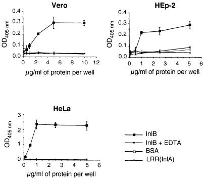
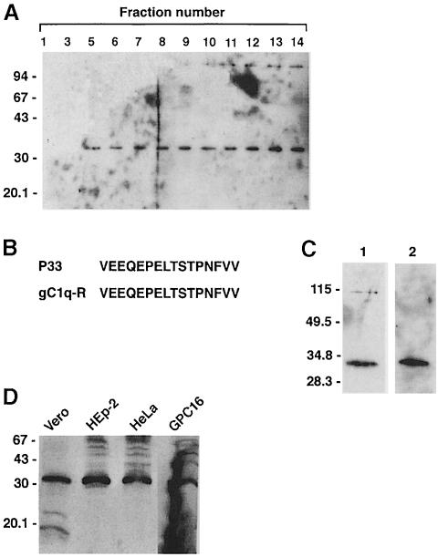
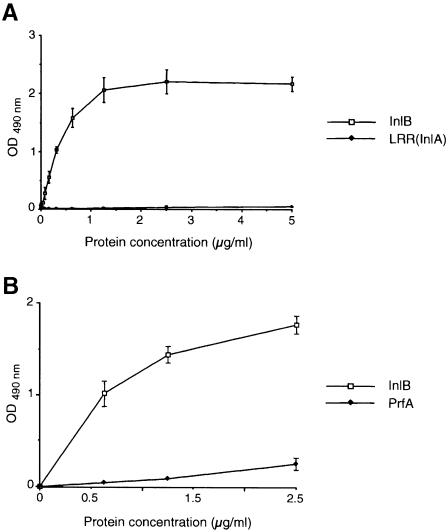
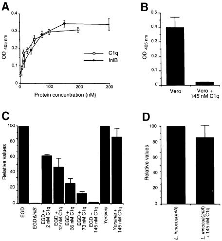
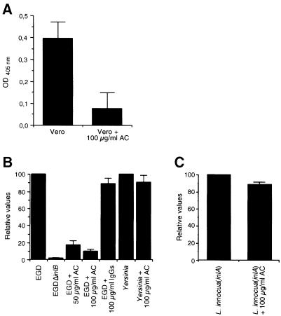
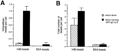
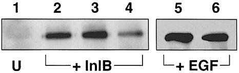
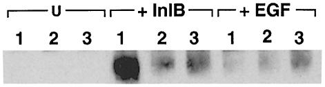
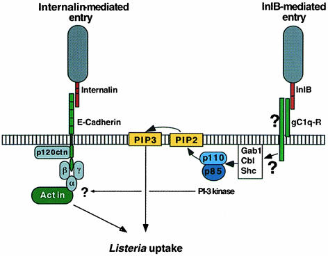
InlB is a Listeria monocytogenes protein that promotes entry of the bacterium into mammalian cells by stimulating tyrosine phosphorylation of the adaptor proteins Gab1, Cbl and Shc, and activation of phosphatidyl- inositol (PI) 3-kinase. Using affinity chromatography and enzyme-linked immunosorbent assay, we demonstrate a direct interaction between InlB and the mammalian protein gC1q-R, the receptor of the globular part of the complement component C1q. Soluble C1q or anti-gC1q-R antibodies impair InlB-mediated entry. Transient transfection of GPC16 cells, which are non-permissive to InlB-mediated entry, with a plasmid-expressing human gC1q-R promotes entry of InlB-coated beads. Furthermore, several experiments indicate that membrane recruitment and activation of PI 3-kinase involve an InlB-gC1q-R interaction and that gC1q-R associates with Gab1 upon stimulation of Vero cells with InlB. Thus, gC1q-R constitutes a cellular receptor involved in InlB-mediated activation of PI 3-kinase and tyrosine phosphorylation of the adaptor protein Gab1. After E-cadherin, the receptor for internalin, gC1q-R is the second identified mammalian receptor promoting entry of L. monocytogenes into mammalian cells.


Figures

None
Fig. 1. Mammalian cells bind to purified InlB. Microtiter plates were coated with increasing concentrations of InlB, LRR(InlA) or BSA, and a suspension of Vero, HEp-2 or HeLa cells was added in either the absence or the presence of 10 mM EDTA. After incubation for 1 h, wells were washed and the number of bound cells was determined by assaying for lysosomal hexosaminidase. Values represent the mean ± SEM of three independent experiments.
None
Fig. 2. InlB binding to electrophoretically separated soluble proteins from Vero cells by ligand overlay assay. Soluble proteins from Vero cells were separated on an 8% polyacrylamide gel and transferred onto a nitrocellulose membrane. Proteins on nitrocellulose were then probed with 50 μg/ml InlB and subsequently detected with the InlB-specific monoclonal antibody H15.1 (Braun et al., 1999).
None
Fig. 3. gC1q–R is a ligand for InlB. (A) N-octyl glucoside extracts were prepared from surface biotin-labeled Vero cell extracts and loaded onto an InlB affinity column. After extensive washing, proteins were eluted with 10 mM EDTA, and 200 μl fractions were collected. Fraction samples (10 μl) were analyzed by SDS–PAGE, transferred onto nitrocellulose and probed with streptavidin coupled to peroxidase to detect biotinylated proteins by chemiluminescence. (B) Amino acid sequence comparison of internal sequences from P33 isolated on the displayed column and gC1q–R encoded by human lymphocytes (Ghebrehiwet et al., 1994). (C) Western blot analysis of an elution fraction (15 μl) with streptavidin (1) or with a polyclonal antibody directed against gC1q–R (2). (D) Western blot analysis of the cellular expression of gC1q–R. A 100 μg aliquot of solubilized Vero, HEp-2, HeLa or GPC16 cell membrane proteins was separated by SDS–PAGE and transferred onto nitrocellulose. gC1q–R was revealed with an anti-gC1q–R polyclonal antibody.
None
Fig. 4. InlB binds to gC1q–R. Wells of a microtiter plate were coated with a solution of 1 μg/ml gC1q–R. After blocking with a 1% BSA solution, wells were incubated with increasing concentrations of purified proteins, either InlB, LRR(InlA) (A) or PrfA (B), and then analyzed by ELISA as described in Materials and methods.
None
Fig. 5. C1q inhibits entry of EGD into Vero cells. (A) Comparison of the binding of Vero cells to wells coated with increasing concen- trations of InlB or C1q using the colorimetric hexosaminidase assay. (B) Effect of C1q on the binding of Vero cells to InlB. Microtiter wells coated with a 10 μg/ml concentration of InlB were incubated with a Vero cell suspension that had been treated or not for 5 min at 37°C with 145 nM C1q. After allowing 1 h for attachment of the Vero cells to immobilized InlB, wells were washed, and cell attachment was quantified using the colorimetric hexosaminidase assay. (C and D) Effect of C1q on entry of L.monocytogenes EGD and Yersinia into Vero cells (C) or on entry of L.innocua(inlA) into Caco-2 cells (D). The inhibitory effect of C1q was tested by incubating the cells with different concentrations of C1q, 5 min before adding the bacteria. The number of intracellular bacteria was determined using gentamicin survival assays as described in Materials and methods. Values along the vertical axis are given relative to efficiencies of invasion of L.monocytogenes EGD in Vero cells or L.innocua(inlA) in Caco-2 cells (D), which are fixed arbitrarily at 100.
None
Fig. 6. Anti-gC1q–R antibodies inhibit entry of EGD into Vero cells. (A) Effect of anti-gC1q–R antibodies on the binding of Vero cells to InlB. Microtiter wells coated with a 10 μg/ml concentration of InlB were incubated with a Vero cell suspension that had been treated or not with 100 μg/ml antibodies (AC) for 60 min at 37°C. Cell attachment to InlB was quantitated using the colorimetric hexos- aminidase assay. (B and C) Effect of anti-gC1q–R antibodies on entry of EGD and Yersinia into Vero cells (B) or L.innocua(inlA) into Caco-2 cells (C). Vero or Caco-2 cells were incubated with anti-gC1q–R antibodies or pre-immune rabbit IgGs for 60 min before adding the bacteria. The inhibitory effect of the antibodies was tested by incubating the cells with the antibodies for 60 min before adding the bacteria. The number of intracellular bacteria was determined using gentamicin survival assays as described in Materials and methods. Values along the vertical axis are given relative to efficiencies of invasion of EGD in Vero cells or L.innocua(inlA) in Caco-2 cells (C), which are the reference (value of 100).
None
Fig. 7. InlB-dependent entry into gC1q–R-transfected GPC16 cells. GPC16 cells were transiently transfected with a control plasmid (hatched bars) or with a plasmid carrying a GFP–gC1q–R fusion protein (black bars) at a multiplicity of ∼100 beads per cell for 1 h. Internalized (A) and associated (B) beads were identified and quantified using immunofluorescence as described previously (Braun et al., 1998). The efficiency of entry is expressed as the average number of intracellular beads per cell. Association corresponds to the number of beads either bound or internalized. The results are expressed as the mean ± SD of three independent experiments.
None
Fig. 8. Anti-gC1q–R antibodies decrease the InlB-dependent association of p85 with tyrosine-phosphorylated proteins. Vero cells were pre-treated for 1 h with DMEM (lanes 1, 2 and 5), with 100 μg/ml rabbit IgGs (lane 3) or with 100 μg/ml anti-gC1q–R antibodies (lanes 4 and 6). Cells were then left untreated (U) (lane 1), or treated with 3 nM InlB (lanes 2–4) or 17 nM EGF for 1 min (lanes 5 and 6). The cells were then washed with PBS and lysed in the immunoprecipitation buffer. Tyrosine-phosphorylated proteins were immunoprecipitated with anti-P-tyr antibodies, separated by SDS–PAGE, and p85 was detected by immunoblotting with anti-p85 antibodies. The anti-gC1q–R antibodies decrease the InlB-induced association of p85 with tyrosine-phosphorylated proteins (lane 4), but had no effect on EGF-induced association of p85 with tyrosine-phosphorylated proteins (lane 6). Similar amounts of material were used for each immunoprecipitation. The results are representative of three experiments.
None
Fig. 9. Association of gC1q–R with tyrosine-phosphorylated Gab1. Vero cells were left untreated (U), or were treated with 3 nM InlB or 17 nM EGF for 1 min. Solubilized lysates were then prepared by addition of immunoprecipitation buffer, and 2.5 mg of lysate were incubated for 2 h at 4°C in the presence of 5 μg His6-gC1q–R (1), 5 μg LRR(InlA)-His6 (2) or buffer alone (3). After addition of Ni-NTA–agarose beads, the adsorbed proteins were dissociated by boiling in the presence of 0.5% SDS/5 mM dithiothreitol, diluted in re-immunoprecipitation buffer and re-precipitated with antibodies against Gab1. Tyrosine-phosphorylated proteins in the final immunoprecipitates were detected with anti-P-Tyr antibodies. Similar amounts of starting material were used for each re-precipitation. The results are represen- tative of two experiments.
None
Fig. 10. The two pathways of entry of L.monocytogenes into mammalian cells.

References

    1. Bobak D., Gaither, T., Frank, M. and Tenner, A.J. (1987) Modulation of FcR function by complement subcomponent C1q enhances the phagocytosis of IgG-opsonized targets by human monocytes and culture-derived macrophages. J. Immunol., 138, 1150–1156. - PubMed
    1. Braun L., Dramsi, S., Dehoux, P., Bierne, H., Lindahl, G. and Cossart, P. (1997) InlB: an invasion protein of Listeria monocytogenes with a novel type of surface association. Mol. Microbiol., 25, 285–294. - PubMed
    1. Braun L., Ohayon, H. and Cossart, P. (1998) The InlB protein of Listeria monocytogenes is sufficient to promote entry into mammalian cells. Mol. Microbiol., 27, 1077–1087. - PubMed
    1. Braun L., Nato, F., Payrastre, B., Mazié, J.-C. and Cossart, P. (1999) The 213-amino acid leucine-rich repeat region of the Listeria monocytogenes InlB protein is sufficient for entry into mammalian cells, stimulation of PI 3-kinase and membrane ruffling. Mol. Microbiol., 34, 10–23. - PubMed
    1. Cossart P. and Lecuit, M. (1998) Interactions of Listeria monocytogenes with mammalian cells during entry and actin-based movement: bacterial factors, cellular ligands and signaling. EMBO J., 17, 3797–3806. - PMC - PubMed

Publication types

MeSH terms