. 2000 Apr 3;19(7):1476-85.
doi: 10.1093/emboj/19.7.1476.

Highly purified glycosylphosphatidylinositols from Trypanosoma cruzi are potent proinflammatory agents

Affiliations

Highly purified glycosylphosphatidylinositols from Trypanosoma cruzi are potent proinflammatory agents

I C Almeida et al. EMBO J. .

Abstract

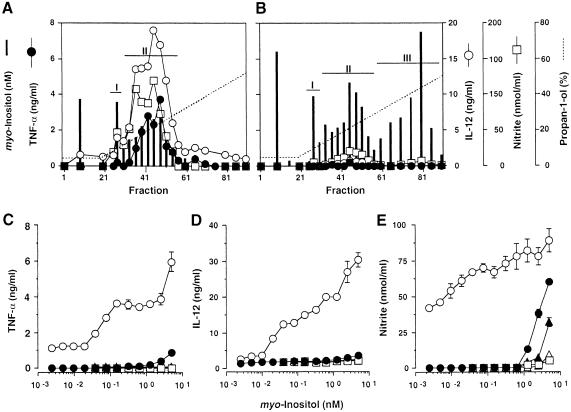
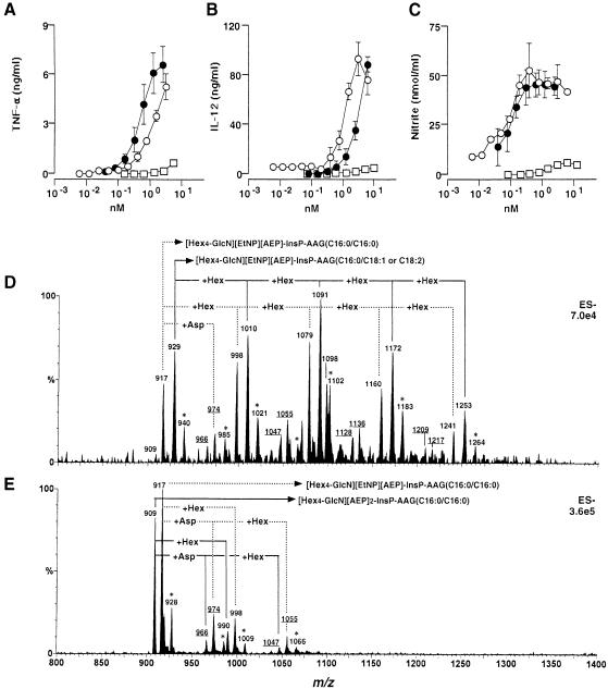
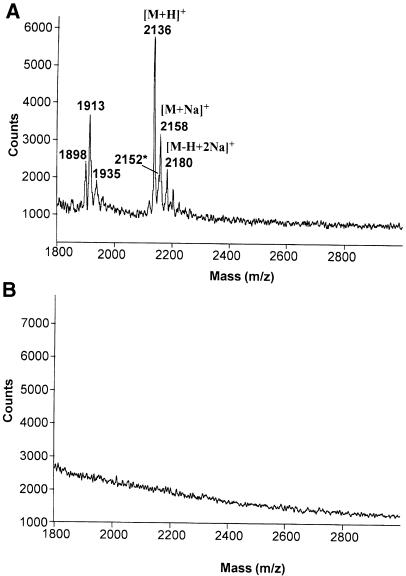
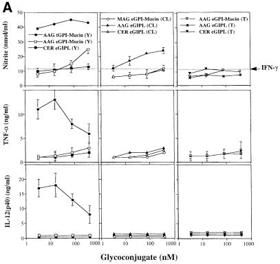
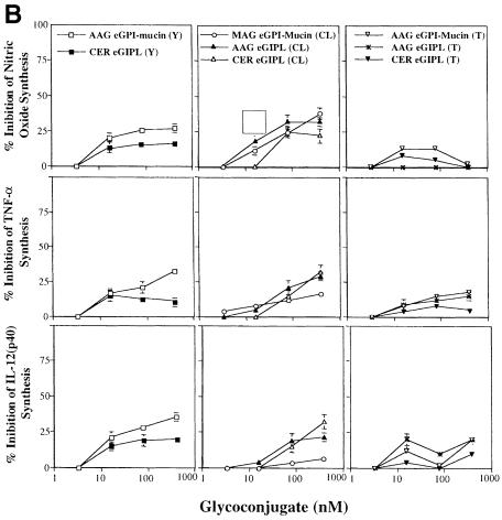
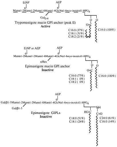
Intracellular protozoan parasites are potent stimulators of cell-mediated immunity. The induction of macrophage proinflammatory cytokines by Trypanosoma cruzi is considered to be important in controlling the infection and the outcome of Chagas' disease. Here we show that the potent tumour necrosis factor-alpha-, interleukin-12- and nitric oxide-inducing activities of T.cruzi trypomastigote mucins were recovered quantitatively in a highly purified and characterized glycosylphosphatidylinositol (GPI) anchor fraction of this material. The bioactive trypomastigote GPI fraction was compared with a relatively inactive GPI fraction prepared from T. cruzi epimastigote mucins. The trypomastigote GPI structures were found to contain additional galactose residues and unsaturated, instead of saturated, fatty acids in the sn-2 position of the alkylacylglycerolipid component. The latter feature is essential for the extreme potency of the trypomastigote GPI fraction, which is at least as active as bacterial endotoxin and Mycoplasma lipopeptide and, therefore, one of the most potent microbial proinflammatory agents known.


Figures

None
Fig. 1. Fractionation and bioactivity of T.cruzi GPI-anchored glycoconjugates. Octyl-Sepharose chromatograms of T.cruzi trypomastigote (A) and epimastigote (B) glycoconjugate extracts from 1.5 × 10¹⁰ and 2.0 × 10¹⁰ cells, respectively. Aliquots of each fraction were presented to 106 thioglycollate-elicited, IFN–γ-primed, murine peritoneal macrophages and the amounts of TNF–α (•), IL–12(p40) (○) and NO (□) (measured as nitrite) produced in 24 h were measured. Vertical bars indicate the molarity of the glycoconjugate used in each assay, based on the myo-inositol content of each fraction eluted from the column. The data shown in (A) and (B) are representative of three different fractionation experiments. Peak fractions were pooled as indicated and titrated for TNF–α- (C), IL–12(p40)- (D) and NO– (E) inducing activity. Trypomastigote-derived peak I and II materials (•, ○); epimastigote-derived peak I, II and III materials (□, ▴, ▵). The data shown in (C–E) are representative of three separate titration experiments with three different sets of fractions. Each cytokine and NO determination was carried out in duplicate, and the error bars (where visible) indicate the standard error of the mean.
None
Fig. 2. Bioactivity and mass spectra of purified GPI anchors released from T.cruzi mucins. Samples were presented to 106 thioglycollate-elicited, IFN–γ-primed, murine peritoneal macrophages and titrations of the TNF–α- (A), IL–12(p40)- (B) and NO- (C) inducing activities of intact trypomastigote mucin (•), purified GPI released from trypomastigote mucin (○) and purified GPI released from epimastigote mucin (□) are shown. The data shown in (A–C) are representative of four separate titration experiments. Each cytokine and NO determination was carried out in duplicate, and the error bars (where visible) indicate the standard error of the mean. The purified trypomastigote GPI (D) and epimastigote GPI (E) fractions were analysed by negative-ion ES–MS. The compositions corresponding to each [M–2H]2– pseudomolecular ion are indicated. Ions marked with asterisks are [M+Na–3H]2– pseudomolecular ions, and the underlined ions correspond to GPI anchors attached to Asp. EtNP, ethanolamine phosphate; AEP, 2–aminoethylphosphonate; InsP, inositol-phosphate; AAG, alkylacylglycerol. The numbers following the AAG abbreviation correspond to the chain length and degree of unsaturation of the alkyl/acyl chains. Note: m/z differences of 81 correspond to mass differences of 162 [equivalent to a hexose (Hex), i.e. mannose or galactose] because the charge state of the ions (z) is 2.
None
Fig. 3. Purified trypomastigote GPIs are free of lipopeptides. A sample of 10 pmol of synthetic Mycoplasma lipopeptide (A) and 10 pmol of purified T.cruzi trypomastigote mucin GPI fraction (B) were analysed by positive-ion MALDI-TOF under identical conditions. The absence of ions in (B) reflects the absence of any lipopeptides or peptides in the preparation and the inability of GPIs to produce positive ions under the conditions used. In (A), the ions at m/z 2136, 2158 and 2180 are the [M+H]+, [M+Na]+ and [M–H+2Na]+ ions of S–(2,3–bishexa- decanoyloxypropyl)cysteine-GNNDESNISFKEK, respectively and the minor ion at m/z 2152* is most probably the [M+H]+ ion of the corresponding sulfoxide. The ions at lower m/z values may be due to degradation products.
None
Fig. 4. Bioactivities of Y, CL and Tulahuen strain epimastigote GPI-mucins and GIPLs. (A) Titration of NO (top panels), TNF–α (middle panels) and IL–12(p40) (bottom panels) induction by intact GPI-mucins or GIPLs purified from the epimastigote forms of Y, CL and Tulahuen (T) strains of T.cruzi. The partial structure and purity of each glycoconjugate were evaluated by ES–MS and ES-MS–CID-MS (Table II); AAG, MAG and CER indicate the presence of alkylacylglycerol, monoalkylglycerol or ceramide lipids in the PI moieties of the GPI structures. Macrophages were primed with IFN–γ (100 U/ml) overnight and then stimulated with 3.3, 16, 80 and 400 nM epimastigote-derived glycoconjugate. As a positive control, Y strain trypomastigote GPI-mucin was used at the same concentrations. TNF–α was measured in the macrophage culture supernatants 24 h after stimulation, and IL–12(p40) and nitrite were measured 48 h after stimulation. Horizontal lines indicate the macrophage response to IFN–γ alone. (B) Inhibitory activities of the epimastigote GPI-mucins and GIPLs. Titration of the inhibition of NO (top panels), TNF–α (middle panels) and IL–12(p40) (bottom panels) induction by 1 nM trypomastigote GPI-mucin by pre-incubation (2 h) with epimastigote GPI-mucins and GIPLs.
None
Fig. 5. Comparison of active and representative relatively inactive T.cruzi GPI structures. The epimastigote mucin (peak II) GPI structure (Previato et al., 1995; Serrano et al., 1995) is consistent with the ES–MS spectrum in Figure 2E and the ES–MS analysis of the PI moiety (Table I). Only the identity and linkage position of the additional hexose residue (±Hex) are unknown (Previato et al., 1995; Serrano et al., 1995). The proposed trypomastigote peak II GPI structure is based on the epimastigote structure and is consistent with the ES–MS spectrum in Figure 2D, the GC–MS monosaccharide analysis and the ES–MS analysis of the PI moiety (Table I). The dotted line indicates that the linkage position(s) of the uncharacterized galactose substituents (Gal0–4) is unknown. The types and proportions of the major alkyl and acyl (fatty acid) chains are indicated. The exact isomer of the C18:2 fatty acid (shown here as linoleic acid) has not been determined. The ceramide-containing epimastigote (peak III) GIPL structure is taken from De Lederkremer et al. (1991) and Previato et al. (1990), and is consistent with an ES–MS spectrum of this fraction (data not shown) and the ES–MS analysis of the PI moiety (Table I).

References

    1. Abbas A.K., Murphy, K.M. and Sher, A. (1996) Functional diversity of helper T lymphocytes. Nature, 383, 787–793. - PubMed
    1. Aliberti J.C.S., Cardoso, M.A., Martins, G.A., Gazzinelli, R.T., Vieira, L.Q. and Silva, J.S. (1996) Interleukin-12 mediates resistance to Trypanosoma cruzi in mice and is produced by murine macrophages in response to live trypomastigotes. Infect. Immun., 64, 1961–1967. - PMC - PubMed
    1. Almeida I.C., Milani, S.R., Gorin, P.A. and Travassos, L.R. (1991) Complement-mediated lysis of Trypanosoma cruzi trypomastigotes by human anti-α-galactosyl antibodies. J. Immunol., 146, 2394–2400. - PubMed
    1. Almeida I.C., Ferguson, M.A.J., Schenkman, S. and Travassos, L.R. (1994) Lytic anti-α-galactosyl antibodies from patients with chronic Chagas' disease recognize novel O-linked oligosaccharides on mucin-like glycosyl-phosphatidylinositol-anchored glycoproteins of Trypanosoma cruzi. Biochem. J., 304, 793–802. - PMC - PubMed
    1. Alves M.J.M. and Colli, W. (1975) Glycoproteins from Trypanosoma cruzi: partial purification by gel chromatography. FEBS Lett., 52, 188–198. - PubMed

Publication types

MeSH terms