. 2000 Apr 3;19(7):1545-54.
doi: 10.1093/emboj/19.7.1545.

Arrest of spermatogenesis in mice expressing an active heat shock transcription factor 1

Affiliations

Arrest of spermatogenesis in mice expressing an active heat shock transcription factor 1

A Nakai et al. EMBO J. .

Abstract

In mammals, testicular temperature is lower than core body temperature, and the vulnerable nature of spermatogenesis to thermal insult has been known for a century. However, the primary target affected by increases in temperature is not yet clear. We report here that male mice expressing an active form of heat shock transcription factor 1 (HSF1) in the testis are infertile due to a block in spermatogenesis. The germ cells entered meiotic prophase and were arrested at pachytene stage, and there was a significant increase in the number of apoptotic germ cells in these mice. In wild-type mice, a single heat exposure caused the activation of HSF1 and similar histological changes such as a stage-specific apoptosis of pachytene spermatocytes. These results suggest that male infertility caused by thermal insult is at least partly due to the activation of HSF1, which induces the primary spermatocytes to undergo apoptosis.


Figures

None
Fig. 1. Structure and expression of a truncated form of human HSF1 in cells. (A) Schematic representation of hHSF1ΔRD (Δ221–315) transgene. Expression of the transgene was driven by the human β–actin promoter region, 5′ untranslated region (5′nt) and the first intron (int). SV40 late region polyadenylation signal (poly A) was located downstream of the hHSF1ΔRD cDNA insert. (B) Analysis of expression of the transgene by Western blotting using anti-HSF1β antibody. Expression vector of the hHSF1ΔRD was transfected into human erythroblast K562 cells, and cells stably expressing hHSF1ΔRD were isolated. The levels of endogenous hHSF1 (open arrow) and hHSF1ΔRD (closed arrow) in these cells (clones a–d) were compared with those of parental K562 cells (K562) and mock-transfected cells (clone x). (C) DNA binding activities in whole cell extracts of cells expressing various amounts of hHSF1ΔRD. Gel shift assay was performed using a 32P-labeled HSE probe. The magnitude of DNA binding activity was dependent on the level of hHSF1ΔRD protein. Non-specific binding is indicated by ns. (D) Analysis of expression of major heat shock genes by Northern blotting. Levels of heat shock genes in normally growing cell lines were compared with those in parental K562 cells (cont). For comparison, K562 cells were heat shocked at 42°C for 1 h and mRNA levels of heat shock genes were analyzed (hs). Probes used were human cDNAs for Hsp90α, Hsp40 and Hsp27, human genomic DNAs for Hsp70 and 28S rRNA, and a mouse cDNA for Hsp110. (E) Growth curves of cells expressing hHSF1ΔRD. A total of 5 × 10⁴ cells were inoculated into 90 mm dishes and cell numbers were counted for up to 96 h. Each experiment was performed in triplicate.
None
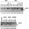
Fig. 2. Generation of mice expressing a truncated form of human HSF1. Analysis of expression of the transgene in adult mice by Western blotting. Whole tissue extracts from the brain (lanes 1 and 8), heart (2 and 9), lung (3 and 10), liver (4 and 11), spleen (5 and 12), stomach (6 and 13) and kidney (7 and 14) in two strains of transgenic mice (B4–6 and B6–6) were prepared, and expression of hHSF1ΔRD (arrows) was examined using antiserum against HSF1. As a control, K562 cells expressing hHSF1ΔRD were analyzed (lanes 15 and 16). To compare the levels of hHSF1ΔRD in male reproductive organs including the testis (lanes 20–22) and epididymis (lanes 23–25) and in the heart (lanes 17–19) in wild-type (WT) and transgenic mice, the same amounts of protein were analyzed as described above. Asterisks indicate non-specific bands.
None
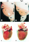
Fig. 3. Gross anatomy of male reproductive organs (A and B) and the heart (C and D) of 4–month-old wild-type (+/+) (A and C) and transgenic B6–6 (Tg/+) (B and D) mice. Note the ∼50% reduction in size of testis in transgenic mice. T, testis; Eh, head of epididymis; Et, tail of epididymis; Ad, adipose tissues.
None
Fig. 4. Transgenic mice showed defective spermatogenesis. Histological analysis of testis sections from 4–month-old wild-type (A) and transgenic mice (B–F). The diameter of seminiferous tubules in transgene-positive mice was reduced by 20–30%, and the number of interstitial Leydig cells was markedly increased (A and B). Many degenerating pachytene spermatocytes with condensed nuclei and eosinophilic cytoplasm (C and D) and giant cells (E) were seen. (G and H) Immunostaining of endogenous HSF1 and ectopically expressed hHSF1ΔRD. Cryosections of testis from wild-type (G) and transgenic (H) adult mice were immunostained with antiserum against HSF1 as the first antibody and peroxidase-conjugated goat anti-rabbit IgG as the second antibody. Signals were detected using a DAB substrate kit. Magnification: A and B, ×200; C–H, ×400.
None
Fig. 5. In situ detection of apoptosis by TUNEL staining. Apoptotic cells were stained brown. Sections were counterstained with methyl green. Apoptotic cells were rare in the testis of wild-type mice (A and C). In contrast, apoptotic cells were abundant in 20–30% of seminiferous tubules (B and D), which are indicated by arrowheads. Magnification: A and B, ×100; C and D, ×400.
None
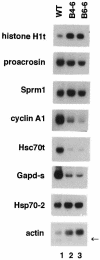
Fig. 6. Analysis of expression of genes specific to spermatogenic cells. Total RNAs were isolated from the testes of 16–week-old wild-type mice and two transgenic lines. Northern blotting was performed using specific probes as described (Dix et al., 1997). The arrow indicates a band of testis-specific isoform of β–actin.
None
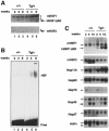
Fig. 7. Spermatogenesis in juvenile mice. (A) Transgene expression in testis of mice at 2 (lanes 1 and 4), 4 (lanes 2 and 5) and 8 (lanes 3 and 6) weeks after birth. Whole testis extracts were prepared and aliquots of proteins were subjected to Western blot analysis using antiserum against HSF1 (upper panel) or HSF2 (lower panel). Asterisks indicate non-specific bands. (B) Acquisition of DNA binding activities in juvenile mice. Tissue extracts prepared as described above were subjected to gel shift assay using a 32P-labeled HSE oligonucleotide as a probe. Free probe (Free) is indicated at the bottom. (C) Analysis of expression of heat shock genes as well as HSF genes during testis development. Total RNAs were isolated from the testes of 2-, 4- and 16–week-old wild-type (+/+) and transgenic (Tg/+) mice. Northern blot analysis was performed using mouse cDNA probes for Hsp110, Hsp70.1 or β–actin, and human cDNA probes for HSF1, HSF2, Hsp90α, Hsp40 or Hsp27. The transcript of the hHSF1ΔRD transgene was expressed at a low level at 2 weeks and showed marked increases during testis development. Testis-specific isoforms of Hsp40 (arrow) and actin (star) are indicated. (D) Histological analysis of testis sections from mice at 2 (a and b), 3 (c and d) and 4 weeks (e and f). At 2 weeks, spermatocytes at pachytene stage were abundant in seminiferous tubules in testes of both wild-type and transgenic mice (a and b). Round spermatids that accomplished meiotic cell division appeared in tubules of 3–week-old wild-type mice (c). Apoptotic pachytene spermatocytes with dense nuclei (arrowheads) were seen in the testis of transgenic mice (12% of tubules) (d). At 4 weeks in transgenic mice, clusters of apoptotic pachytene spermatocytes were observed in 34% of tubules (f). Magnification: ×400.
None
Fig. 8. Germ cell death induced by single exposure to heat. (A) Acquisition of HSF1 DNA binding activity. Testicles of anesthetized wild-type mice were submerged in a water bath at 22°C (lanes 1, 2, 5, 6, 9 and 10) or 43°C (lanes 3, 4, 7, 8, 11 and 12) for 15 min and whole tissue extracts were prepared immediately after heat shock. Gel shift assay was performed using a 32P-labeled HSE oligonucleotide as a probe in the presence of preimmune serum (lanes 1–4), anti-HSF1 serum (lanes 5–8) or anti-HSF2 serum (lanes 9–12). (B) Histological analysis of germ cell death in wild-type (a and c) and transgenic (b and d) mice induced by heat shock. At 24 h after heat treatment at 43°C for 15 min, testes were dissected, fixed and embedded in paraffin. Sections 5 μm thick were stained with hematoxylin and eosin (a and b) or stained for apoptotic cells by the TUNEL method (c and d). Non-specific TUNEL staining was observed in the interstitial Leydig cells. An arrowhead in b indicates a tubulus having many vacuoles. Seminiferous tubules containing a cluster of apoptotic cells are indicated by arrows in c and d. Magnification: a, b and d, ×200; c, ×100.

References

    1. Alastalo T.-P., Lönnström, M., Leppä, S., Kaarniranta, K., Pelto-Huikko, M., Sistonen, L. and Parvinen, M. (1998) Stage-specific expression and cellular localization of the heat shock factor 2 isoforms in the rat seminiferous epithelium. Exp. Cell Res., 240, 16–27. - PubMed
    1. Andersen B., Pearse, R.V., Schlegel, P.N., Cichon, Z., Schonemann, M.D., Bardin, C.W. and Rosenfeld, M.G. (1993) Sperm 1: a POU-domain gene transiently expressed immediately before meiosis I in the male germ cells. Proc. Natl Acad. Sci. USA, 90, 11084–11088. - PMC - PubMed
    1. Baler R., Dahl, G. and Voellmy, R. (1993) Activation of human heat shock genes is accompanied by oligomerization, modification and rapid translocation of heat shock transcription factor HSF1. Mol. Cell. Biol., 13, 2486–2496. - PMC - PubMed
    1. Bartell J.G., Davis, T., Kremer, E.J., Dewey, M.J. and Kistler, W.S. (1996) Expression of the rat testis-specific histone H1t gene in transgenic mice. J. Biol. Chem., 23, 4046–4054. - PubMed
    1. Chowdhury A.K. and Steinberger, E. (1970) Early changes in the germinal epithelium of rat testes following exposure to heat. J. Reprod. Fert., 22, 205–212. - PubMed

Publication types

MeSH terms

LinkOut - more resources