

Review
doi: 10.1083/jcb.149.1.13.
Pores in the wall: claudins constitute tight junction strands containing aqueous pores
Affiliations
- PMID: 10747082
- PMCID: PMC2175101
- DOI: 10.1083/jcb.149.1.13
Item in Clipboard
Review
Pores in the wall: claudins constitute tight junction strands containing aqueous pores
J Cell Biol.
.
No abstract available
Figures
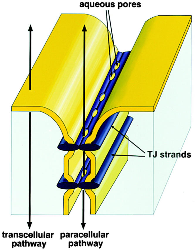
Schematic drawing of TJs. At the most apical region of lateral membranes, TJs occur between two adjacent cells. TJs are composed of paired TJ strands, in which each TJ strand laterally and tightly associates with that in the apposing membrane of adjacent cells. Paired TJ strands are thought to contain aqueous pores. In transcellular and paracellular pathways, materials move across plasma membranes and TJs, respectively.

Membrane folding model of claudin-1. Both NH2 and COOH termini are located in the cytoplasm. Each circle corresponds to each amino acid residue.
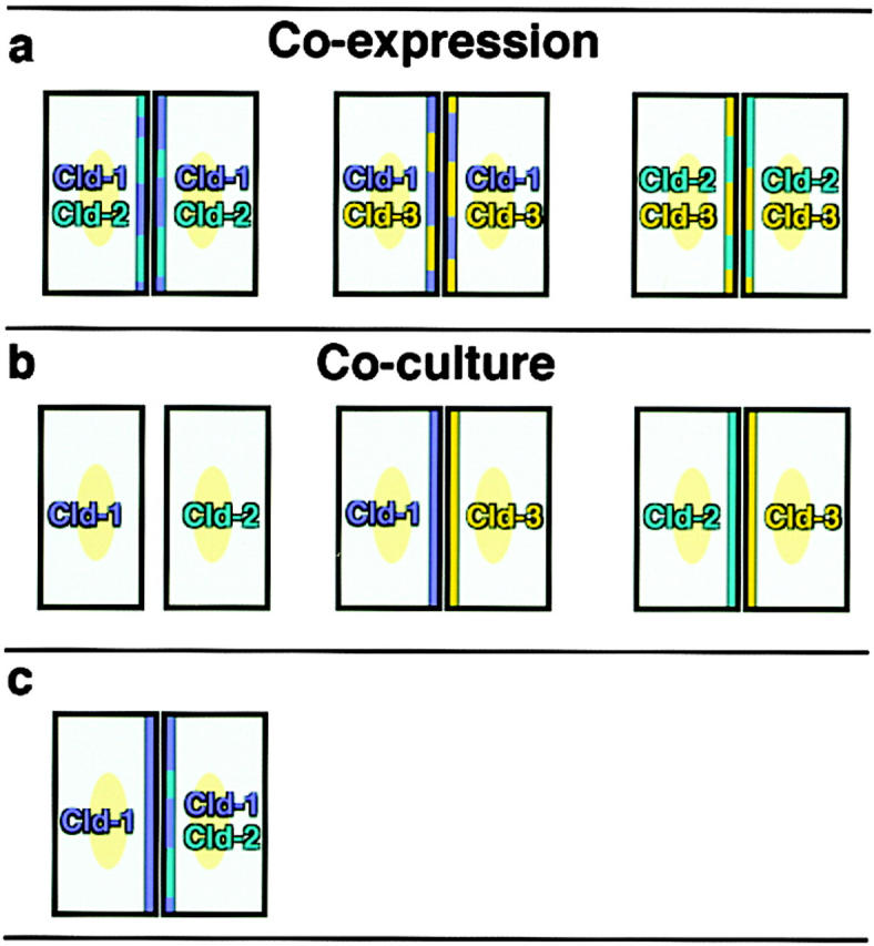
Manner of interaction of heterogeneous claudin species within and between TJ strands. (a) L transfectants coexpressing two of claudin-1, -2, and -3 were cultured. (b) Two of the L transfectants singly expressing claudin-1, -2, or -3 were cocultured. (c) L transfectants expressing claudin-1 were cocultured with those coexpressing claudin-1 and -2.
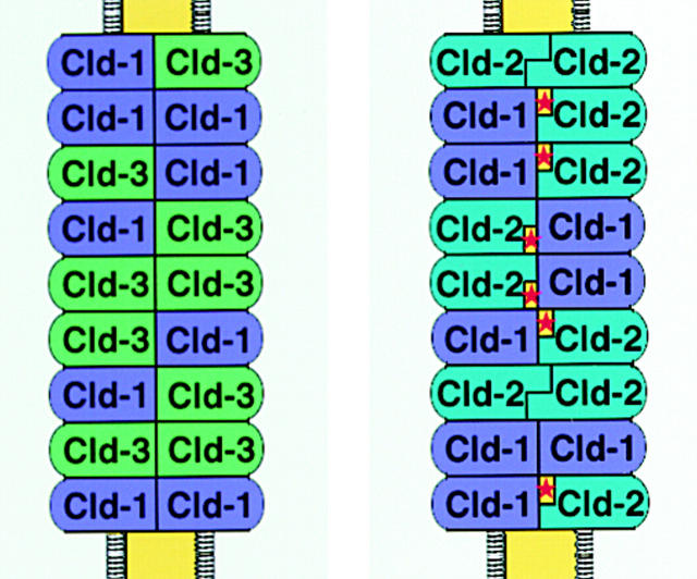
Two types of paired strands: one composed of claudin-1 and -3 (left), and the other composed of claudin-1 and -2 (right). Asterisks, putative pores. See text for details.
References
-
- Anderson J.M., van Itallie C.M. Tight junctions and the molecular basis for regulation of paracellular permeability. Am. J. Physiol. 1995;269:G467–G475. - PubMed
-
- Balda M.S., Whitney J.A., Flores C., González S., Cereijido M., Matter K. Functional dissociation of paracellular permeability and transepithelial electrical resistance and disruption of the apical-basolateral intramembrane diffusion barrier by expression of a mutant tight junction membrane protein. J. Cell Biol. 1996;134:1031–1049. - PMC - PubMed
-
- Briehl M.M., Miesfeld R.L. Isolation and characterization of transcripts induced by androgen withdrawal and apoptotic cell death in the rat ventral prostate. Mol. Endocrinol. 1991;5:1381–1388. - PubMed
-
- Bronstein J.M., Popper P., Micevych P.E., Farber D.B. Isolation and characterization of a novel oligodendrocyte-specific protein. Neurology. 1996;47:772–778. - PubMed
Publication types
MeSH terms
Substances
LinkOut - more resources
Full Text Sources
Other Literature Sources
Molecular Biology Databases
