. 2000 Apr 3;149(1):17-22.
doi: 10.1083/jcb.149.1.17.

Keratin-dependent, epithelial resistance to tumor necrosis factor-induced apoptosis

Affiliations

Keratin-dependent, epithelial resistance to tumor necrosis factor-induced apoptosis

C Caulin et al. J Cell Biol. .

Abstract

Tumor necrosis factor (TNF) is a cytokine produced by macrophages and T lymphocytes that acts through two distinct receptors, TNFR1 (60 kD, CD120a) and TNFR2 (80 kD, CD120b), to affect cellular proliferation, differentiation, survival, and cell death. In addition to its proinflammatory actions in mucosal tissue, TNF is important for liver regeneration. Keratin 8 (K8) and keratin 18 (K18) form intermediate filaments characteristic of liver and other single cell layered, internal epithelia and their derivative cancers. K8-deficient (K8(-)) mice, which escape embryonic lethality, develop inflammatory colorectal hyperplasia, mild liver abnormalities, and tolerate hepatectomy poorly. We show that normal and malignant epithelial cells deficient in K8 and K18 are approximately 100 times more sensitive to TNF-induced death. K8 and K18 both bind the cytoplasmic domain of TNFR2 and moderate TNF-induced, Jun NH(2)-terminal kinase (JNK) intracellular signaling and NFkappaB activation. Furthermore, K8(-) and K18(-) mice are much more sensitive to TNF dependent, apoptotic liver damage induced by the injection of concanavalin A. This moderation of the effects of TNF may be the fundamental function of K8 and K18 common to liver regeneration, inflammatory bowel disease, hepatotoxin sensitivity, and the diagnostic, persistent expression of these keratins in many carcinomas.


Figures

Figure 1
Figure 1
Keratin-dependent TNF sensitivity. Cells were incubated for 12 h with the indicated amounts of TNF in the presence of 5 μg/ml CHX (a), or for 24 h with TNF alone (b), CHX alone (c), or daunomycin (DNM; d). Cell survival is expressed as the mean of triplicate samples ± SD. Filled circles and diamonds represent HR9 and a stable HR9 transfectant expressing wild-type K18, respectively. Open triangles and squares are HR-1 and HR-7 clones, respectively, that express dominant negative K18 (Kulesh et al. 1989).
Figure 2
Figure 2
TNF sensitivity of MDA-MB-435 and CMK8 epithelial cells. a, K8-transfected MDA-MB-435 cells (filled squares) and K8 negative cells (open circles) were treated for 12 h with the indicated amounts of TNF in the presence of 15 μg/ml of CHX. Cell survival was determined with the MTT assay. Data represent the mean ± SD of three independent K8 and three K8+ clones. b, the K8 CMK8 epithelial cell line was transfected with the mouse K8 gene. TNF sensitivity was determined in the presence of 2.5 μg/ml of CHX. Open circles represent the mean of three keratin negative clones. Filled squares represent the mean of three keratin positive clones.
Figure 3
Figure 3
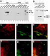
Binding of K8 and K18 to members of the TNFR family. a, GST fusion proteins (5 μg) of the cytoplasmic domains of different members of the TNFR family were incubated with 35S-methionine labeled, in vitro translated, K8 (lanes 1–7) or K18 (lanes 8–14). Input samples (lanes 15 and 16) represent 1/10 of the amount incubated with the fusion proteins. b, In vitro translated K18 (lanes 1 and 2) or caspase-6–cleaved K18 (lanes 3 and 4) was incubated with GST-TNFR2 (lanes 2 and 4) or GST-Fas (lane 1). Binding products were analyzed by SDS-PAGE and fluorography. c–h, Double immunofluorescent staining of K18 and TNFR2. HR9 (c–e) and SW13 cells stably transfected with K8 and K18 (f–h) were transiently transfected with TNFR2 and stained with polyclonal antiserum (c) or mAb for K18 (f) and mAb for TNFR2 (d and g). Colocalization of K18 and TNFR2 (e and h) was visualized in single confocal sections.
Figure 4
Figure 4
Downstream signaling induced by TNF. a, JNK activity after stimulation with TNF (10 ng/ml) in HR9 and HR-7 cells. Autoradiographic exposures of GST-c-jun after incubation with immunoprecipitated JNK and radioactive ATP are shown. b, Densitometric analysis of the films shown in a. Filled bars, HR9; hatched bars, HR-7. c, Normalized luciferase activity of NFκB and Ets2 (d) reporter genes in HR9 (black bars) and HR-7 (hatched bars) after stimulation with 1 or 10 ng/ml of TNF. Results are represented as a percentage of the maximum activity.
Figure 5
Figure 5
ConA-induced liver damage and apoptosis. a and b, The activity of liver transaminases in the serum of K18 (a) or K8 (b) mice. Serum activity of aspartate aminotransferase (black bars) and aspartate aminotransferase (hatched bars) was determined 8 h after i.v injection of 30 mg/kg ConA. c–h, Apoptosis in livers from K18 mice (c), K8 (d), and control mice (e and f) challenged with ConA was detected by terminal deoxynucleotidyl transferase TUNEL method. f, Represents a magnification of the insert area in e, g and h. Hematoxylin and eosin staining. f and g, Represent serial sections of the same liver. Note the presence of pyknotic nuclei in g, in areas that are TUNEL positive (f). Infiltrating lymphocytes in this area are indicated by arrows in h.

References

    1. Akerman P., Cote P., Yang S.Q., McClain C., Nelson S., Bagby G.J., Diehl A.M. Antibodies to tumor necrosis factor-alpha inhibit liver regeneration after partial hepatectomy. Am. J. Physiol. 1992;263:G579–G585. - PubMed
    1. Ashkenazi A., Dixit V.M. Death receptorssignaling and modulation. Science. 1998;281:1305–1308. - PubMed
    1. Baribault H., Price J., Miyai K., Oshima R.G. Mid-gestational lethality in mice lacking keratin 8. Genes Dev. 1993;7:1191–1202. - PubMed
    1. Baribault H., Penner J., Iozzo R.V., Wilson-Heiner M. Colorectal hyperplasia and inflammation in keratin 8-deficient FVB/N mice. Genes Dev. 1994;8:2964–2973. - PubMed
    1. Blazka M.E., Wilmer J.L., Holladay S.D., Wilson R.E., Luster M.I. Role of proinflammatory cytokines in acetaminophen hepatotoxicity. Toxicol. Appl. Pharmacol. 1995;133:43–52. - PubMed

Publication types

MeSH terms