. 2000 Apr 3;149(1):125-40.
doi: 10.1083/jcb.149.1.125.

Dynamic localization of protein phosphatase type 1 in the mitotic cell cycle of Saccharomyces cerevisiae

Affiliations

Dynamic localization of protein phosphatase type 1 in the mitotic cell cycle of Saccharomyces cerevisiae

A Bloecher et al. J Cell Biol. .

Abstract

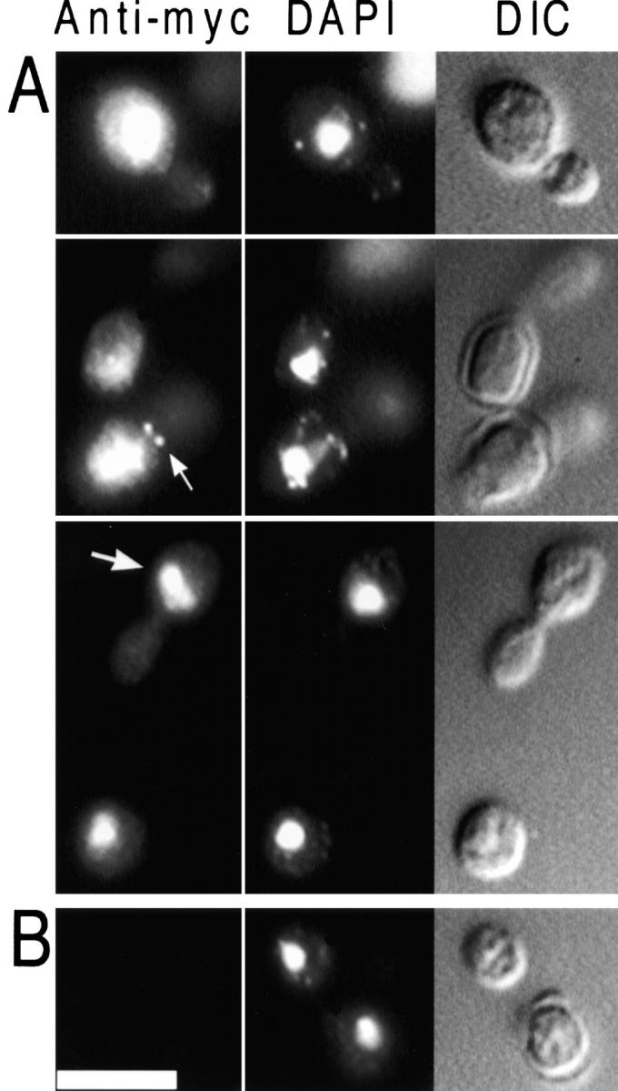
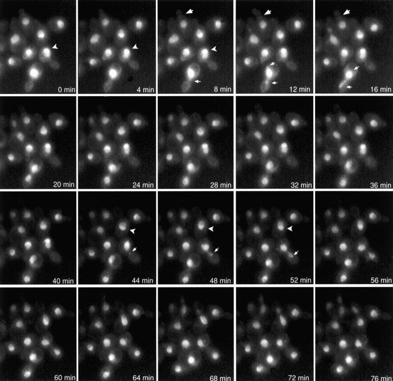
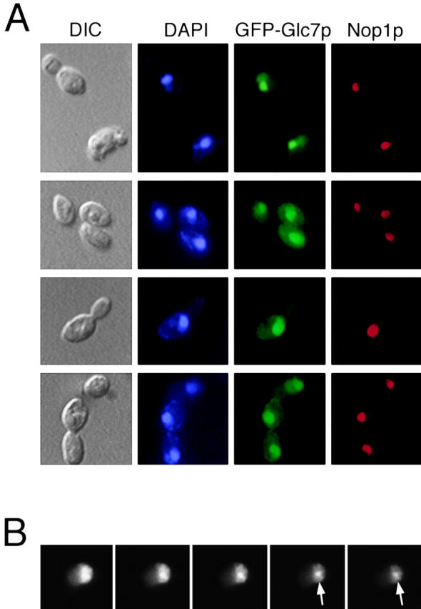
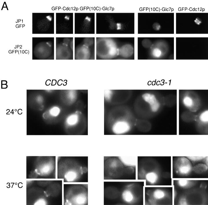
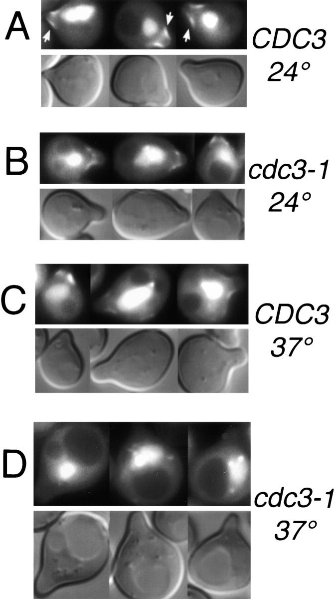
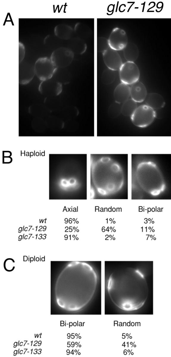
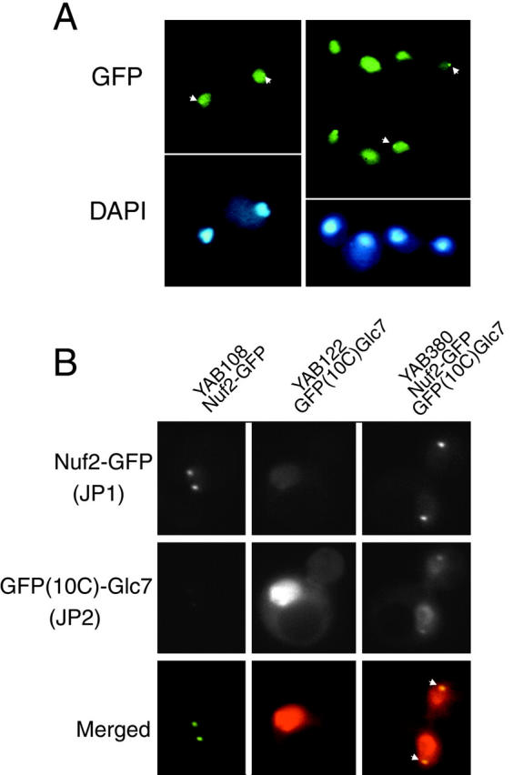
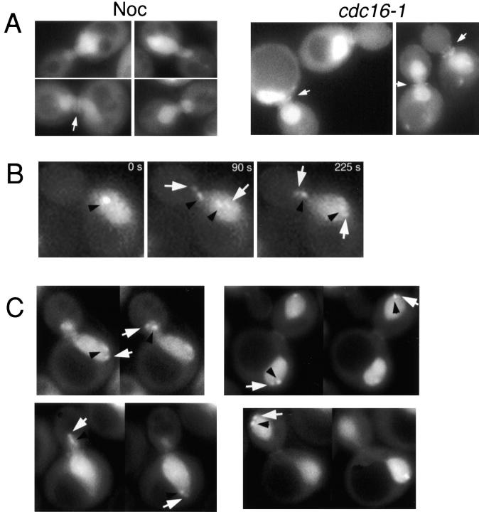
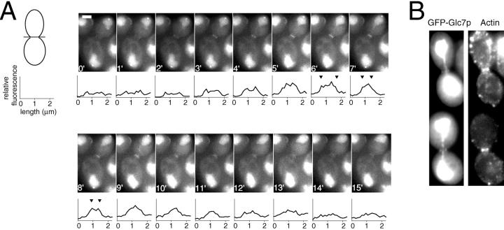
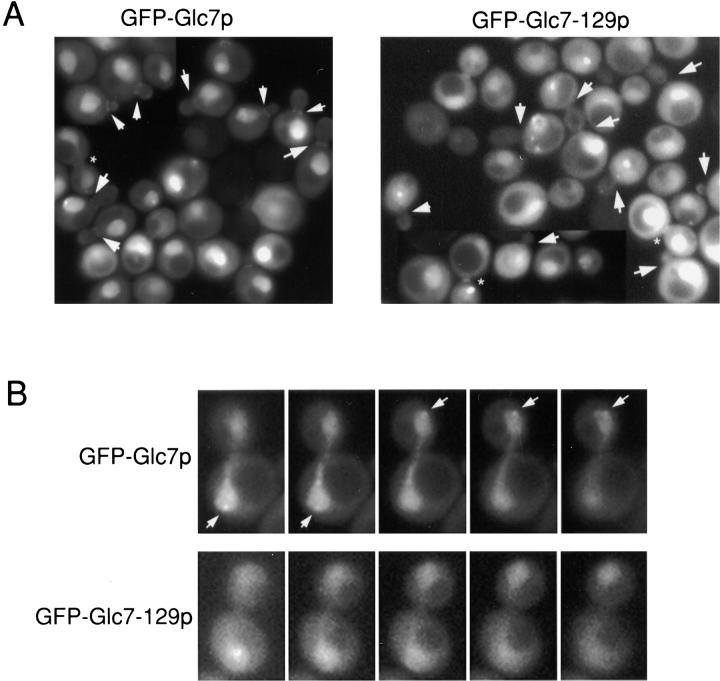
Protein phosphatase type I (PP1), encoded by the single essential gene GLC7 in Saccharomyces cerevisiae, functions in diverse cellular processes. To identify in vivo subcellular location(s) where these processes take place, we used a functional green fluorescent protein (GFP)-Glc7p fusion protein. Time-lapse fluorescence microscopy revealed GFP-Glc7p localizes predominantly in the nucleus throughout the mitotic cell cycle, with the highest concentrations in the nucleolus. GFP-Glc7p was also observed in a ring at the bud neck, which was dependent upon functional septins. Supporting a role for Glc7p in bud site selection, a glc7-129 mutant displayed a random budding pattern. In alpha-factor treated cells, GFP-Glc7p was located at the base of mating projections, again in a septin-dependent manner. At the start of anaphase, GFP-Glc7p accumulated at the spindle pole bodies and remained there until cytokinesis. After anaphase, GFP-Glc7p became concentrated in a ring that colocalized with the actomyosin ring. A GFP-Glc7-129 fusion was defective in localizing to the bud neck and SPBs. Together, these results identify sites of Glc7p function and suggest Glc7p activity is regulated through dynamic changes in its location.


Figures

Figure 1
Figure 1
Localization of myc-Glc7p. Diploid strain KT1208/1210 (A), which contains myc-GLC7, and the control strain SB90/91 (B), which lacks myc-GLC7, were harvested from log phase in YPD medium, fixed, and prepared for indirect immunofluorescence microscopy as described in the Materials and Methods. myc-Glc7p was visualized using 9E10 primary antibody (left), DNA was visualized with DAPI (middle) and cells were visualized with DIC optics (right). Bar, 5 μm.
Figure 2
Figure 2
Localization of GFP–Glc7p throughout the mitotic cell cycle. YAB4 cells were placed on an agarose pad containing synthetic complete medium and visualized by fluorescence microscopy as described (Waddle et al. 1996). Each image is a 2D projection from 10 z-axis planes, each offset by 0.5 μm. Images are shown at 4-min intervals. Large arrows denote bud neck fluorescence. Carets indicate subnuclear fluorescence. Small arrows denote spots observed at nuclear periphery of cells in anaphase.
Figure 3
Figure 3
GFP–Glc7p is highly concentrated in the nucleolus. A, GFP–Glc7p cells (YAB4) were fixed and stained for DNA (DAPI) and Nop1p (a known nucleolar protein) as described in Materials and Methods. GFP–Glc7p was visualized by its native fluorescence. B, Serial z-sections through a live cell (YAB4) reveal the nonuniform nucleolar fluorescence pattern of GFP–Glc7p. Arrows indicate a bright spot commonly associated with the nucleus/nucleolus.
Figure 4
Figure 4
The GFP–Glc7p ring at the mother/bud neck is dependent upon septins. A, Images of cells containing Cdc12-GFPm2 (YAB453), GFP(10C)–Glc7p (YAB122), or Cdc12-GFPm2 and GFP (10C)–Glc7p (YAB441) were captured using the JP1 or JP2 filter sets as described in Materials and Methods. The top row shows images of Cdc12-GFP fluorescence and the bottom row shows images of GFP(10C)–Glc7p fluorescence. The first four columns show images of Cdc12-GFP, GFP(10C)–Glc7p cells in different stages of the cell cycle. The fifth and sixth columns include control images of a GFP(10C)–Glc7p cell and a Cdc12-GFP cell showing lack of significant bleed-through into the JP1 and JP2 filter sets, respectively. B, Images of wild-type GFP–GLC7 (KT1556) or cdc3-1 GFP–GLC7 (YAB58) cells growing logarithmically at the permissive temperature (24°C) or after a shift to the nonpermissive temperature (37°C) for 50 min.
Figure 5
Figure 5
GFP–Glc7p localizes to the base of the mating projection. Images of GFP–Glc7p or DIC in wild-type (YAB1556; A and C) or cdc3-1 cells (YAB58; B and D) that were arrested in α-factor (8 μg/ml) for 3 h at the permissive temperature (24°C; A and B), or after a 40-min shift to the nonpermissive temperature (37°C; C and D). Arrows in A indicate GFP–Glc7p fluorescence at the base of the mating projection.
Figure 6
Figure 6
glc7-129 mutants exhibits a random budding pattern. A, Images of wild-type and glc7-129/glc7-129 diploid cells stained with calcofluor. Note that the normalization values for these two images are identical, illustrating the increased level of calcofluor staining in glc7-129 mutants. B, Haploid wild-type (KT1112) and glc7-129 strains (YAB305) were scored for their budding pattern. n = 210 for wild-type cells, n = 211 for glc7-129 cells. C, Diploid wild-type (YAB107) and glc7-129/glc7-129 strains (YAB86) were scored as in A. n = 198 for wild-type cells, n = 186 for glc7-129 cells.
Figure 7
Figure 7
GFP–Glc7p localizes to SPBs in late mitosis. A, GFP–Glc7p and DAPI fluorescence of live, logarithmically growing cells of the rho0strain, YAB6. B, Images of logarithmically growing cells containing Nuf2–GFP (YAB108), GFP(10C)–Glc7p (YAB122), or both Nuf2–GFP and GFP(10C)–Glc7p (YAB380) captured with JP1 and JP2 filters as described in Materials and Methods. The top row shows images of Nuf2–GFP fluorescence, the middle row shows images of GFP(10C)–Glc7p fluorescence, and in the bottom row are merged images of Nuf2–GFP and GFP(10C)–Glc7p fluorescence. Areas of colocalization are yellow (arrows) in these pseudocolored images.
Figure 8
Figure 8
GFP–Glc7p accumulates at the SPBs in early anaphase. A, Images of nocodazole (Noc)-arrested GFP–Glc7p cells (YAB4) or GFP–Glc7p cells containing cdc16-1 (YAB381) after incubation at the nonpermissive temperature for 3 h. Arrows indicate the double ring of GFP–Glc7p often observed in these arrested cells. B, Logarithmically growing GFP–Glc7p cells containing lacI-GFP/lacO marking centromeres (YAB439) were placed on an agarose pad containing synthetic complete media and imaged using a custom script written for IPLab Spectrum software (see Materials and Methods). The black arrowheads denote the lacI-GFP spots (marking the centromeres), whereas the white arrows mark the GFP–Glc7p spots at SPBs. C, Serial z-sections were taken through YAB439 cells in various stages of anaphase. Black arrowheads denote centromeres, white arrows denote GFP–Glc7p at SPBs.
Figure 9
Figure 9
GFP–Glc7p colocalizes with the actomyosin ring in late mitosis. A, Logarithmically growing GFP–Glc7p cells (YAB4) in YPD media were imaged at 1-min intervals as described in Materials and Methods. The schematic of a yeast cell is drawn to demonstrate how the line was drawn across the bud neck to quantify fluorescence. The size bar in the 0′ image represents the actual length of the line drawn across the bud neck (2.25 μm). Carets in the graphs at 6′, 7′, and 8′ mark outer edges of the GFP–Glc7p fluorescence at the bud neck. B, GFP–Glc7p cells (YAB4) were fixed and stained with rhodamine-phalloidin to visualize the actomyosin ring. Shown are two cells displaying colocalization of the GFP–Glc7p ring with the actomyosin ring.
Figure 10
Figure 10
GFP–Glc7-129p is defective in localization to the bud neck and SPBs. A, Images of wild-type strain KT1357 transformed with either pAB26 (GFP–GLC7) or pAB18 (GFP–glc7-129) after inoculation into YPD media for 4 h at 30°C. Arrows indicate bud neck localization of GFP–Glc7p and the lack of this localization for GFP–Glc7-129p. Asterisks denote GFP–Glc7p or GFP–Glc7-129p at the presumed actomyosin ring. For ease of comparison, grayscale values for GFP–Glc7-129p were increased to approximately the same levels as those of GFP–Glc7p. B, Serial z-sections are shown through anaphase KT1357 cells containing pAB26 or pAB18. Arrows indicate GFP–Glc7p spots at SPBs. This localization is not observed for GFP–Glc7-129p.

References

    1. Adams A., Pringle J. Relationship of actin and tubulin distribution to bud growth in wild-type and morphogenetic-mutant Saccharomyces cerevisiae . J. Cell Biol. 1984;98:934–945. - PMC - PubMed
    1. Alexandru G., Zachariae W., Schleiffer A., Nasmyth K. Sister chromatid separation and chromosome re-duplication are regulated by different mechanisms in response to spindle damage. EMBO (Eur. Mol. Biol. Organ.) J. 1999;18:2707–2721. - PMC - PubMed
    1. Andreassen P.R., Lacroix F.B., Villa-Moruzzi E., Margolis R.L. Differential subcellular localization of protein phosphatase-1 alpha, gamma1, and delta isoforms during both interphase and mitosis in mammalian cells. J. Cell Biol. 1998;141:1207–1215. - PMC - PubMed
    1. Andrews P.D., Stark M.J. Type 1 protein phosphatase is required for maintenance of cell wall integrity, morphogenesis and cell cycle progression in Saccharomyces cerevisiae . J Cell Sci. 2000;113:507–520. - PubMed
    1. Aris J.P., Blobel G. Identification and characterization of a yeast nucleolar protein that is similar to a rat liver nucleolar protein. J. Cell Biol. 1988;107:17–31. - PMC - PubMed

Publication types

MeSH terms