. 2000 Apr;38(4):1331-8.
doi: 10.1128/JCM.38.4.1331-1338.2000.

New Ehrlichia species closely related to Ehrlichia chaffeensis isolated from Ixodes ovatus ticks in Japan

Affiliations

New Ehrlichia species closely related to Ehrlichia chaffeensis isolated from Ixodes ovatus ticks in Japan

S Shibata et al. J Clin Microbiol. 2000 Apr.

Abstract

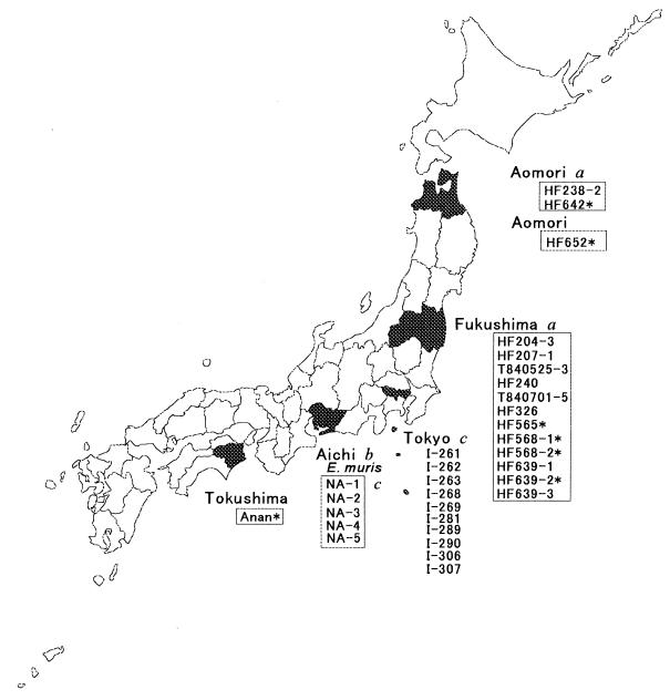
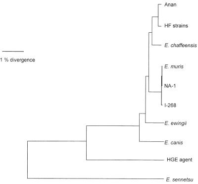
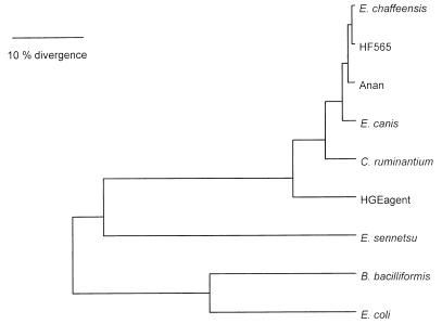
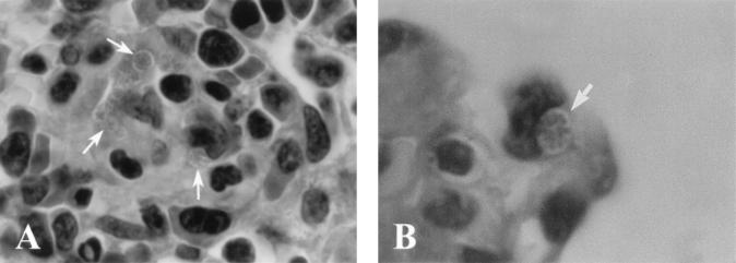
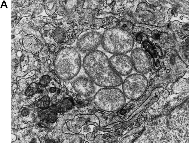
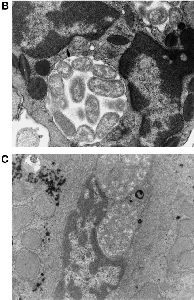
Seven Ehrlichia strains (six HF strains and one Anan strain) that were obtained from laboratory mice by intraperitoneally inoculating homogenates of adult Ixodes ovatus collected in Japan were characterized. 16S rRNA sequences of all six HF strains were identical, and the sequences were 99.7, 98.2, and 97.7% identical to those of Anan strain, Ehrlichia chaffeensis (human monocytic ehrlichiosis agent), and E. muris, respectively. Partial GroEL amino acid sequencing also revealed that the six HF strains had identical sequences, which were 99.0, 98.5, and 97.3% identical to those of E. chaffeensis, the Anan strain, and E. canis, respectively. All HF strains were lethal to mice at higher dosages and intraperitoneal inoculation, whereas the Anan or E. muris strain induced only mild clinical signs. Light and electron microscopy of moribund mice inoculated with one of the HF strains revealed severe liver necrosis and the presence of numerous ehrlichial inclusions (morulae) in various organs. The study revealed that members of E. canis genogroup are naturally present in Ixodes ticks. HF strains that can cause severe illness in immunocompetent laboratory mice would be valuable in studying the pathogenesis and the roles of both cellular and humoral immune responses in ehrlichiosis caused by E. canis genogroup.


Figures

FIG. 1
FIG. 1
Geographic region where Ehrlichia spp. were isolated from ticks or wild mice. Tick isolates are boxed. An asterisk indicates an isolate used for this study. Letters designate sources as follows: a, Fujita and Watanabe (7); b, Kawahara et al. (12); and c, Kawahara et al. (14).
FIG. 2
FIG. 2
Phylogenetic relationships between Anan, HF, and other Ehrlichia strains based on 16S rRNA gene sequence comparisons. HF strains included HF565, HF568-1, HF568-2, HF639-2, HF642, and HF652.
FIG. 3
FIG. 3
Phylogenetic relationships generated with UPGMA based on an alignment of the first 409 amino acid sequences of GroEL of HF and Anan strains and other members of the tribe Ehrlichieae.
FIG. 4
FIG. 4
Light micrographs of paraffin-embedded section of the thymus (A) and the lung (B) from the mice intraperitoneally inoculated with HF565 strain at day 9 p.i. Note several morulae packed with many ehrlichial organisms (colonies of ehrlichiae) along the capillary (arrows) (A) [magnification, ×1,070]) and in the capillary of the alveolar wall (arrows) (B) [magnification, ×1,700]). Hemtoxylin and eosin staining was used.
FIG. 5
FIG. 5
Electron micrographs of morulae of HF565 in the cytoplasm of a monocyte (A) and an eosinophil (B) in the blood vessel in the spleen and in a Kupffer cell in the liver (C) from the mouse at day 7 p.i. Note the numerous pleomorphic coccobacilli enveloped in two layers of membranes embedded in a fine filamentous matrix in the membrane-bound inclusion in panel A. An intramorular tubule is indicated by the arrow in panel B. The inclusions are tightly packed with ehrlichiae without intramorular space in the Kupffer cells adhered to the endothelial layer lining the sinusoid. Magnifications: ×24,200 (A), ×21,000 (B), and ×16,400 (C).
FIG. 5
FIG. 5
Electron micrographs of morulae of HF565 in the cytoplasm of a monocyte (A) and an eosinophil (B) in the blood vessel in the spleen and in a Kupffer cell in the liver (C) from the mouse at day 7 p.i. Note the numerous pleomorphic coccobacilli enveloped in two layers of membranes embedded in a fine filamentous matrix in the membrane-bound inclusion in panel A. An intramorular tubule is indicated by the arrow in panel B. The inclusions are tightly packed with ehrlichiae without intramorular space in the Kupffer cells adhered to the endothelial layer lining the sinusoid. Magnifications: ×24,200 (A), ×21,000 (B), and ×16,400 (C).

References

    1. Anderson B E, Dawson J E, Jones D C, Wilson K H. Ehrlichia chaffeensis, a new species associated with human ehrlichiosis. J Clin Microbiol. 1991;29:2838–2842. - PMC - PubMed
    1. Anderson B E, Greene C E, Jones D C, Dawson J E. Ehrlichia ewingii sp. nov., the etiologic agent of canine granulocytic ehrlichiosis. Int J Syst Bacteriol. 1992;42:299–302. - PubMed
    1. Anderson B E, Sims K G, Olson J G, Childs J M, Piesman J F, Happ C M, Maupin G O, Johnson B J B. Amblyomma americanum: a potential vector of human ehrlichiosis. Am J Trop Med Hyg. 1993;49:239–244. - PubMed
    1. Buller R S, Arens M, Hmiel S P, Paddock C, Sumner J W, Rikihisa Y, Unver A, Graudreauls-Keener M, Marinian F A, Liddell A M, Schmulewitz N, Storch G. Ehrlichia ewingii, a newly recognized agent of human ehrlichiosis. New Engl J Med. 1999;341:148–155. - PubMed
    1. Chen S-M, Dumler J S, Bakken J S, Walker D. Identification of granulocytic Ehrlichia species as the etiologic agent of human disease. J Clin Microbiol. 1994;32:585–595. - PMC - PubMed

Publication types

MeSH terms

Associated data

LinkOut - more resources