. 2000 Apr 1;524 Pt 1(Pt 1):279-92.
doi: 10.1111/j.1469-7793.2000.t01-2-00279.x.

Regional blood flow and nociceptive stimuli in rabbits: patterning by medullary raphe, not ventrolateral medulla

Affiliations

Regional blood flow and nociceptive stimuli in rabbits: patterning by medullary raphe, not ventrolateral medulla

W W Blessing et al. J Physiol. .

Abstract

1. Regional blood flow was measured with Doppler ultrasonic probes in anaesthetized rabbits. We used focal microinjections of pharmacological agents to investigate medullary pathways mediating ear pinna vasoconstriction elicited by electrical stimulation of the spinal tract of the trigeminal nerve or by pinching the lip, and pathways mediating mesenteric vasoconstriction elicited by electrical stimulation of the afferent abdominal vagus nerve. 2. Bilateral injection of kynurenate into the rostral ventrolateral medulla reduced arterial pressure and prevented the mesenteric vasoconstriction and the rise in arterial pressure elicited by abdominal vagal stimulation. However, kynurenate did not prevent ear pinna vasoconstriction or the fall in pressure elicited by trigeminal tract stimulation. Similar injections of muscimol also failed to prevent the trigeminally elicited cardiovascular changes. 3. Injections of kynurenate into the raphe-parapyramidal area did not diminish trigeminally elicited ear vasoconstriction or the depressor response. However, injections of muscimol substantially reduced or abolished the trigeminally elicited ear vasoconstriction, without affecting the depressor response. Raphe-parapyramidal muscimol injections also entirely abolished ear vasoconstriction elicited by pinching the rabbit's lip. 4. The trigeminal depressor response does not depend on either the rostral ventrolateral medulla or the raphe-parapyramidal region. 5. Mesenteric vasoconstriction elicited by stimulation of the afferent abdominal vagus nerve is mediated via the rostral ventrolateral medulla, but ear vasoconstriction elicited by lip pinch or by stimulation of the trigeminal tract is mediated by the raphe-parapyramidal region. Our study is the first to suggest a brainstem pathway mediating cutaneous vasoconstriction elicited by nociceptive stimulation.


Figures

Figure 1
Figure 1. RVLM kynurenate
AP, ear and mesenteric (Mes) blood flow and mean vascular conductance (cond) at rest and in response to electrical stimulation of the trigeminal tract or of the afferent abdominal vagus nerve, before (A and B) and after (C and D) bilateral injection of kynurenate into the rostral ventrolateral medulla (RVLM). The vascular conductance signal was normalized to 100 % at the beginning of the recordings in each panel. Between the recordings in A and B an increase in the inspired halothane concentration restored ear blood flow to a higher baseline level. Kynurenate abolished the mesenteric constriction and the rise in AP elicited by abdominal vagal stimulation, but it did not reduce the ear pinna vasoconstriction or the fall in AP elicited by stimulation of the trigeminal tract.
Figure 2
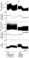
Figure 2. RVLM L-glutamate and GABA
AP, ear pinna and mesenteric blood flow and mean vascular conductance at rest and after unilateral injection of L-glutamate (A) or GABA with high baseline flow (B) into the RVLM. The vascular conductance signal was normalized to 100 % at the beginning of the recordings in each panel.
Figure 3
Figure 3. RVLM muscimol
AP and ear pinna blood flow at rest and in response to electrical stimulation of the trigeminal tract before (A) and after (B and C) bilateral injection of muscimol into the RVLM. Between the records shown in A and B ear blood flow suddenly decreased, apparently spontaneously. Muscimol restored ear blood flow and reduced AP, but it did not abolish the ear vasoconstriction or the fall in AP elicited by stimulation of the trigeminal tract.
Figure 4
Figure 4. raphe–parapyramidal kynurenate
AP and ear pinna blood flow at rest and in response to electrical stimulation of the trigeminal tract before (A) and after (B) injection of kynurenate into the raphe–parapyramidal region. Kynurenate did not abolish the ear vasoconstriction or the fall in AP elicited by stimulation of the trigeminal tract.
Figure 5
Figure 5. raphe–parapyramidal muscimol
A and B, AP, and ear blood flow and vascular conductance at rest and in response to electrical stimulation of the trigeminal tract (A) and pinching of the lip with pliers (B) before injection of muscimol into the raphe–pyramidal region (1 injection of 1 nmol in 100 nl). C and D, when muscimol was injected ear blood flow increased (C). Trigeminal tract stimulation and lip pinching still caused a fall in AP but the fall in ear conductance was abolished (D). The insets in A and Dshow expansions of the ear blood flow signal (vertical scale the same as the Y-axis in A) demonstrating that after injection of muscimol the trigeminally elicited decrease in pulse amplitude was changed to an increase. The vascular conductance signal was normalized to 100 % at the beginning of the recordings shown in each panel.
Figure 6
Figure 6. Injection and stimulation sites
Nissl stains of transverse sections of the medulla oblongata after perfusion of the brain with aldehyde solution. A, reaction for Prussian Blue after electrical stimulation of the trigeminal tract (horizontal and vertical arrows indicate site of stimulation). In B-D, the dark areas are HRP-DAB reaction product obtained after a single injection of muscimol with HRP into the RVLM (B), or after mid-line (1) and 1 mm lateral (2 and 3) injections into the raphe–pyramidal region (C) or after a single injection into the mid-line raphe region (D). Scale bars represent 1.0 mm. Abbreviations: AP, area postrema; Cu, cuneate nucleus; Gr, gracile nucleus; ICP, inferior cerebellar peduncle; IO, inferior olive; mVe, medial vestibular nucleus; nA, nucleus ambiguus; nTS, nucleus tractus solitarii; P, pyramidal tract; RVLM, rostral ventrolateral medulla; Vsp, spinal nucleus of the trigeminal nerve; Vspt, spinal tract of the trigeminal nerve; VII, facial nucleus; XII, hypoglossal nucleus.
Figure 7
Figure 7. Proposed neural pathways
Suggested medullary neural circuitry mediating ear pinna vasoconstriction in response to facial pinch or to stimulation of the spinal tract of the trigeminal nerve (A) and mesenteric vasoconstriction in response to stimulation of the abdominal vagus nerve (B). Trigeminal tract stimulation also causes a fall in AP which results from vasodilatation in mesenteric, renal and skeletal muscle vascular beds, following inhibition of the relevant sympathetic preganglionic neurones by presently unknown pathways, not via RVLM or raphe–parapyramidal presympathetic motoneurones. Abbreviations: EAA, excitatory amino acid; nTS, nucleus tractus solitarii; RVLM, rostral ventrolateral medulla; Vsp, spinal nucleus of the trigeminal nerve.

References

    1. Basbaum AI, Clanton CH, Fields HL. Three bulbospinal pathways from the rostral medulla of the cat: an autoradiographic study of pain modulating systems. Journal of Comparative Neurology. 1978;178:209–224. - PubMed
    1. Blessing WW. Distribution of glutamate decarboxylase-containing neurons in rabbit medulla oblongata with attention to intramedullary and spinal projections. Neuroscience. 1990;37:171–185. - PubMed
    1. Blessing WW. The Lower Brainstem and Bodily Homeostasis. New York: Oxford University Press; 1997.
    1. Blessing WW, Yu YH, Nalivaiko E. Raphe pallidus and parapyramidal neurons regulate ear pinna vascular conductance in the rabbit. Neuroscience Letters. 1999;270:33–36. - PubMed
    1. Bowker RM, Abbott LC, Dilts RP. Peptidergic neurons in the nucleus raphe magnus and the nucleus gigantocellularis: their distributions, interrelationships, and projections to the spinal cord. Progress in Brain Research. 1988;77:95–127. - PubMed

Publication types

MeSH terms

LinkOut - more resources