Review
. 2000 Apr;80(2):767-852.
doi: 10.1152/physrev.2000.80.2.767.

Synaptic control of motoneuronal excitability

Affiliations
Review

Synaptic control of motoneuronal excitability

J C Rekling et al. Physiol Rev. 2000 Apr.

Abstract

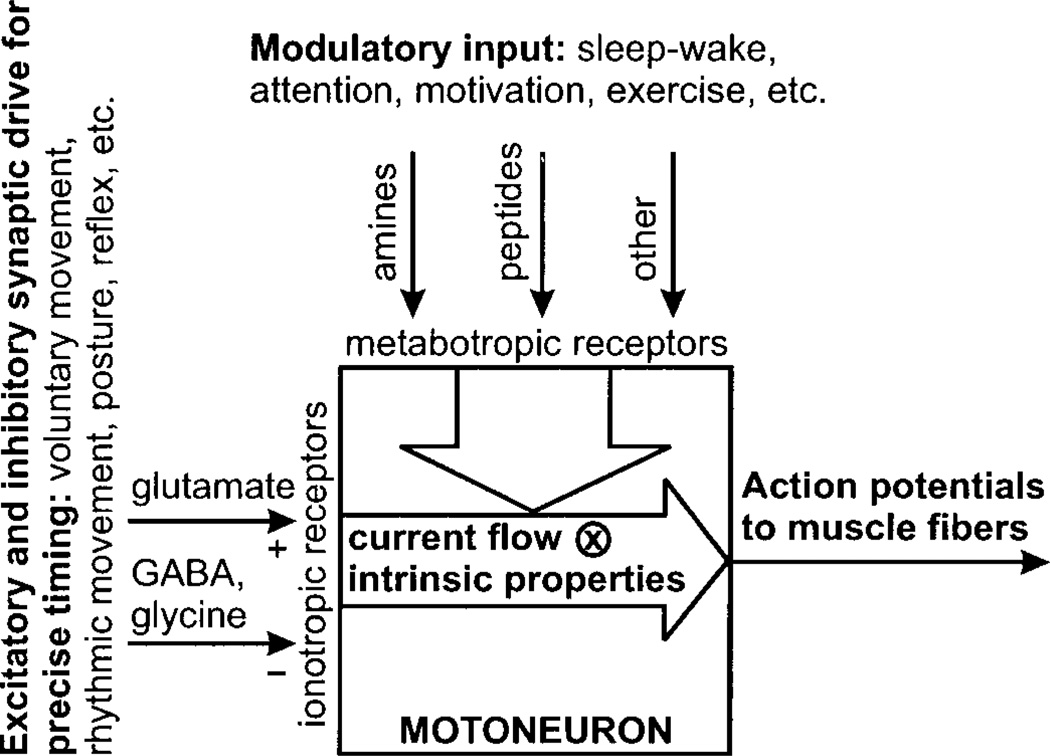
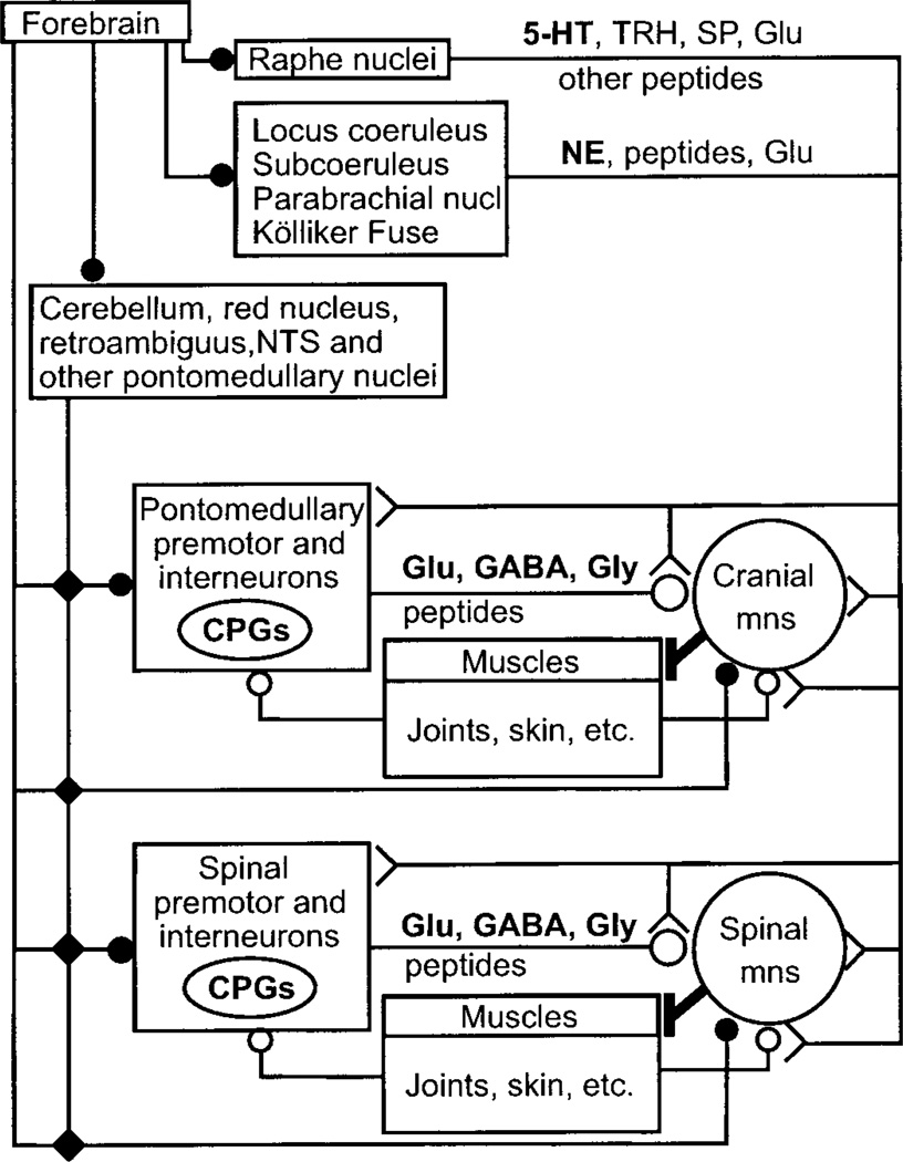
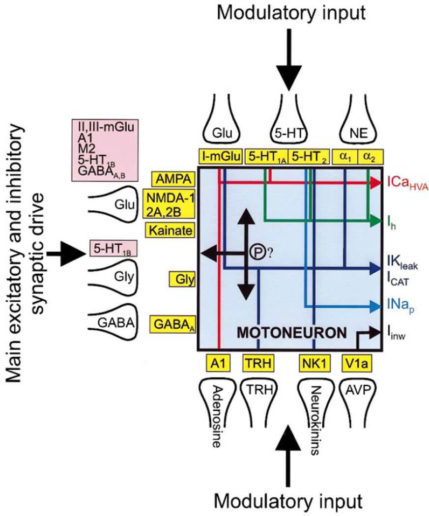
Movement, the fundamental component of behavior and the principal extrinsic action of the brain, is produced when skeletal muscles contract and relax in response to patterns of action potentials generated by motoneurons. The processes that determine the firing behavior of motoneurons are therefore important in understanding the transformation of neural activity to motor behavior. Here, we review recent studies on the control of motoneuronal excitability, focusing on synaptic and cellular properties. We first present a background description of motoneurons: their development, anatomical organization, and membrane properties, both passive and active. We then describe the general anatomical organization of synaptic input to motoneurons, followed by a description of the major transmitter systems that affect motoneuronal excitability, including ligands, receptor distribution, pre- and postsynaptic actions, signal transduction, and functional role. Glutamate is the main excitatory, and GABA and glycine are the main inhibitory transmitters acting through ionotropic receptors. These amino acids signal the principal motor commands from peripheral, spinal, and supraspinal structures. Amines, such as serotonin and norepinephrine, and neuropeptides, as well as the glutamate and GABA acting at metabotropic receptors, modulate motoneuronal excitability through pre- and postsynaptic actions. Acting principally via second messenger systems, their actions converge on common effectors, e.g., leak K(+) current, cationic inward current, hyperpolarization-activated inward current, Ca(2+) channels, or presynaptic release processes. Together, these numerous inputs mediate and modify incoming motor commands, ultimately generating the coordinated firing patterns that underlie muscle contractions during motor behavior.


Figures

FIG. 1
FIG. 1
Overview of control of motoneuronal excitability. Precise timing of voluntary movements, rhythmic movements, afferent reflexes, and other motor acts is mediated primarily by excitatory and inhibitory synaptic drive to motoneurons using glutamate, GABA, and glycine. These transmitters activate ionotropic receptors generating synaptic current in motoneurons, which is convolved with intrinsic membrane properties to produce action potentials, which trigger muscle contraction. Modulatory systems, using amines, peptides, and other transmitters, act (mostly) through metabotropic receptors to modify excitability via changes in postsynaptic ion channel function and presynaptic release processes. These various modulatory systems produce changes in excitability related to the sleep-wake cycle, motivation, and exercise.
FIG. 2
FIG. 2
Attenuation of synaptic potentials along a dendrite of a motoneuron in culture. Top traces: simultaneous recordings from 2 electrodes of spontaneous excitatory postsynaptic potentials (EPSP) from the soma and a dendrite. Bottom trace: difference of the 2 recordings. Note that early and late parts of the trace are dominated by somatic and dendritic EPSP, respectively. [Adapted from Larkum et al. (706).]
FIG. 3
FIG. 3
Supra- and subthreshold membrane behavior of motoneurons. A: ionic currents underlying the action potential waveform. B: ionic currents underlying subthreshold membrane behavior, in this case, elicited by a short-lasting depolarizing/hyperpolarizing square current pulse. C: different phases of adaptation during repetitive firing and postdischarge hyperpolarization after a long-lasting current pulse. Unless noted, currents are activated at times indicated. For definitions, see Table 1 and section iiC.
FIG. 4
FIG. 4
Anatomical organization of synaptic input to motoneurons. Main synaptic input to both cranial and spinal motoneurons comes from premotor and interneurons located close to the brain stem and spinal motoneuron pools; the few notable exceptions include direct corticospinal and rubrospinal inputs to motoneurons controlling the distal musculature, especially the digits, vestibulospinal projections to postural muscles, and bulbospinal projections transmitting inspiratory drive to phrenic motoneurons. Several cranial and spinal central pattern generators (CPG) are embedded in these premotor systems. The local premotor and interneurons also form the main gateway for relaying and integrating multisensorial afferent input from muscle, joints, skin, and descending synaptic information from forebrain, cerebellum, some brain stem nuclei, and the raphe, locus coeruleus, and other pontine/brain stem regions. Long projections from brain stem and pontine nuclei, both from diffusely projecting premotor groups, e.g., raphe and locus coeruleus, and from premotor groups involved in specialized motor tasks, e.g., respiration, equilibrium, posture, project directly to motoneurons, and to the local premotor and interneurons. Glutamate, GABA, and glycine are the principal transmitters of local premotor and interneurons but are also used in certain brain stem/pontine projections. 5-Hydroxytryptamine (5-HT), norepinephrine (NE), thyrotropin-releasing hormone (TRH), substance P (SP), and a host of other peptides are the main transmitters in the projections originating in brain stem/pontine nuclei, subserving modulatory functions in control of motoneuronal excitability. Symbols (solid circle, small open circle, large open circle, and fork shape) indicate different anatomical projection systems. Mns, motoneurons.
FIG. 5
FIG. 5
dl-α-Amino-3-hydroxy-5-methylisoxazole-propionic acid (AMPA) and N-methyl-d-aspartate (NMDA) receptor components of monosynaptic EPSP, elicited by stimulation of dorsal root filaments, in a L5 motoneuron in a hemisected spinal cord from neonatal rat. A: EPSP are shortened by bath application of 20 µM dl-2-amino-5-phosphonovaleric acid (APV; an NMDA antagonist). The remaining fast rising EPSP is blocked by addition of 10 µM 6-cyano-7-nitroquinoxaline-2,3-dione (CNQX; a non-NMDA antagonist) to the bath. B: addition of CNQX (10 µM) to the bathing medium eliminates a predominant non-NMDA receptor-mediated component of the monosynaptic EPSP, revealing a slow-rising EPSP, which is blocked by 20 mM APV. Bathing media contained 1 mM mephenesin (to reduce polysynaptic transmission), 5 µM strychnine, and 10 µM bicuculline. [Adapted from Pinco and Lev-Tov (991).]
FIG. 6
FIG. 6
Corelease of glycine and GABA. Simultaneous whole cell patch-clamp recordings from a spinal interneuron and a (putative) motoneuron in a spinal cord slice. A: unitary inhibitory postsynaptic currents (IPSC) in this neuron pair before antagonist application. B: IPSC is partially blocked by 400 nM strychnine (glycine antagonist). C: remaining inhibitory synaptic current is blocked by adding 5 µM bicuculline (GABAA antagonist), demonstrating corelease of glycine and GABA in this motoneuronal synapse. Bottom traces in B and C are shown at an expanded amplitude scale. [From Jonas et al. (582). Copyright 1998 American Association for the Advancement of Science.]
FIG. 7
FIG. 7
5-HT depolarizes neonatal rat phrenic motoneurons (postsynaptic effect) and reduces inspiratory synaptic drive (presynaptic effect). Current and voltage-clamp recording from a phrenic motoneuron in a brain stem-spinal cord in vitro preparation, which generates spontaneous respiratory-like motor activity. One burst of inspiratory synaptic drive is captured in each trace. Traces show the following, left to right: control condition, after exposure to 20 µM DOI (5-HT2A,1C agonist), and after addition of 20 µM 5-HT (25 min after DOI). Note that DOI depolarizes the membrane and induces an inward current, and addition of 5-HT reduces the amplitude of the inspiratory synaptic drive. Inspiratory drive is transmitted by glutamate. This dual action of 5-HT may play a role in ensuring transmission of inspiratory drive regardless of variations in 5-HT release as part of the sleep-wake cycle (355). Vm, membrane potential; Im, membrane current. [Adapted from Lindsay and Feldman (744).]
FIG. 8
FIG. 8
Different neuromodulators can affect motoneuronal excitability via similar mechanisms. These records show the response of hypoglossal motoneurons to phenylephrine (PE), an α1-adrenoceptor agonist [top trace; from Parkis et al. (958)]; thyrotropin-releasing hormone [TRH; middle trace, from Bayliss et al. (75)]; and substance P (SP; bottom trace). All 3 transmitters induce a membrane depolarization, which can reach threshold for repetitive firing, e.g., see middle trace, spikes at the peak of the TRH response are truncated. The negative deflections in the sample traces, which represent responses to constant-amplitude current pulses, are enhanced by the transmitters, reflecting the transmitter-induced decrease in a resting K+ current. A second component, involving activation of a cationic current, also contributes to membrane depolarization by all 3 transmitters (data not shown). −d.c. indicates negative bias current used to bring the membrane potential back to control level.
FIG. 9
FIG. 9
Major transmitters (labeled in presynaptic terminals), presynaptic (red boxes) and postsynaptic (yellow boxes) receptors, and ion channel effectors involved in conveying main excitatory/inhibitory synaptic drive and modulatory input to motoneurons. Transmitter systems converge on 3 major effectors: ICa HVA, Ih, and IK leak-ICAT. Although graphically separated, several of the listed transmitters are colocalized in synaptic boutons. (P)?, possibility that phosphorylation of receptors or associated synaptic proteins play a role in regulating motoneuronal excitability; Glu, glutamate; Gly, glycine; AVP, arginine vasopressin.

References

    1. Abbracchio MP, Burnstock G. Purinoceptors: are there families of P2X and P2Y purinoceptors? Pharmacol. Ther. 1994;64:445–475. - PubMed
    1. Abbracchio MP, Burnstock G. Purinergic signalling: pathophysiological roles. Jpn. J. Physiol. 1998;78:113–145. - PubMed
    1. Abe T, Sugihara H, Nawa H, Shigemoto R, Mizuno N, Nakanishi S. Molecular characterization of a novel metabotropic glutamate receptor mGluR5 coupled to inositol phosphate/Ca2+ signal transduction. J Biol. Chem. 1992;267:13361–13368. - PubMed
    1. Aggelopoulos N, Burton M, Clarke R, Edgley S. Characterization of a descending system that enables crossed group II inhibitory reflex pathways in the cat spinal cord. J Neurosci. 1996;16:723–729. - PMC - PubMed
    1. Aghajanian GK. Modulation of a transient outward current in serotonergic neurones by alpha 1-adrenoceptors. Nature. 1985;315:501–503. - PubMed

Publication types

Substances

LinkOut - more resources