. 2000 Apr 3;191(7):1241-6.
doi: 10.1084/jem.191.7.1241.

Differentiating between memory and effector CD8 T cells by altered expression of cell surface O-glycans

Affiliations

Differentiating between memory and effector CD8 T cells by altered expression of cell surface O-glycans

L E Harrington et al. J Exp Med. .

Abstract

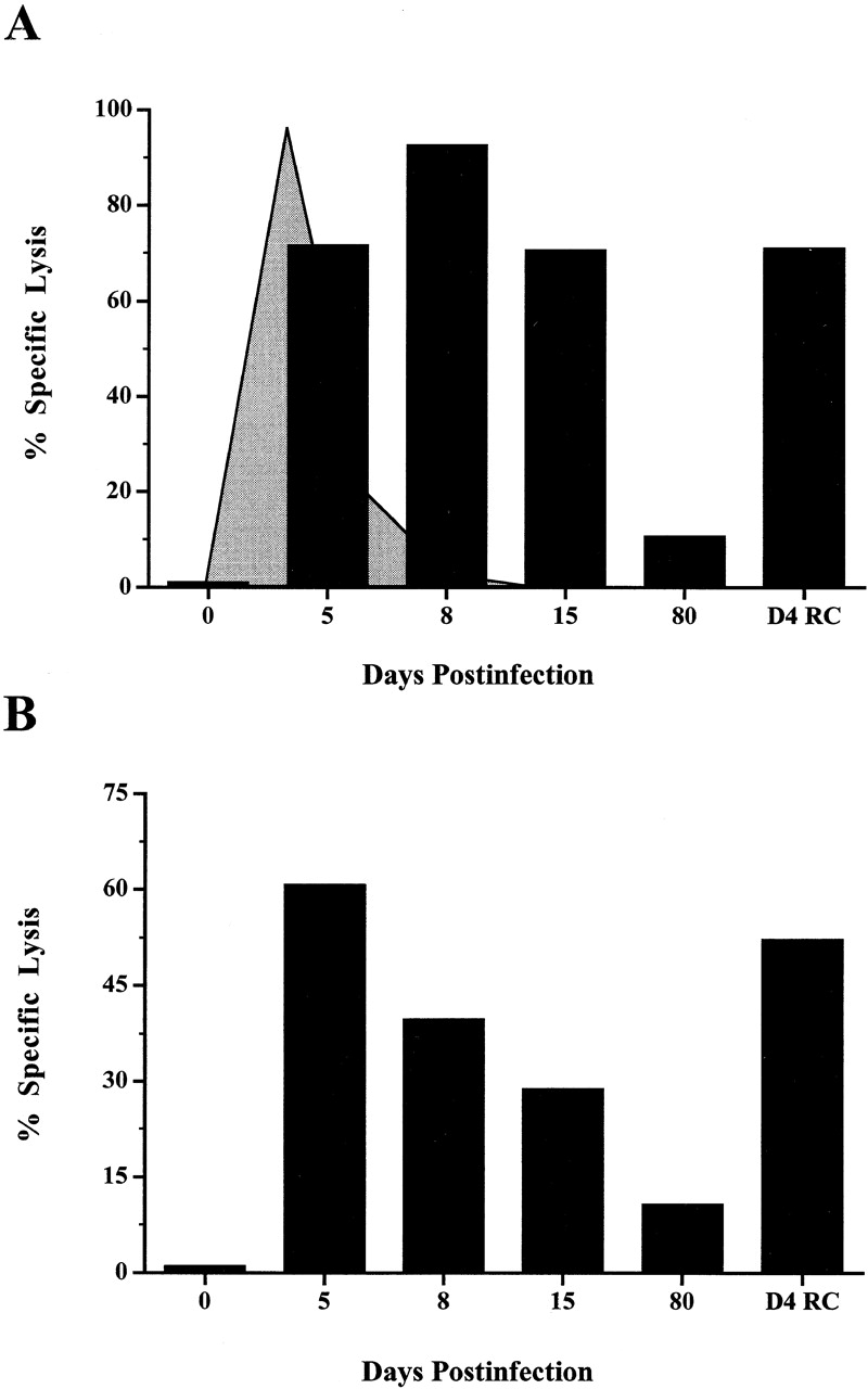
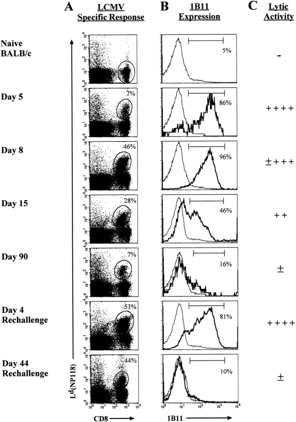
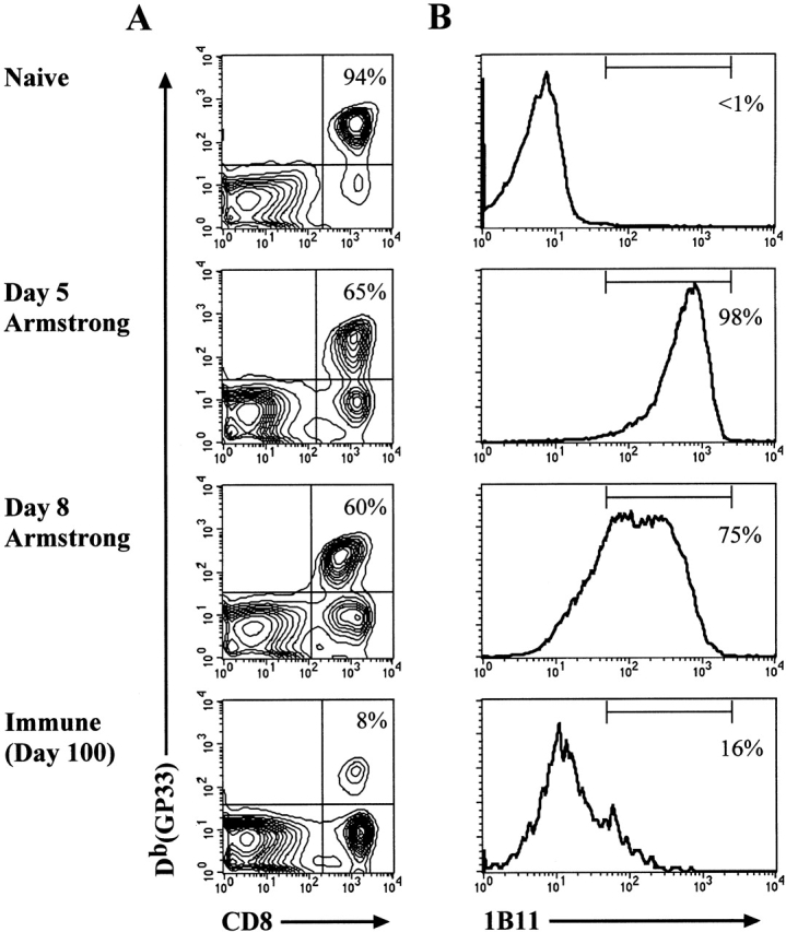
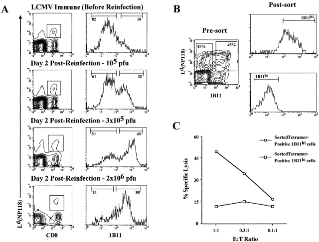
Currently there are few reliable cell surface markers that can clearly discriminate effector from memory T cells. To determine if there are changes in O-glycosylation between these two cell types, we analyzed virus-specific CD8 T cells at various time points after lymphocytic choriomeningitis virus infection of mice. Antigen-specific CD8 T cells were identified using major histocompatibility complex class I tetramers, and glycosylation changes were monitored with a monoclonal antibody (1B11) that recognizes O-glycans on mucin-type glycoproteins. We observed a striking upregulation of a specific cell surface O-glycan epitope on virus-specific CD8 T cells during the effector phase of the primary cytotoxic T lymphocyte (CTL) response. This upregulation showed a strong correlation with the acquisition of effector function and was downregulated on memory CD8 T cells. Upon reinfection, there was again increased expression of this specific O-glycan epitope on secondary CTL effectors, followed once more by decreased expression on memory cells. Thus, this study identifies a new cell surface marker to distinguish between effector and memory CD8 T cells. This marker can be used to isolate pure populations of effector CTLs and also to determine the proportion of memory CD8 T cells that are recruited into the secondary response upon reencounter with antigen. This latter information will be of value in optimizing immunization strategies for boosting CD8 T cell responses.


Figures

Figure 1
Figure 1
Kinetics of direct ex vivo virus-specific CTL activity during primary LCMV infection and after rechallenge. (A) Splenocytes from LCMV-infected mice were analyzed for direct ex vivo CTL activity (D4 RC = day 4 after LCMV rechallenge). Data shown are for an E/T ratio of 200:1. Lysis of noninfected target cells at the same E/T ratio was <5% (data not shown). The kinetics of viral clearance during acute LCMV infection are indicated by the shaded graph. The virus levels in the spleen peak between days 1 and 3 (at 107 pfu per spleen) and then drop precipitously between days 5 and 8 to <102 pfu per spleen. (B) The direct ex vivo CTL activity at different time points after infection after normalizing the number of antigen-specific CD8 T cells present in each sample. There were 104 NP118-specific CD8 T cells present in each sample, giving an E/T ratio of 1:1 based on virus-specific T cells. The data shown are representative of three experiments.
Figure 2
Figure 2
Kinetic analysis of 1B11 binding to antigen-specific CD8 T cells. (A) Splenocytes from LCMV-infected mice were stained with the Ld(NP118) tetramer and anti-CD8 and the percentage of CD8 T cells that were tetramer positive is noted. (B) Expression of cell surface O-glycans was examined with the mAb 1B11. Within the histograms, the bold line represents the 1B11 staining gated on the Ld(NP118)+ CD8 T cells at each time point, and the thin line corresponds to the 1B11 staining on naive BALB/c CD8 T cells. The percentage of 1B11hi cells within the gated population is noted. (C) This panel shows the direct ex vivo CTL activity as assessed on LCMV-infected targets.
Figure 3
Figure 3
The 1B11 O-glycan epitope is upregulated on antigen-specific effector T cells. (A) LCMV-specific CD8 T cells were obtained from naive P14-transgenic mice or at days 5, 8, and 100 after adoptive transfer and infection. Splenocytes from these various groups of mice were stained with anti-CD8 and the Db(GP33) tetramer. The percent of CD8 T cells that are GP33 specific is noted in the upper right quadrant. (B) 1B11 staining of the GP33-specific CD8 T cells. The percent of cells which are 1B11hi within the gated Db(GP33)+CD8+ population is noted.
Figure 4
Figure 4
Using the 1B11 antibody to monitor recruitment of memory CD8 T cells into the secondary response (A) and to isolate antigen-specific effector CTLs (B). (A) LCMV immune mice were rechallenged with varying doses of LCMV-Armstrong intravenously, and recruitment of the antigen-specific CD8 T cells into the secondary response was monitored by 1B11hi staining. The contour plots show the Ld(NP118) tetramer-positive CD8 T cells at day 2 after reinfection with different doses of LCMV. The histograms demonstrate the 1B11 staining on the gated populations from the panels on the left, with the percentage of 1B11hi and 1B11lo tetramer-positive CD8 T cells noted. (B) LCMV immune mice were reinfected with 3 × 10⁵ pfu LCMV-Armstrong intravenously, and 2 d later, Ld(NP118) tetramer-positive CD8 T cells were sorted into 1B11hi and 1B11lo populations. (C) The sorted 1B11hi and 1B11lo Ld(NP118) CD8 T cells were then analyzed for their LCMV-specific direct ex vivo CTL activity. The level of killing on uninfected target cells was <6%.

References

    1. Ahmed R., Gray D. Immunological memory and protective immunityunderstanding their relation. Science. 1996;272:54–60. - PubMed
    1. Zimmerman C., Brduscha-Riem K., Blaser C., Zinkernagel R.M., Pircher H. Visualization, characterization, and turnover of CD8+ memory T cells in virus-infected hosts. J. Exp. Med. 1996;183:1367–1375. - PMC - PubMed
    1. Hamann D., Baars P.A., Rep M.H., Hooibrink B., Kerkhof-Garde S.R., Klein M.R., van Lier R.A. Phenotypic and functional separation of memory and effector human CD8+ T cells. J. Exp. Med. 1997;186:1407–1418. - PMC - PubMed
    1. Murali-Krishna K., Altman J.D., Suresh M., Sourdive D.J., Zajac A.J., Miller J.D., Slansky J., Ahmed R. Counting antigen-specific CD8 T cellsa reevaluation of bystander activation during viral infection. Immunity. 1998;8:177–187. - PubMed
    1. Buchmeier M.J., Zajac A.J. Lymphocytic choriomeningitis virus. In: Ahmed R., Chen I.S.Y., editors. Persistent Viral Infections. John Wiley & Sons; Chichester, UK: 1999. pp. 575–605.

Publication types