. 2000 Apr;105(7):905-13.
doi: 10.1172/JCI8604.

A novel signaling mechanism between gas and blood compartments of the lung

Affiliations

A novel signaling mechanism between gas and blood compartments of the lung

W M Kuebler et al. J Clin Invest. 2000 Apr.

Erratum in

Abstract

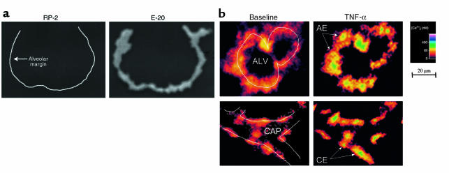
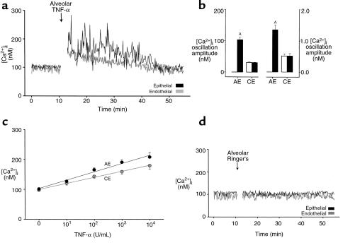
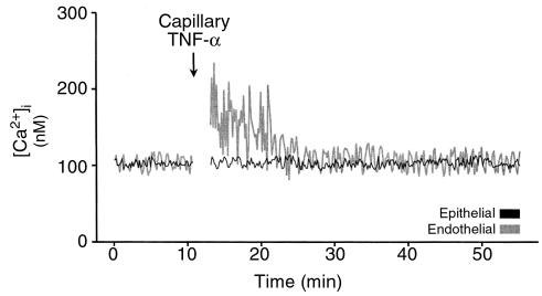
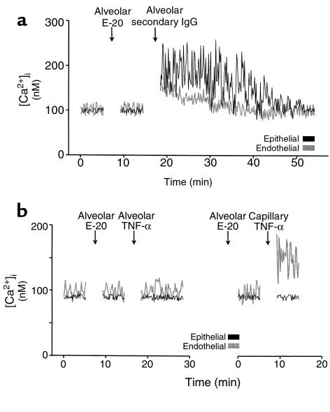
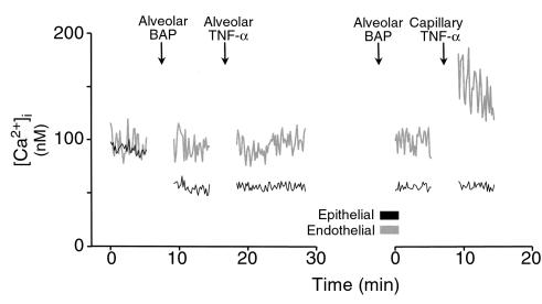
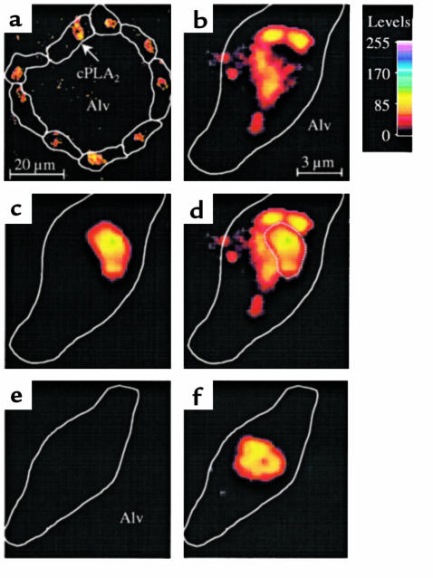
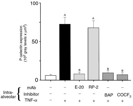
Propagation of inflammatory signals from the airspace to the vascular space is pivotal in lung inflammation, but mechanisms of intercompartmental signaling are not understood. To define signaling mechanisms, we microinfused single alveoli of blood-perfused rat lung with TNF-alpha, and determined in situ cytosolic Ca(2+) concentration ([Ca(2+)](i)) by the fura-2 ratio method, cytosolic phospholipase A(2) (cPLA(2)) activation and P-selectin expression by indirect immunofluorescence. Alveolar TNF-alpha increased [Ca(2+)](i) and activated cPLA(2) in alveolar epithelial cells, and increased both endothelial [Ca(2+)](i) and P-selectin expression in adjoining perialveolar capillaries. All responses were blocked by pretreating alveoli with a mAb against TNF receptor 1 (TNFR1). Crosslinking alveolar TNFR1 also increased endothelial [Ca(2+)](i). However, the endothelial responses to alveolar TNF-alpha were blocked by alveolar preinjection of the intracellular Ca(2+) chelator BAPTA-AM, or the cPLA(2) blockers AACOCF(3) and MAFP. The gap-junction uncoupler heptanol had no effect. We conclude that TNF-alpha induces signaling between the alveolar and vascular compartments of the lung. The signaling is attributable to ligation of alveolar TNFR1 followed by receptor-mediated [Ca(2+)](i) increases and cPLA(2) activation in alveolar epithelium. These novel mechanisms may be relevant in the alveolar recruitment of leukocytes.


Figures

Figure 1
Figure 1
In situ lung imaging. (a) Images of a single alveolus show indirect immunofluorescence for a nonspecific mAb (RP-2) and an anti-TNFR1 mAb (E-20). Line in left image was drawn from parallel bright-field image. Replicated 3 times. (b) Images show the 340:380 ratio color-coded for [Ca2+]i in an alveolus (ALV) and its adjoining capillary (CAP). Line sketches depict alveolar and capillary margins. Individual alveolar epithelial (AE) and capillary endothelial (CE) cells are indicated. Images were obtained in the same alveolus and capillary at baseline (left) and 5 minutes after alveolar infusion of 1,000 U/mL TNF-α (right). Replicated 8 times.
Figure 2
Figure 2
[Ca2+]i responses to intra-alveolar infusion of TNF-α. (a) Tracings are from an epithelial cell of an alveolus and an endothelial cell of an adjoining capillary as indicated. TNF-α injected at 1,000 U/mL. Replicated 8 times. (b) Data show amplitude (left panel) and frequency (right panel) of [Ca2+]i oscillations. Open bars, baseline; filled bars, 5 minutes after alveolar TNF-α infusion (1,000 U/mL). AP < 0.05 vs. baseline; n = 8 experiments. AE, alveolar epithelial cells; CE, capillary endothelial cells. (c) Group [Ca2+]i responses 5 minutes after alveolar TNF-α infusion. Each point is mean ± SEM of 4 experiments. Lines were drawn by linear regression (P < 0.001). (d) Single experiment tracings show [Ca2+]i responses to intra-alveolar infusion of Ringer’s solution. Replicated 4 times.
Figure 3
Figure 3
Single experiment tracings show [Ca2+]i responses to intracapillary infusion of TNF-α (1,000 U/mL). Replicated 5 times.
Figure 4
Figure 4
E-20 ligation in a single alveolocapillary unit. Intra-alveolar infusion of Ab E-20 was followed by alveolar infusion of (a) secondary IgG or (b) TNF-α (1,000 U/mL), given either by alveolar (left panel) or capillary (right panel) infusion. Replicated 4 times.
Figure 5
Figure 5
Ca2+ chelation in a single alveolocapillary unit. BAP (BAPTA-AM), 40 μM. TNF-α, 1,000 U/mL. Alveolar or capillary routes of infusion are indicated. Replicated 4 times.
Figure 6
Figure 6
Translocation of cPLA2 in single alveoli. An alveolus was infused with 1,000 U/mL TNF-α (ad) or with Ringer’s solution (e and f). Then the alveolus was stained for both the immunofluorescence of cPLA2 (a, b, e) and the nuclear fluorescence of HOECHST 33324 (c and f). The alveolus was washed and imaged at low (a) and high (bf) magnifications as indicated by the scale bars. Color code shows fluorescence intensities. Cell margins identified by bright-field microscopy are depicted by line sketches. Alv, alveolar lumen. Images for cPLA2 and nuclear fluorescence were combined by image overlay (d, f). Nuclear outline is indicated by dotted line (d). Replicated 4 times.
Figure 7
Figure 7
Effects of cPLA2 and its products. (a and b) Tracings from single experiments. Alveolar or capillary routes of TNF-α infusion are indicated. Replicated 4 times each. (c) Group data for indicated inhibitors given in 10-minute alveolar infusions before alveolar infusion of TNF-α; n = 4 for each. AP < 0.05 vs. baseline (open bar). BP < 0.05 vs. TNF-α (solid bar). COCF3, AACOCF3; indo, indomethacin; MK, MK886; WEB, WEB 2170BS. (d) Alveoli were permeabilized by 10-minute saponin infusions. Then arachidonate was infused into permeabilized (closed circles) and control (open circle) alveoli. Each point is the mean ± SEM of 4 experiments. Line was drawn by linear regression (P < 0.001).
Figure 8
Figure 8
P-selectin expression in lung capillaries. Capillaries were stained for P-selectin expression 5 minutes after alveolar infusion as indicated. AACOCF3 (COCF3) or mAbs were given 10 minutes before TNF-α infusion; BAP (BAPTA-AM, 40 μM) was given 15 minutes before TNF-α infusion (1,000 U/mL). P-selectin expression was quantified as the product of mean fluorescence intensity and fluorescent area 1 minute after washout of unbound FITC-IgG with blood flow; n = 4 for each. AP < 0.05 vs. baseline (open bar). BP < 0.05 vs. TNF-α alone (black bar).

Comment in

References

    1. Nicod LP. Pulmonary defense mechanisms. Respiration. 1999;66:2–11. - PubMed
    1. Strieter RM, Kunkel SL. Acute lung injury: the role of cytokines in the elicitation of neutrophils. J Investig Med. 1994;42:640–651. - PubMed
    1. Ward PA. Role of complement, chemokines, and regulatory cytokines in acute lung injury. Ann N Y Acad Sci. 1996;796:104–112. - PubMed
    1. Nelson S, et al. Compartmentalization of intraalveolar and systemic lipopolysaccharide-induced tumor necrosis factor and the pulmonary inflammation response. J Infect Dis. 1989;159:189–194. - PubMed
    1. Wiener Kronish J, Albertine K, Matthay M. Differential responses to the endothelial and epithelial barriers of the lung in sheep to Escherichia coli endotoxin. J Clin Invest. 1991;88:864–875. - PMC - PubMed

Publication types

MeSH terms