

The central melanocortin system affects the hypothalamo-pituitary thyroid axis and may mediate the effect of leptin
- PMID: 10749579
- PMCID: PMC377483
- DOI: 10.1172/JCI8857
The central melanocortin system affects the hypothalamo-pituitary thyroid axis and may mediate the effect of leptin
Abstract
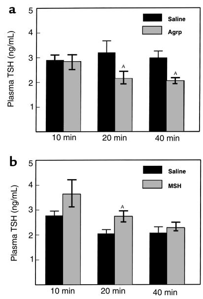
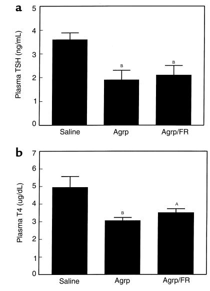
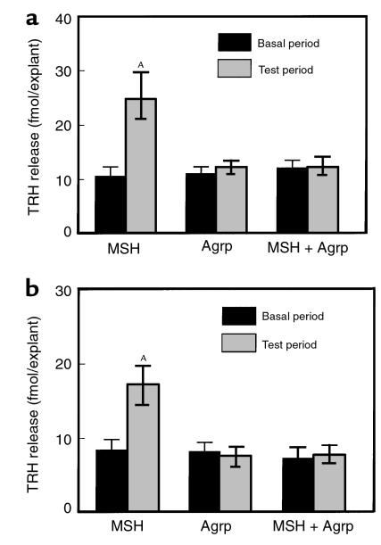
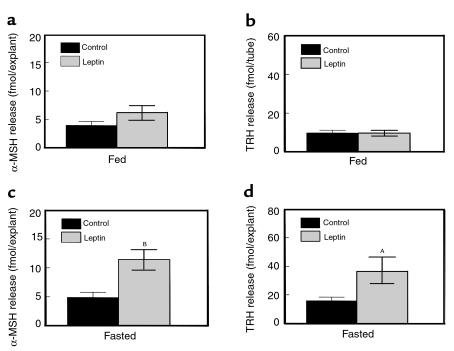
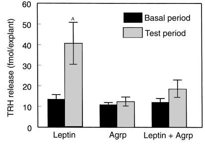
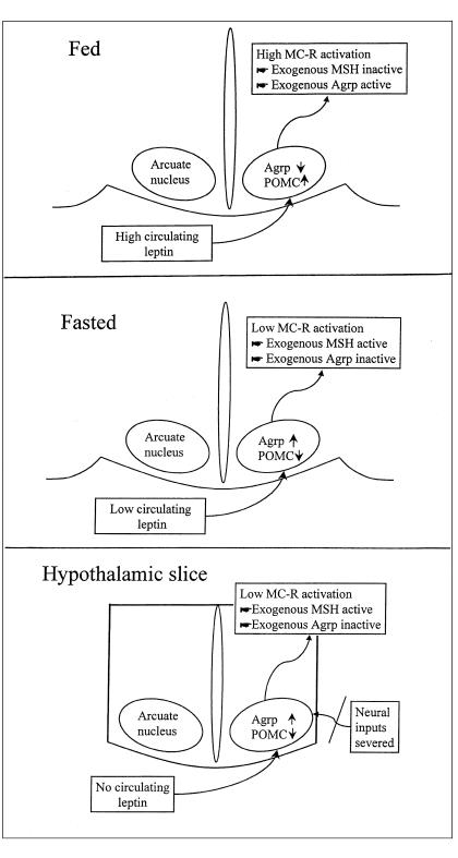
Prolonged fasting is associated with a downregulation of the hypothalamo-pituitary thyroid (H-P-T) axis, which is reversed by administration of leptin. The hypothalamic melanocortin system regulates energy balance and mediates a number of central effects of leptin. In this study, we show that hypothalamic melanocortins can stimulate the thyroid axis and that their antagonist, agouti-related peptide (Agrp), can inhibit it. Intracerebroventricular (ICV) administration of Agrp (83-132) decreased plasma thyroid stimulating hormone (TSH) in fed male rats. Intraparaventricular nuclear administration of Agrp (83-132) produced a long-lasting suppression of plasma TSH, and plasma T4. ICV administration of a stable alpha-MSH analogue increased plasma TSH in 24-hour-fasted rats. In vitro, alpha-MSH increased thyrotropin releasing hormone (TRH) release from hypothalamic explants. Agrp (83-132) alone caused no change in TRH release but antagonized the effect of alpha-MSH on TRH release. Leptin increased TRH release from hypothalami harvested from 48-hour-fasted rats. Agrp (83-132) blocked this effect. These data suggest a role for the hypothalamic melanocortin system in the fasting-induced suppression of the H-P-T axis.
Figures
Comment in
-
Leptin, nutrition, and the thyroid: the why, the wherefore, and the wiring.J Clin Invest. 2000 Apr;105(7):859-61. doi: 10.1172/JCI9725. J Clin Invest. 2000. PMID: 10749565 Free PMC article. No abstract available.
References
-
- Silva JE. Thyroid hormone control of thermogenesis and energy balance. Thyroid. 1995;5:481–492. - PubMed
-
- Blake NG, Eckland DJ, Foster OJ, Lightman SL. Inhibition of hypothalamic thyrotropin-releasing hormone messenger ribonucleic acid during food deprivation. Endocrinology. 1991;129:2714–2718. - PubMed
-
- Spencer CA, Lum SM, Wilber JF, Kaptein EM, Nicoloff JT. Dynamics of serum thyrotropin and thyroid hormone changes in fasting. J Clin Endocrinol Metab. 1983;56:883–888. - PubMed
-
- Weigle DS, et al. Effect of fasting, refeeding, and dietary fat restriction on plasma leptin levels. J Clin Endocrinol Metab. 1997;82:561–565. - PubMed
-
- Ahima RS, et al. Role of leptin in the neuroendocrine response to fasting. Nature. 1996;382:250–252. - PubMed
Publication types
MeSH terms
Substances
Grants and funding
LinkOut - more resources
Full Text Sources
Other Literature Sources
