. 2000 Jun 16;275(24):18391-8.
doi: 10.1074/jbc.C000062200.

The gut-enriched Kruppel-like factor (Kruppel-like factor 4) mediates the transactivating effect of p53 on the p21WAF1/Cip1 promoter

Affiliations

The gut-enriched Kruppel-like factor (Kruppel-like factor 4) mediates the transactivating effect of p53 on the p21WAF1/Cip1 promoter

W Zhang et al. J Biol Chem. .

Abstract

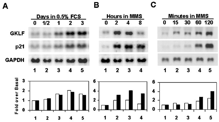
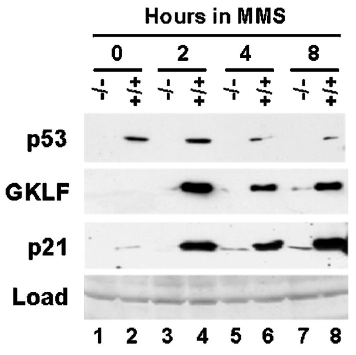
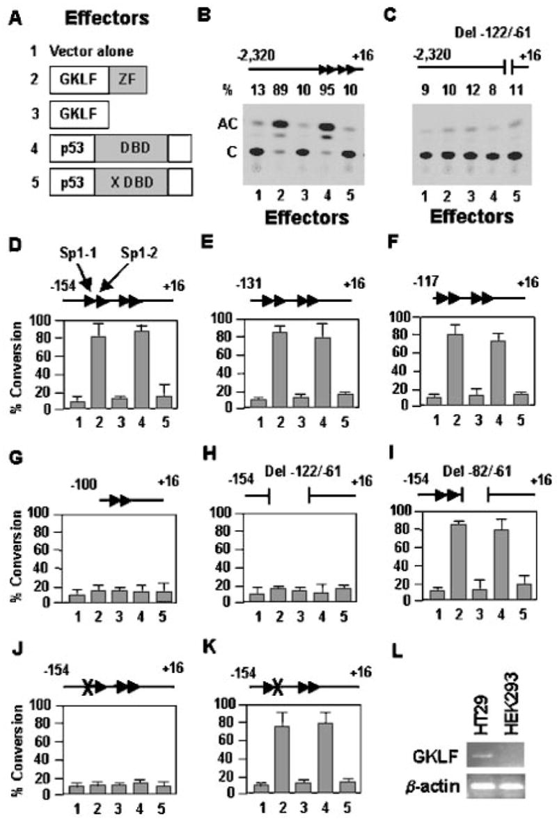
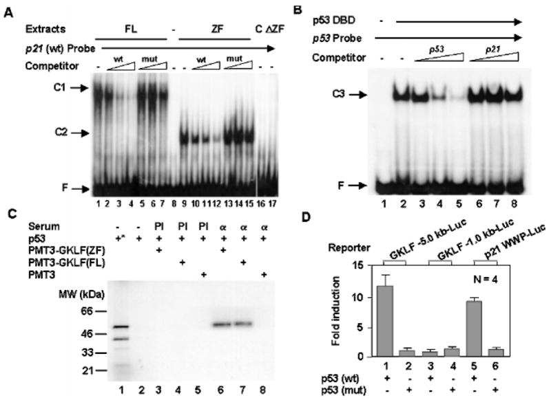
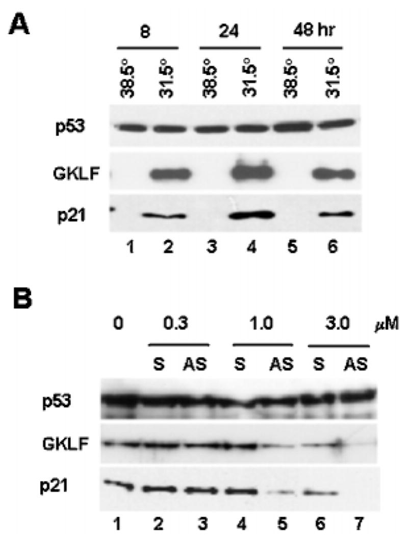
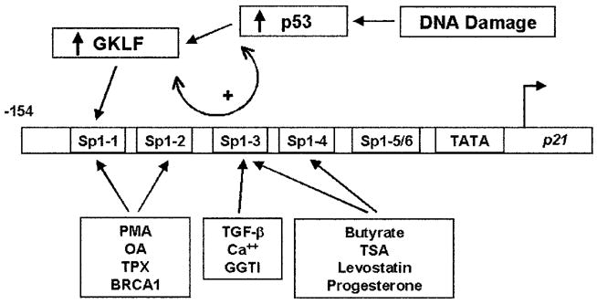
An important mechanism by which the tumor suppressor p53 maintains genomic stability is to induce cell cycle arrest through activation of the cyclin-dependent kinase inhibitor p21(WAF1/Cip1) gene. We show that the gene encoding the gut-enriched Krüppel-like factor (GKLF, KLF4) is concurrently induced with p21(WAF1/Cip1) during serum deprivation and DNA damage elicited by methyl methanesulfonate. The increases in expression of both Gklf and p21(WAF1/Cip1) due to DNA damage are dependent on p53. Moreover, during the first 30 min of methyl methanesulfonate treatment, the rise in Gklf mRNA level precedes that in p21(WAF1/Cip1), suggesting that GKLF may be involved in the induction of p21(WAF1/Cip1). Indeed, GKLF activates p21(WAF1/Cip1) through a specific Sp1-like cis-element in the p21(WAF1/Cip1) proximal promoter. The same element is also required by p53 to activate the p21(WAF1/Cip1) promoter, although p53 does not bind to it. Potential mechanisms by which p53 activates the p21(WAF1/Cip1) promoter include a physical interaction between p53 and GKLF and the transcriptional induction of Gklf by p53. Consequently, the two transactivators cause a synergistic induction of the p21(WAF1/Cip1) promoter activity. The physiological relevance of GKLF in mediating p53-dependent induction of p21(WAF1/Cip1) is demonstrated by the ability of antisense Gklf oligonucleotides to block the production of p21(WAF1/Cip1) in response to p53 activation. These findings suggest that GKLF is an essential mediator of p53 in the transcriptional induction of p21(WAF1/Cip1) and may be part of a novel pathway by which cellular responses to stress are modulated.


Figures

Fig. 1
Fig. 1. Northern blot analysis of Gklf and p21WAF1/Cip1 in NIH 3T3 cells during growth arrest
Growth arrest was induced in actively proliferating NIH 3T3 cells maintained in a medium containing 10% fetal calf serum (FCS) by the reduction of serum content to 0.5% (A) or by the addition of 100 μg/ml MMS to the medium (B and C). RNA was isolated at the indicated time points, and 20 μg were loaded in each lane and analyzed for the message content of Gklf, p21WAF1/Cip1, or glyceraldehyde-3-phosphate dehydrogenase (GAPDH). The bar graphs show the quantitative information of -fold induction of Gklf (open bars) and p21WAF1/Cip1 (closed bars) at each treatment time point over the untreated (Basal) value for each experiment. The calculation was performed first by normalizing the band intensity of the Gklf or p21WAF1/Cip1 transcript to that of glyceraldehyde-3-phosphate dehydrogenase at each time point and then comparing the normalized value of Gklf or p21WAF1/Cip1 at each treatment time point with that of untreated cells (time 0).
Fig. 2
Fig. 2. Western blot analysis of GKLF and p21WAF1/Cip1 in MEFs proficient or deficient in p53
MEFs were prepared from p53-deficient (−/−) mouse embryos (34) or their wild-type littermate control (+/+) and treated with 100 μg/ml MMS for the time periods indicated. Proteins were isolated and analyzed for the content of p53, GKLF, or p21WAF1/Cip1 by Western blot analysis. Load represents a portion of the gel stained with Coomassie Blue before electrophoretic transfer.
Fig. 3
Fig. 3. GKLF and p53 transactivate the p21WAF1/Cip1 promoter
A depicts the five effectors used throughout the cotransfection studies. Effector 1 is the PMT3 expression vector alone. Effectors 2 and 4 are expression constructs of wild-type GKLF and p53, respectively. Effector 3 is a mutant GKLF that does not have its zinc fingers (ZF) (30). Effector 5 is a mutant p53 with a missense mutation at codon 143 (X) in the DBD of p53 (32). Various regions of the p21WAF1/Cip1 promoter were linked to the CAT reporter (BK) and cotransfected with an equivalent quantity of the various effectors into HEK 293 cells. The four Sp1-binding sites (27) between nt −122 and −61 of the promoter are represented by the four arrowheads. The locations for Sp1-1 and Sp1-2 are identified in D. The × in J and K represents a 3-bp mutation in the first and second Sp1-binding sites, respectively. The numbers on the x axis in DK correspond to the five effectors shown in A. C is the substrate chloramphenicol, and AC represents the acetylated product. % Conversion = (AC/(AC + C)) × 100. Shown in DK are the means of three independent experiments. Bars represent S.D. L shows the results of reverse transcription-PCR of the mRNA levels of Gklf and β-actin in HT29 and HEK 293 cells.
Fig. 4
Fig. 4. Relationship among p53, GKLF, and the Sp1-1 site of the p21WAF1/Cip1 promoter
A, GKLF binds to the Sp1-1 site. EMSAs were performed using nuclear extracts prepared from COS-1 cells transfected with an expression construct containing the full-length (FL) GKLF (lanes 1–7) or the zinc finger (ZF) region of GKLF (lanes 9–15) and a radiolabeled oligonucleotide probe containing the sequence between nt −129 and −99 of the p21WAF1/Cip1 promoter, which includes both Sp1-1 and Sp1-2 sites (27). Where indicated, increasing amounts of unlabeled oligonucleotides representing either the wild-type (wt) sequence or a mutated (mut) sequence that contains a 3-bp substitution in the Sp1-1 site were included. Lane 8 contains the probe alone without added proteins. Lanes 16 and 17 contain nuclear extracts obtained from COS-1 cells transfected with the PMT3 vector alone (C) and the GKLF construct that lacks the zinc fingers (ΔZF) as in Fig. 3A, respectively. C1 is the complex formed between full-length GKLF and the probe, and C2 is formed between the zinc fingers and the probe. F is free probe. B, p53 does not interact with the sequence between nt −129 and −99 of the p21WAF1/Cip1 promoter. EMSAs were performed with the purified DBD of p53 (40) and a labeled probe representing an established p53-binding site. Competitors include unlabeled p53-binding sequence (lanes 3–5) and unlabeled wild-type p21WAF1/Cip1 sequence between nt −129 and −99 (lanes 6–8). C3 indicates the complex between p53 DBD and the probe. C, GKLF interacts with p53. 35S-Labeled p53 synthesized by in vitro transcription and translation was mixed with nuclear extracts from COS-1 cells transfected with PMT3-GKLF(ZF), PMT3-GKLF(FL), or PMT3 vector alone and precipitated with either preimmune (PI) serum or anti-GKLF serum (α). Lane 1 (*) contains the input p53, and lane 2 is p53 mixed with protein A-Sepharose beads without added serum. The precipitated materials were resolved by denaturing polyacrylamide gel electrophoresis and visualized by autoradiography. D, p53 transactivates the Gklf promoter. Either 5.0 or 1.0 kb of the 5′-flanking sequence of the mouse Gklf gene was linked to a luciferase reporter and cotransfected into Chinese hamster ovary cells with an expression construct containing either wild-type p53 or mutant p53 that no longer binds DNA (see Fig. 3A). Included was a p21 WWP-Luc construct containing 2.4 kb of the p21WAF1/Cip1 promoter sequence linked to the luciferase reporter as a control (7). Shown are the means of four experiments. Bars are S.D.
Fig. 5
Fig. 5. Synergistic activation of the p21WAF1/Cip1 promoter by GKLF and p53
Cotransfection experiments were performed in HEK 293 cells with a luciferase reporter linked to 2.4 or 2.2 kb of the p21WAF1/Cip1 promoter sequence (WWP-Luc, which contains an upstream p53-binding site at nt −2301, or DM-Luc, which does not, respectively ( and 33)) and subsaturating amounts of expression constructs containing GKLF, p53, or both. Shown are the means of four independent experiments. Bars represent S.D.
Fig. 6
Fig. 6. GKLF mediates the inductive effect of p53 on p21WAF1/Cip1
A, induction of Gklf and p21WAF1/Cip1 in 10(1) cells (36) containing a temperature-sensitive mutant p53 protein. 10(1) cells stably expressing the temperature-sensitive p53 val135 mutant (35, 37) were propagated at either the nonpermissive temperature of 38.5 °C or the permissive temperature of 31.5 °C for the time periods indicated. Proteins were harvested and analyzed for p53, GKLF, or p21WAF1/Cip1 by Western blot analysis. Both GKLF and p21WAF1/Cip1 were absent at time 0 when cells were maintained at 38.5 °C (data not shown). B, inhibition of p21WAF1/Cip1 formation in the 10(1)-p53 val135 cell line by antisense oligonucleotides to GKLF. Cells were transfected by lipofection with increasing amounts of sense (S) or antisense (AS) oligonucleotides to GKLF at 38.5 °C for 5 h and shifted to 31.5 °C for an additional 24 h before being harvested for quantification of p53, GKLF, or p21WAF1/Cip1 by Western blot analysis.
Fig. 7
Fig. 7. Model for the regulation of the p21WAF1/Cip1 proximal promoter by p53 and GKLF
The locations of the six Sp1-like elements within −154 bp of the p21WAF1/Cip1 promoter are designated according to a previous report (27). The model illustrates that the activation of p53 by DNA damage leads to both an increase in GKLF synthesis and an interaction between p53 and GKLF (double arrow), which cumulates in the binding of GKLF to the Sp1-1 element of the p21WAF1/Cip1 promoter. The various Sp1 cis-elements that mediate the functions of other physiological stimuli are also indicated. They include the phorbol ester phorbol 12-myristate 13-acetate (PMA) and okadaic acid (OA) (23); trapoxin (TPX), a histone deacetylase inhibitor (44); BRCA1, the breast cancer tumor suppressor gene (45); transforming growth factor-β (TGF-β) (24); Ca2+, which is important in keratinocyte differentiation (28); a geranylgeranyltransferase I (GGTI) inhibitor (46); butyrate (27) and trichostatin A (TSA) (29), both also histone deacetylase inhibitors; levostatin, a 3-hydroxy-3-methylglutaryl-coenzyme A reductase inhibitor (47); and progesterone (43).

References

    1. Agarwal ML, Taylor WR, Chernov MV, Chernova OB, Stark GR. J Biol Chem. 1998;273:1–4. - PubMed
    1. Ko LJ, Prives C. Genes Dev. 1996;10:1054–1072. - PubMed
    1. Levine AJ. Cell. 1997;88:323–331. - PubMed
    1. Giaccia AJ, Kastan MB. Genes Dev. 1998;12:2973–2983. - PubMed
    1. Prives C. Cell. 1998;95:5–8. - PubMed

Publication types