. 2000 Apr;11(4):1153-67.
doi: 10.1091/mbc.11.4.1153.

A novel helicase-type protein in the nucleolus: protein NOH61

Affiliations
Free PMC article

A novel helicase-type protein in the nucleolus: protein NOH61

R F Zirwes et al. Mol Biol Cell. 2000 Apr.
Free PMC article

Abstract

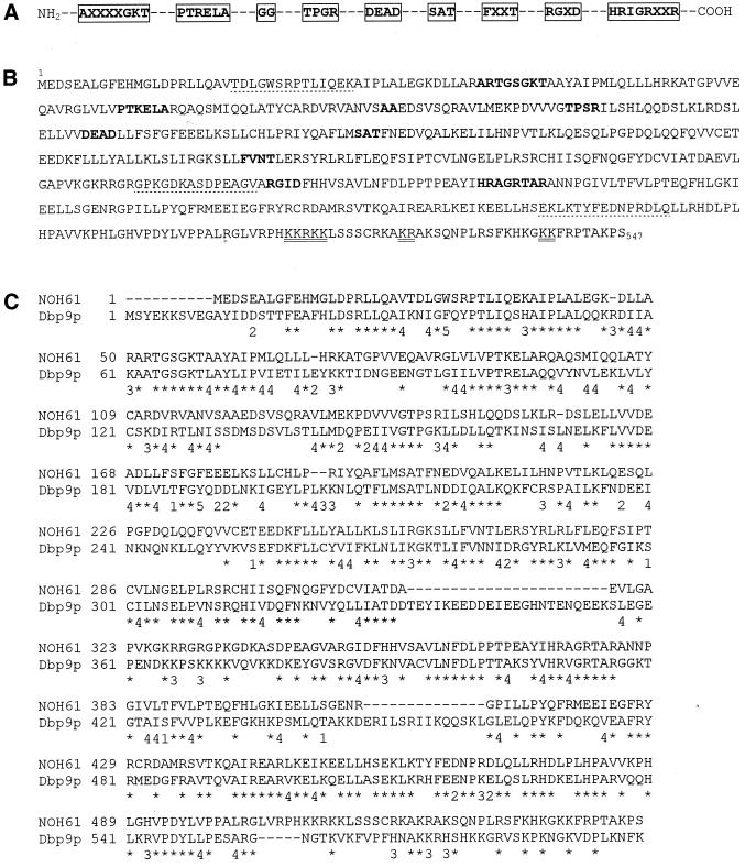
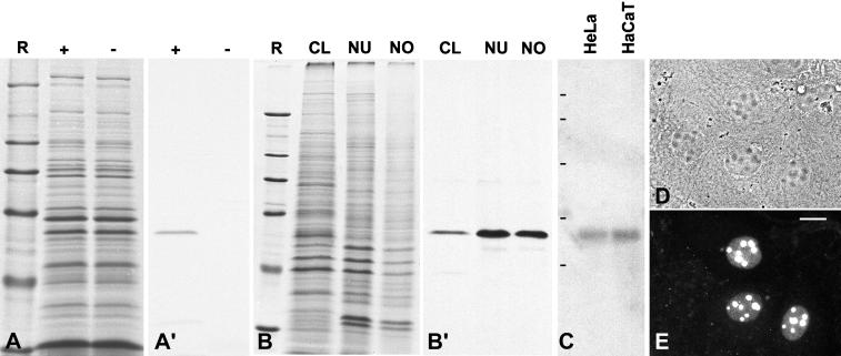
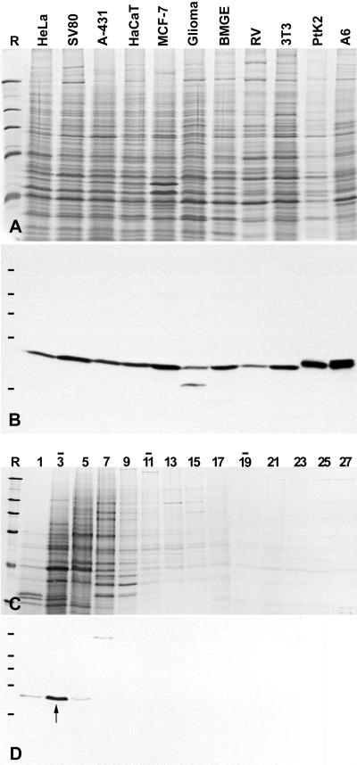
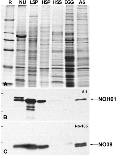
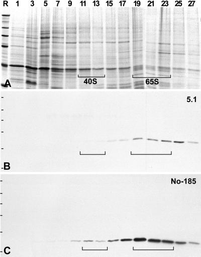
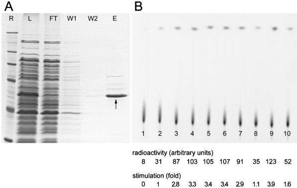
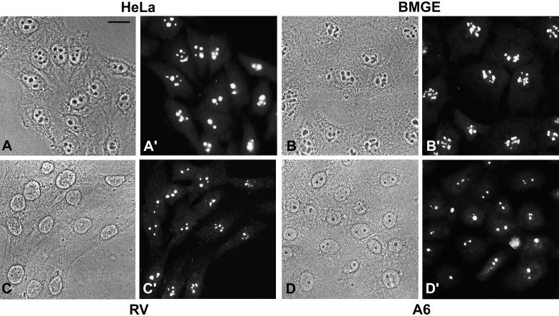
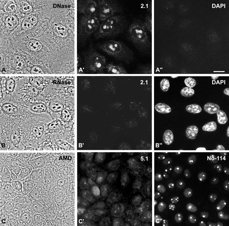
We report the identification, cDNA cloning, and molecular characterization of a novel, constitutive nucleolar protein. The cDNA-deduced amino acid sequence of the human protein defines a polypeptide of a calculated mass of 61.5 kDa and an isoelectric point of 9.9. Inspection of the primary sequence disclosed that the protein is a member of the family of "DEAD-box" proteins, representing a subgroup of putative ATP-dependent RNA helicases. ATPase activity of the recombinant protein is evident and stimulated by a variety of polynucleotides tested. Immunolocalization studies revealed that protein NOH61 (nucleolar helicase of 61 kDa) is highly conserved during evolution and shows a strong accumulation in nucleoli. Biochemical experiments have shown that protein NOH61 synthesized in vitro sediments with approximately 11.5 S, i.e., apparently as homo-oligomeric structures. By contrast, sucrose gradient centrifugation analysis of cellular extracts obtained with buffers of elevated ionic strength (600 mM NaCl) revealed that the solubilized native protein sediments with approximately 4 S, suggestive of the monomeric form. Interestingly, protein NOH61 has also been identified as a specific constituent of free nucleoplasmic 65S preribosomal particles but is absent from cytoplasmic ribosomes. Treatment of cultured cells with 1) the transcription inhibitor actinomycin D and 2) RNase A results in a complete dissociation of NOH61 from nucleolar structures. The specific intracellular localization and its striking sequence homology to other known RNA helicases lead to the hypothesis that protein NOH61 might be involved in ribosome synthesis, most likely during the assembly process of the large (60S) ribosomal subunit.


Figures

Figure 1
Figure 1
Amino acid sequence of the human helicase-type protein NOH61 and comparison with its putative yeast homologue Dbp9p. (A) Schematic representation of highly conserved amino acid regions characteristic of putative ATP-dependent RNA helicases of the DEAD-box protein family (Schmid and Linder, 1992; Pause and Sonenberg, 1993). The nine conserved motifs are boxed (X indicates any amino acid). (B) Amino acid sequence of the protein NOH61 deduced from the cDNA insert of pBT-NOH61 (EMBL accession number AJ131712, 1926 bp). The helicase core region extends from aa 51 to 378. The nine conserved motifs characteristic of the family of DEAD-box proteins are all present in the primary sequence of NOH61 (indicated by bold letters). Putative nuclear localization signals are double underlined, and sequences used for generating antibodies are marked by dotted lines. (C) Sequence comparison of human protein NOH61 with the yeast protein Dbp9p (EMBL accession number Q06218). Horizontal bars are omissions introduced to optimize the alignment. The symbols below the primary sequences are defined as follows: *, identical amino acids; 1–5, conservative exchanges (1, aliphatic, polar; 2, acidic; 3, basic; 4, aliphatic, nonpolar; 5, aromatic).
Figure 2
Figure 2
Molecular characterization of the cDNA clone encoding the helicase-type protein NOH61. (A) Coomassie blue staining of SDS-PAGE–separated rabbit reticulocyte lysates after in vitro transcription and translation in the presence (+) or absence (−) of the pBT-NOH61 template. R, reference proteins: 205, 116, 97.4, 66, 45, and 29 kDa (from top to bottom). (A′) Corresponding autoradiograph of translation products. (B) Coomassie blue staining of various cellular fractions of HeLa cells separated by SDS-PAGE (CL, total cell lysate; NU, fraction enriched in nuclei; NO, fraction enriched in nucleoli). Reference proteins (R) are the same as in A. (B′) Corresponding immunoblot probed with antibody NOH61-5.1, which reacts specifically with a 61-kDa protein. This protein is highly enriched in the nuclear and nucleolar fractions. (C) Identification of specific mRNAs by Northern blot analysis. Poly(A)+ RNA from HeLa and HaCaT cells, respectively, separated by agarose gel electrophoresis was hybridized with a random prime–labeled NOH61-specific probe. Note the reaction of a single band corresponding to an ∼2-kb RNA. RNA size markers of 9.5, 7.4, 4.4, 2.4, and 1.4 kb are indicated on the left (from top to bottom). (D) Phase-contrast microscopy of methanol-fixed human hepatocellular carcinoma (PLC) cells after transfection with a chimeric cDNA encoding protein NOH61 fused to GFP. (E) Corresponding immunofluorescence micrograph. The expressed GFP-NOH61 fusion protein localizes almost exclusively in nucleoli. Some cells show in addition a weak staining of the nucleoplasm. Bar, 15 μm.
Figure 3
Figure 3
Identification of protein NOH61 in different cultured cell lines and analysis of its native state by sucrose gradient centrifugation. (A) Coomassie blue–stained total cellular proteins of cultured cells of different origin (see MATERIALS AND METHODS). (B) Corresponding autoradiogram showing ECL detection of the antigenic polypeptide using antibody NOH61-5.1. (C) Cellular extracts of HeLa cells were fractionated after sucrose gradient centrifugation, separated by SDS-PAGE, and stained with Coomassie blue. Fraction numbers are indicated on top of the lanes (the top of the gradient is on the left). Bars indicate the peak positions of the reference proteins BSA (4.3S; fraction 3), catalase (11.3S; lane 11), and thyroglobulin (16.5S; fraction 19). (D) Corresponding immunoblot using antibody NOH61-2.1. The bulk of protein NOH61 is recovered in fraction 3 with a sedimentation coefficient of ∼4.3 S (marked by an arrow). Reference proteins (R) are the same as in Figure 2.
Figure 4
Figure 4
Identification of NOH61 in different nuclear fractions from Xenopus oocytes. (A) Coomassie blue staining of various nuclear fractions of Xenopus oocytes and somatic cells, respectively, separated by SDS-PAGE. Total mass-isolated oocyte nuclei (NU), proteins of the LSP, HSP, and HSS fractions of fractionated oocyte nuclei, Xenopus egg extract (Egg), and total cellular proteins of Xenopus XLKE-A6 cells (A6) are shown. (B) Corresponding immunoblot probed with antibody NOH61-5.1, which shows a strong cross-reactivity with the putative Xenopus homologue in Western blots. Protein NOH61 is detectable in all fractions analyzed, except the egg extract. The identity of the slightly larger polypeptide band recognized by the antibody in the lane containing total oocyte nuclei is unknown. (C) To ascertain the fractionation procedure, the immunoblot shown in B was reprobed with mAb No-185 directed against the well-characterized nucleolar protein NO38. Reference (R) proteins are the same as in Figure 2.
Figure 5
Figure 5
Identification of NOH61 in free nucleoplasmic preribosomal particles separated by sucrose gradient centrifugation of HSP fractions from Xenopus oocyte nuclei. (A) Coomassie blue staining of the resulting gradient fractions. The fraction numbers are indicated at the top of the lanes. Brackets mark the positions of the preribosomal presursor molecules. (B) Corresponding immunoblot after incubation with antibody NOH61-5.1. Protein NOH61 can be detected in the 65S preribosomal particle but not in the 40S precursor for the small ribosomal subunit. (C) In comparison, reprobing of the identical immunoblot with mAb No-185 against nucleolar protein NO38 discloses the association of the latter protein with both 40S and 65S preribosomal particles. Reference (R) proteins are the same as in Figure 2.
Figure 6
Figure 6
Expression and purification of His6-NOH61 exhibiting ATPase activity. (A) His6-NOH61 was purified as described in MATERIALS AND METHODS. The resulting protein fractions, analyzed by SDS-PAGE, are as follows: R, reference proteins are the same as in Figure 2; L, total cell lysate (soluble fraction) from bacteria after induction with isopropyl-1-thio-β-d-galactopyranoside; FT, flow-through fraction of the Ni-agarose column; W1, proteins washed from the column with 10 mM imidazole; W2, proteins washed from the column with 20 mM imidazole; E, proteins eluted from the column in the presence of 250 mM imidazole. The position of the recombinant protein is indicated by an arrow. (B) ATPase activity of purified recombinant protein His6-NOH61. An autoradiograph of a polyethyleneimine-cellulose thin-layer chromatography plate shows the separation of radioactive phosphate released from [γ32P]ATP in ATP hydrolysis assays. Lane 1 shows the ATP input; in lane 2 no polynucleotide was added; in lanes 3–10, ATP hydrolysis was stimulated in the presence of the polynucleotides poly(A), poly(C), poly(G), poly(U), poly(I), HeLa total RNA, dsDNA, and ssDNA. Reactions shown in lanes 2–10 contain 200 ng His6-NOH61. The signal intensity and fold-stimulation of ATPase activity are indicated. Each value represents the average of four independent experiments. A representative experiment is shown. Quantitations were performed on a phosphorimager.
Figure 7
Figure 7
Immunolocalization studies in cultured cells from different species. Phase-contrast micrographs are shown in A–D, and the corresponding immunofluorescence micrographs are shown in A′–D′. (A and A′) Human cervical adenocarcinoma cells of line HeLa. (B and B′) Bovine mammary gland–derived cells, line BMGE+H. (C and C′) Rat vascular smooth muscle–derived cells, line RV. (D and D′) Xenopus kidney epithelial cells, line A6. Antibody NOH61-5.1 shows a strong and specific labeling of nucleoli in all cell lines analyzed. Sometimes, a few cells show additional weak labeling of the nucleoplasm and the cytoplasm. Bar, 20 μm
Figure 8
Figure 8
Intracellular distribution of NOH61 in cells treated with nucleases and transcription inhibitors, respectively. (A–A") HeLa cells (phase-contrast image shown in A) were treated with DNase and subsequently incubated with antibody NOH61-2.1 (A′). The protein was still associated with nucleoli, although some weak staining in the cytoplasm was visible. (A") Corresponding DAPI staining. (B–B") HeLa cells (phase-contrast image shown in B) were incubated with RNase A and analyzed by indirect immunofluorescence microscopy using antibody NOH61-2.1 (B′) and DAPI (B"), respectively. NOH61 was no longer detectable in nucleoli of the treated cells. (C–C") Xenopus kidney epithelial cells, line A6, were treated with AMD (phase-contrast image shown in C) and subsequently stained with antibody NOH61-5.1 (C′) and mAb No-114 (C"). While the 180-kDa nucleolar protein recognized by mAb No-114 (cf. Schmidt-Zachmann et al., 1984) was specifically retained in the dense fibrillar component of the segregated nucleoli, protein NOH61 was released and distributed throughout the cells. Bar, 15 μm.
Figure 9
Figure 9
Immunoelectron microscopy of protein NOH61 within the nucleolus of HeLa cells. Antibody NOH61-2.1 was detected by secondary antibodies coupled to nanogold particles. The granular component (GC) is specifically labeled, whereas fibrillar centers (FC) and the dense fibrillar component (DFC) are practically free of gold particles. Bar, 0.5 μm.

References

    1. Alber T. Structure of the leucine zipper. Curr Opin Genet Dev. 1992;2:205–210. - PubMed
    1. Busch H. Nucleolar and nucleolonemal proteins of cancer cells. J Tumor Marker Oncol. 1997;12:4–68.
    1. Cairns C, McStay B. Identification and cDNA cloning of a Xenopus nucleolar phosphoprotein, xNopp180, that is the homolog of the rat nucleolar protein Nopp140. J Cell Sci. 1995;108:3339–3347. - PubMed
    1. Chan PK. Characterization and cellular localization of nucleophosmin/B23 in HeLa cells treated with selected cytotoxic agents (studies of B23-translocation mechanism) Exp Cell Res. 1992;203:174–181. - PubMed
    1. Chan PK, Chan FY. Nucleophosmin/B23 (NPM) oligomer is a major and stable entity in HeLa cells. Biochim Biophys Acta. 1995;1262:37–42. - PubMed

Publication types

MeSH terms

Associated data

LinkOut - more resources