. 2000 Apr;11(4):1241-55.
doi: 10.1091/mbc.11.4.1241.

A family of ADP-ribosylation factor effectors that can alter membrane transport through the trans-Golgi

Affiliations
Free PMC article

A family of ADP-ribosylation factor effectors that can alter membrane transport through the trans-Golgi

A L Boman et al. Mol Biol Cell. 2000 Apr.
Free PMC article

Abstract

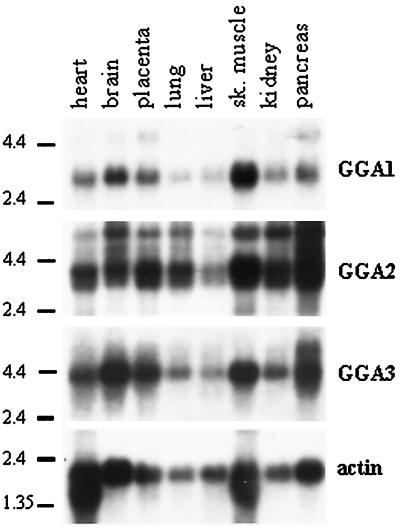
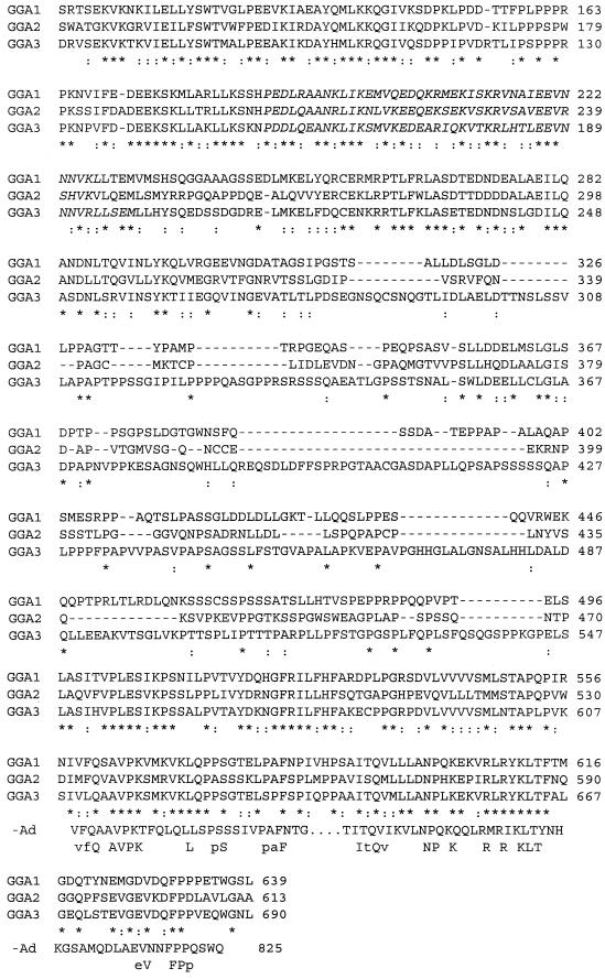
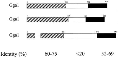
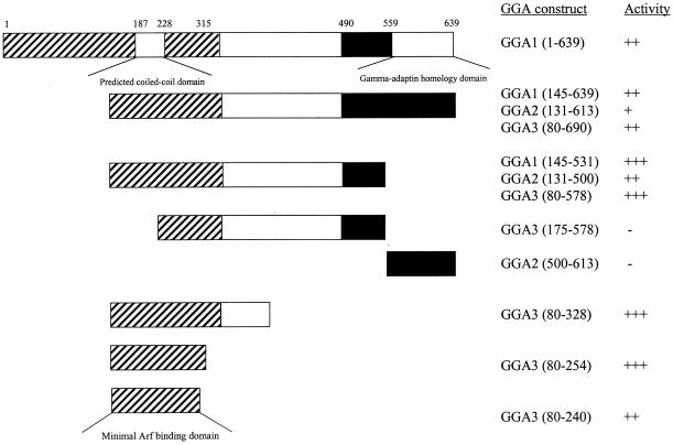
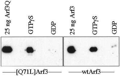
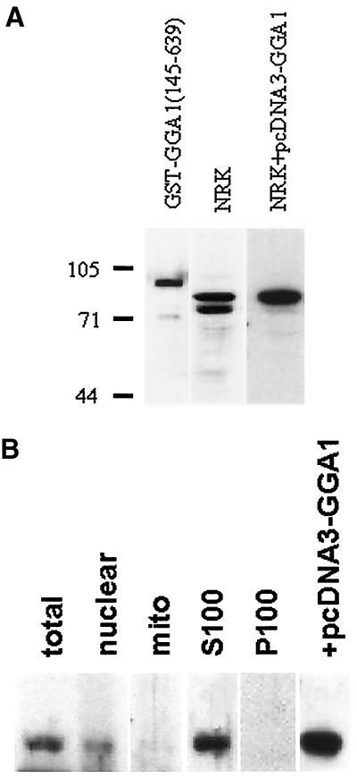
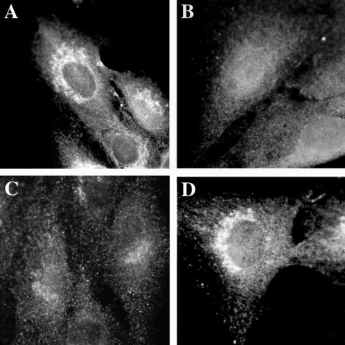
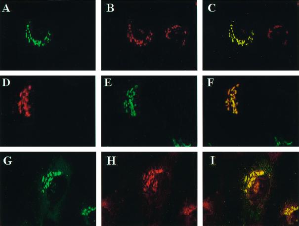
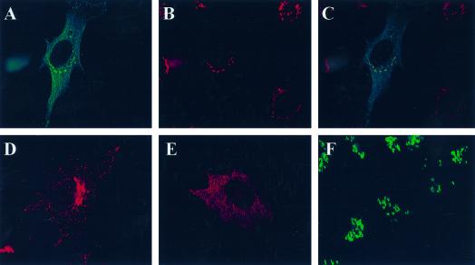
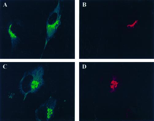
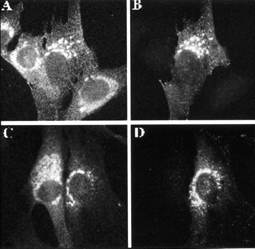
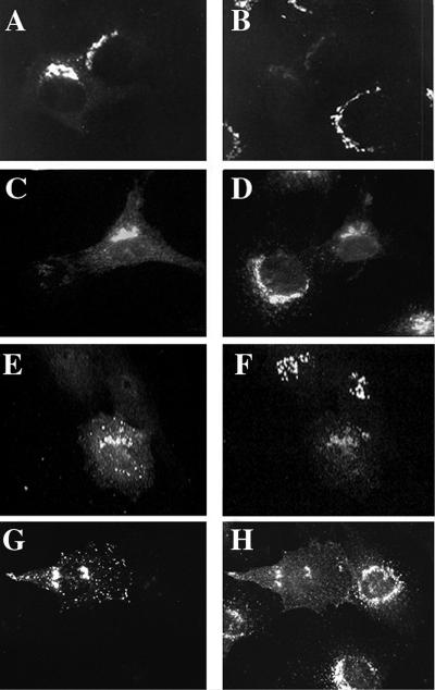
A family of three structurally related proteins were cloned from human cDNA libraries by their ability to interact preferentially with the activated form of human ADP-ribosylation factor 3 (ARF3) in two-hybrid assays. The specific and GTP-dependent binding was later confirmed through direct protein binding of recombinant proteins. The three proteins share large ( approximately 300 residues) domains at their N termini that are 60-70% identical to each other and a shorter (73 residues) domain at their C termini with 70% homology to the C-terminal "ear" domain of gamma-adaptin. Although GGA1 is found predominantly as a soluble protein by cell fractionation, all three proteins were found to localize to the trans-Golgi network (TGN) by indirect immunofluorescence. The binding of GGAs to TGN was sensitive to brefeldin A, consistent with this being an ARF-dependent event. Thus, these proteins have been named Golgi-localizing, gamma-adaptin ear homology domain, ARF-binding proteins, or GGAs. The finding that overexpression of GGAs was sufficient to alter the distribution of markers of the TGN (TGN38 and mannose 6-phosphate receptors) led us to propose that GGAs are effectors for ARFs that function in the regulation of membrane traffic through the TGN.


Figures

Figure 1
Figure 1
All three GGAs are ubiquitously expressed in adult human tissues. Northern blotting was performed sequentially, as described in MATERIALS AND METHODS, with the use of probes derived from GGA1, GGA2, or GGA3 and a multiple-tissue blot that contained mRNA from eight tissues. The locations of RNA size markers are indicated on the left of each blot. Actin was used as a control for RNA integrity and was the last probe used.
Figure 2
Figure 2
Alignment of the protein sequences of GGA1, GGA2, and GGA3 showing regions of homology to γ-adaptin and predicted coiled-coil domains. (A) The amino acid sequences for GGA1, GGA2, and GGA3 were aligned with the use of CLUSTAL. Identical residues are indicated by asterisks (*) in the line below, and conservative substitutions are indicated by colons (:). Residues predicted to be in a coiled-coil configuration are italicized (begins at residue 188 for GGA1). The alignment with the C terminus of γ-adaptin (-Ad) is also shown, with the line below that showing identities in all four sequences (uppercase) or in three of the four sequences (lowercase). (B) The three domains with different levels of sequence conservation between GGAs are shown schematically. Bars representing the protein domains are scaled to reflect the relative size of each domain. The percent pairwise identity within each domain is indicated, as determined by Bestfit (Genetics Computer Group, Madison, WI).
Figure 2
Figure 2
Alignment of the protein sequences of GGA1, GGA2, and GGA3 showing regions of homology to γ-adaptin and predicted coiled-coil domains. (A) The amino acid sequences for GGA1, GGA2, and GGA3 were aligned with the use of CLUSTAL. Identical residues are indicated by asterisks (*) in the line below, and conservative substitutions are indicated by colons (:). Residues predicted to be in a coiled-coil configuration are italicized (begins at residue 188 for GGA1). The alignment with the C terminus of γ-adaptin (-Ad) is also shown, with the line below that showing identities in all four sequences (uppercase) or in three of the four sequences (lowercase). (B) The three domains with different levels of sequence conservation between GGAs are shown schematically. Bars representing the protein domains are scaled to reflect the relative size of each domain. The percent pairwise identity within each domain is indicated, as determined by Bestfit (Genetics Computer Group, Madison, WI).
Figure 3
Figure 3
ARF binds to the N-terminal, most highly conserved region of GGAs. A variety of deletion constructs of all three GGA ORFs were tested in two-hybrid assays when paired with [Q71L]ARF3, as described in MATERIALS AND METHODS. The three domains of GGAs, shown in Figure 2B, are indicated, along with the specific GGA fragments that were cloned into the two-hybrid expression vector and the results of β-galactosidase assays. The minimal predicted fragment that includes the ARF-binding domain, determined by consensus from all three GGAs, is shown at the bottom.
Figure 4
Figure 4
GST-GGA1(145–639) binds to ARF3 in a GTP-dependent manner. Purified recombinant ARF3 or [Q71L]ARF3 was preincubated with GTPγS or GDP and then added to glutathione-agarose beads containing bound GST-GGA1(145–639), as described in MATERIALS AND METHODS. After extensive washing of the beads, the proteins that remained bound were analyzed on immunoblots with the use of antisera specific to ARFs. Note that both ARF3 and [Q71L]ARF3 bound to GST-GGA1(145–639) only when in the activated, GTPγS-bound state, and not when bound to GDP. Purified ARF3 proteins were used as a positive control for the Western blots and are shown on the left in each case.
Figure 5
Figure 5
GGA1 antibodies detect the soluble endogenous or overexpressed GGA1 proteins on immunoblots. (A) Rabbit polyclonal antiserum R-79709 was used as the primary antibody at a dilution of 1:2000 to detect GGA1 in immunoblots of purified GST-GGA1(145–639) antigen (1 ng; left lane), NRK cell lysate (20 μg; center lane), or an NRK cell lysate from cells overexpressing GGA1 (20 μg; right lane). The GGA1 was identified as the upper band in the doublet seen in NRK cells. Note that the immunoreactivity of the lower band is the same in the two right lanes but that the right lane was exposed to film for a shorter time to allow clear visualization of the upper band. (B) NRK cells were fractionated, as described in MATERIALS AND METHODS, and fractions were probed with the GGA1 antiserum to reveal a predominantly soluble location.
Figure 6
Figure 6
Endogenous and overexpressed GGA1 proteins localize to the perinuclear region in NRK cells. NRK cells were cultured, fixed, and permeabilized as described in MATERIALS AND METHODS. (A) The rabbit polyclonal GGA1 antiserum (R-79709) was used as the primary antibody at a dilution of 1:500 to increase the signal from endogenous protein. (B) Specific staining was prevented by previous incubation of the primary antibody with antigen. (C) Exposure of the cells to BFA (30 μM) for 1 min. (D) Staining very similar to that of endogenous protein was seen after the GGA1-GST fusion protein was introduced into cells by microinjection.
Figure 7
Figure 7
GGA1 localizes to the TGN and colocalizes with ARFs. Full-length wild-type or HA-tagged GGA1 was expressed in NRK cells after transient transfection, and cells were fixed and permeabilized as described in MATERIALS AND METHODS. Staining of GGA1 is shown in A, D, and G; A and G show GGA1 staining with rabbit polyclonal antiserum (R-79709), and D shows the staining profile of GGA1-HA revealed with the mAb to the HA epitope (12CA5). Cells in B were stained with the TGN38 mAb, and those in E were stained with the polyclonal mannosidase II antiserum. The primary antibody in H was the pan-ARF antibody 1D9. The images in the left and middle panels were merged and are shown on the right in each case. Note the near colocalization of GGA1 with the marker of the medial-Golgi (mannosidase II) and the improved colocalization of GGA1 with TGN38 and ARFs.
Figure 8
Figure 8
Like GGA1, GGA2 and GGA3 each localize to the trans-Golgi region and are sensitive to BFA. HA-GGA2 (A–C) or HA-GGA3 (D–F) was transiently transfected into NRK cells, and indirect immunofluorescence was performed as described in MATERIALS AND METHODS. The top panels show the colocalization of HA-GGA2 (A) with TGN38 (B), and the overlaid images are shown in C. The bottom panels show the staining of NRK cells expressing HA-GGA3. The typical pattern of staining for GGA3 is shown in D. The doubly stained images in E and F reveal the BFA sensitivity of the perinuclear localized GGA3. Cells treated with 10 μM BFA for 2 min show much more diffuse staining of HA-GGA3 (E), whereas the staining of mannosidase II in the same cells is unchanged (F) at this early time point.
Figure 9
Figure 9
GGA1, GGA2, and GGA3 localize to the same membranes. NRK cells were transiently transfected with plasmids directing the overexpression of GGA1 and HA-GGA2 (A and B) or GGA1 and HA-GGA3 (C and D), as described in MATERIALS AND METHODS. Cells in A and C were stained with the GGA1 antiserum R-79709, and cells in B and D were stained with the HA antibody 12CA5.
Figure 10
Figure 10
Overexpression of GGA1 prevents [Q71L]ARF1-induced expansion of the Golgi. A/B and C/D show two different examples of doubly stained images revealing that GGA1 expression blocks the Golgi vesiculation resulting from [Q71L]ARF1 expression. GGA1 was transiently transfected into NRK cells stably transfected with inducible human [Q71L]ARF1. Three hours after transfection with the GGA1 plasmid, cells were induced with interferon to express human [Q71L]ARF1. After 21 h of induction, cells were labeled with antibodies to ARF (1D9; A and C) or GGA1 (R-79709; B and D).
Figure 11
Figure 11
Markers of the TGN compartment, TGN38 and M6PR, are redistributed in cells overexpressing GGA1 or HA-GGA3. GGA1 (A and B), HA-GGA1 (C and D), or HA-GGA3 (E–H) were transiently transfected into NRK cells, as described in MATERIALS AND METHODS. Doubly labeled images are shown of cells stained for GGA1 (A) and TGN38 (B); HA-GGA1 (C) and M6PR (D); HA-GGA3 (E) and TGN38 (F); and HA-GGA3 (G) and M6PR. Note the loss of signal from TGN38 and M6PR in cells overexpressing GGAs compared with untransfected cells in the surrounding area.

References

    1. Altschul SF, Gish W, Miller W, Myers EW, Lipman DJ. Basic local alignment search tool. J Mol Biol. 1990;215:403–410. - PubMed
    1. Balch WE, Kahn RA, Schwaninger R. ADP-ribosylation factor is required for vesicular trafficking between the endoplasmic reticulum and the cis-Golgi compartment. J Biol Chem. 1992;267:13053–13061. - PubMed
    1. Barlowe C, Orci L, Yeung T, Hosobuchi M, Hamamoto S, Salama N, Rexach MF, Ravazzola M, Amherdt M, Schekman R. COPII: a membrane coat formed by Sec proteins that drive vesicle budding from the endoplasmic reticulum. Cell. 1994;77:895–907. - PubMed
    1. Boman AL, Kahn RA. ARF proteins: the membrane traffic police? Trends Biochem Sci. 1995;20:147–150. - PubMed
    1. Boman AL, Taylor TC, Melancon P, Wilson KL. A role for ADP-ribosylation factor in nuclear vesicle dynamics. Nature. 1992;358:512–514. - PubMed

MeSH terms

Associated data