. 2000 Apr;11(4):1385-400.
doi: 10.1091/mbc.11.4.1385.

A dibasic motif in the tail of a class XIV apicomplexan myosin is an essential determinant of plasma membrane localization

Affiliations
Free PMC article

A dibasic motif in the tail of a class XIV apicomplexan myosin is an essential determinant of plasma membrane localization

C Hettmann et al. Mol Biol Cell. 2000 Apr.
Free PMC article

Abstract

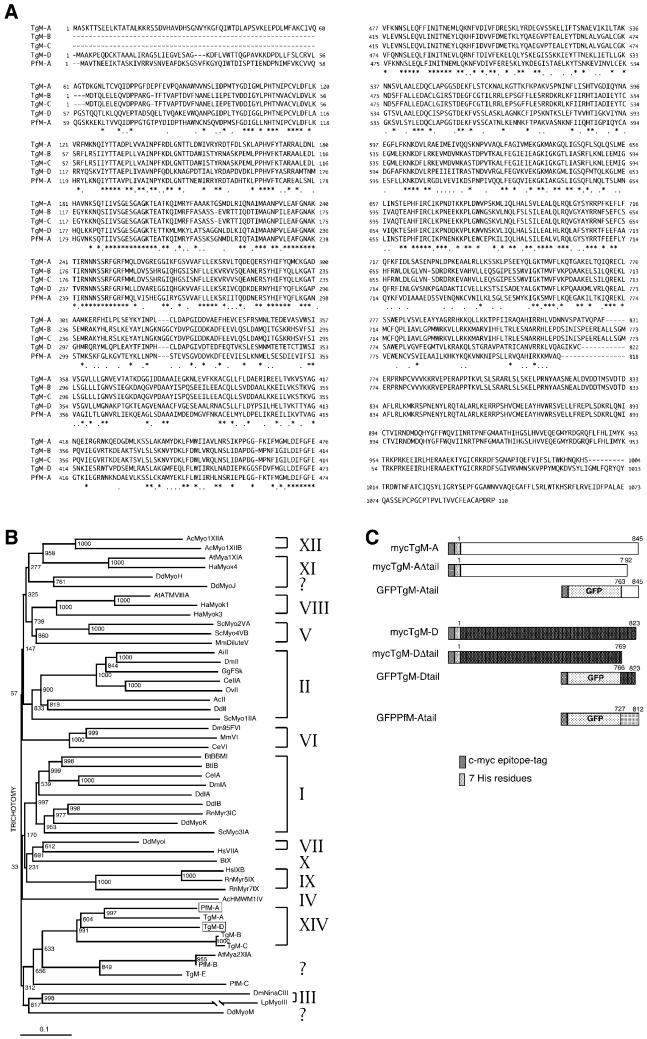
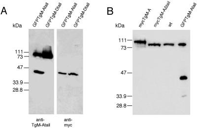
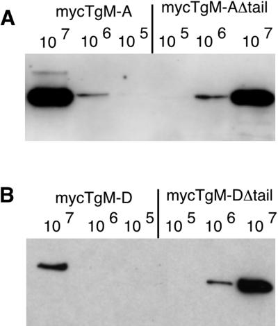
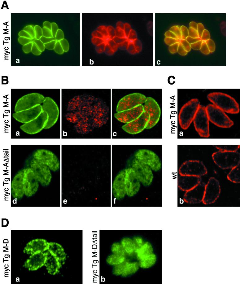
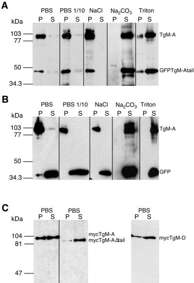
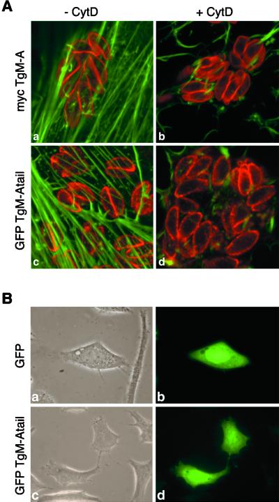
Obligate intracellular parasites of the phylum Apicomplexa exhibit gliding motility, a unique form of substrate-dependent locomotion essential for host cell invasion and shown to involve the parasite actin cytoskeleton and myosin motor(s). Toxoplasma gondii has been shown to express three class XIV myosins, TgM-A, -B, and -C. We identified an additional such myosin, TgM-D, and completed the sequences of a related Plasmodium falciparum myosin, PfM-A. Despite divergent structural features, TgM-A purified from parasites bound actin in an ATP-dependent manner. Isoform-specific antibodies revealed that TgM-A and recombinant mycTgM-A were localized right beneath the plasma membrane, and subcellular fractionation indicated a tight membrane association. Recombinant TgM-D also had a peripheral although not as sharply defined localization. Truncation of their respective tail domains abolished peripheral localization and tight membrane association. Conversely, fusion of the tails to green fluorescent protein (GFP) was sufficient to confer plasma membrane localization and sedimentability. The peripheral localization of TgM-A and of the GFP-tail fusion did not depend on an intact F-actin cytoskeleton, and the GFP chimera did not localize to the plasma membrane of HeLa cells. Finally, we showed that the specific localization determinants were in the very C terminus of the TgM-A tail, and site-directed mutagenesis revealed two essential arginine residues. We discuss the evidence for a proteinaceous plasma membrane receptor and the implications for the invasion process.


Figures

Figure 1
Figure 1
TgM-D is a member of the class XIV myosins. (A) Sequence alignment of the class XIV myosins. Identical amino acids are indicated by a star; homologies are indicated by a dot. The TgM-A glutamine 419 corresponds to the TEDS site, and the serine 693 is found instead of the conserved glycine proposed to act as a pivot point of the lever arm. (B) Phylogenetic tree with 51 myosins representative of each known class. TgM-D and the completed PfM-A belong to the class XIV. (C) Schematic presentation of the constructs used in this study. The numbers on top of the bars indicate the corresponding residues from each myosin (see A). Most accession numbers can be found at http://www.mrc-lmb.cam.ac.uk/myosin/trees/accession.html, and some can be found in an article by Schwarz et al. (1999). Accession numbers for the apicomplexan myosins: TgM-A, AF006626; TgM-B, AF006627; TgM-C, AF006628; a genomic fragment with the correct 5′end of TgM-B and -C cDNAs, AF202585; TgM-D, AF105118; TgM-E, AF221131; PfM-A, AF105117; PfM-B, AF222716; and PfM-C, AF222717.
Figure 2
Figure 2
Immunoblot analysis of wild-type and recombinant parasites. (A and B) The antibody against the tail of TgM-A does not recognize the tail of TgM-D. Extracts of parasites expressing GFP·TgM-Atail or GFP·TgM-Dtail were blotted with anti-myc antibodies (mAb 9E10) and with anti-TgM-A tail antibodies (A). (B) The expression of mycTgM-A down-regulates the level of endogenous protein. Detection of endogenous and recombinant TgM-A with an antibody directed against its tail is shown. The lanes were loaded with similar amounts of freshly lysed wild-type parasites or parasites expressing mycTgM-A, mycTgM-AΔtail, or GFP·TgM-Atail.
Figure 3
Figure 3
The presence of its tail limits the expression level of recombinant TgM-D. Western blot analysis used the anti-myc antibody on parasites expressing recombinant mycTgM-A and mycTgM-AΔtail (A) or mycTgM-D and mycTgM-DΔtail (B). The respective expression levels of each full-length and truncated myosin were judged by comparing the signals resulting from the loading of 105, 106, or 107 parasites, respectively. MycTgM-A and mycTgM-AΔtail,as well as mycTgM-DΔtail, were expressed at comparable levels, whereas full-length mycTgM-D was expressed at an approximately 10-fold reduced level.
Figure 4
Figure 4
Immunofluorescence localization of TgM-A and TgM-D and their respective tail-less constructs. Classical (A and D) and confocal (B and C) immunofluorescence microscopic analysis of wild-type parasites (C, b) or parasites expressing mycTgM-A (A, B, a–c, and C, a), mycTgM-AΔtail (B, d–f), mycTgM-D (D, a), or mycTgM-DΔtail (D, b) is shown. Fixation was performed either by paraformaldehyde-glutaraldehyde (A, B, and D) or ultracold methanol (C). (A) MycTgM-A, visualized by anti-myc antibodies (a), colocalized at the parasite periphery (c) with SAG1, the major glycosylphosphatidylinositol-anchored surface antigen of T. gondii (b). (B and C) The antibody against the tail of TgM-A did not detect TgM-A (B, e) or mycTgM-A (B, b) at the plasma membrane when cells were fixed by paraformaldehyde-glutaraldehyde, even though mycTgM-A was detected at the periphery by anti-myc staining (B, a). The rapid freezing and fixation in ultracold methanol allowed peripheral localization of endogenous TgM-A (C, b) and mycTgM-A (C, a). Myc-TgM-AΔtail was mainly found in the cytoplasm (B, d). The anti-tail antibodies detected a cytosolic pool of mycTgM-A (B, b). (D) As detected by anti-myc antibodies, mycTgM-D was enriched at the parasite periphery (a) but in a way distinct from TgM-A (compare with A–C). MycTgM-DΔtail was mainly cytoplasmic (b).
Figure 5
Figure 5
Distribution of endogenous and recombinant myosins studied by subcellular fractionation. Cells expressing GFP (B) or GFP·TgM-Atail (A) were lysed in the presence of 5 mM ATP under different conditions and separated by high-speed centrifugations in soluble (S) and particulate (P) fractions. Cells were lysed in PBS, in a low-osmolarity buffer (PBS diluted 1:10), in PBS with 1 M NaCl, in 0.1 M Na2CO3, pH 11.5, or in PBS with 1% Triton X-100. The distribution of endogenous TgM-A and either GFP·TgM-Atail or GFP was determined simultaneously by immunoblotting with antibodies against the tail of TgM-A either alone (A) or combined with anti-myc antibodies (B). (C) The distribution of mycTgM-A, mycTgM-AΔtail, and mycTgM-D was detected by anti-myc antibody after lysis in PBS.
Figure 6
Figure 6
MycTgM-AΔtail sediments with F-actin in an ATP-dependent manner. (A) SDS-PAGE and Coomassie blue staining of mycTgM-AΔtail purified from an extract of recombinant parasites. (B) SDS-PAGE analysis and Coomassie blue staining of the supernatant (S) and pellets (P) obtained after cosedimentation of mycTgM-AΔtail with F-actin in the presence (+) or absence (−) of ATP. MycTgM-AΔtail was completely soluble in the absence of F-actin (Control). MycTgM-AΔtail could be released from F-actin by subsequent incubation with ATP. Rabbit muscle F-actin was used at 4 μM and ATP at 10 mM in the presence of 130 mM KCl.
Figure 7
Figure 7
The tails of TgM-A and TgM-D are sufficient to confer to GFP both peripheral localization and association with the particulate fraction. (A) Alignment of three highly related apicomplexan myosin tails. The basic residues are boxed. (B) Localization of GFP (a), GFP·TgM-Atail (b), GFP·TgM-Dtail (c), and GFP·PfM-Atail (d). Fluorescence was observed directly (a and b) or after indirect immunofluorescence with anti-myc antibodies (c and d). (C) Recombinant parasites expressing GFP, GFP·TgM-Atail, GFP·TgM-Dtail, and GFP·PfM-Atail were lysed in PBS and separated by high-speed centrifugations in soluble (S) and particulate (P) fraction. The partitioning of GFP and the tail chimeras reflected their respective intracellular localization observed by imunofluorescence microscopy.
Figure 8
Figure 8
Cytochalasin treatment did not influence the peripheral localization of mycTgM-A and GFP·TgM-Atail. GFP·TgM-Atail did not localize to the plasma membrane of HeLa cells. (A) MycTgM-A (a and b) and GFP·TgM-Atail (c and d) localized at the periphery of parasites after incubation in the presence (b and d) or absence (a and c) of 10 μg/ml cytochalasin D for 3 h, indicating that it did not depend on an intact actin cytoskeleton. To optimally present the host actin cytoskeleton and the peripheral staining in the parasites, one confocal section was recorded in the ventral part of the fibroblasts where the actin stress fibers are most prominent, and another confocal section was recorded through the middle of the parasites, 1 μm higher in the cell. Only the overlay is shown here. (B) Contrary to what was seen in T. gondii, GFP·TgM-Atail did not interact with the plasma membrane of an animal cell (d) and had a distribution similar to GFP (b). (a and c) Phase-contrast pictures corresponding to b and d.
Figure 9
Figure 9
A dibasic motif in the tail of TgM-A is essential for the cell membrane localization of GFP-tail chimera. (A) The wild-type sequence of the tail of TgM-A (wt) is shown aligned with the site-specific mutants (I–III) and the two deletion mutants (Δ14 and Δ22). The mutated residues are indicated in red. (B) Confocal microscopic analysis of parasites stably transformed with the respective mutant tail constructs fused to GFP. Indirect immunofluorescence was performed using the anti-myc antibodies. (C) Anti-myc immunoblot analysis of recombinant parasites expressing the different constructs presented in A.

References

    1. Arhets P, Olivo JC, Gounon P, Sansonetti P, Guillen N. Virulence and functions of myosin II are inhibited by overexpression of light meromyosin in Entamoeba histolytica. Mol Biol Cell. 1998;9:1537–1547. - PMC - PubMed
    1. Baker JP, Titus MA. A family of unconventional myosins from the nematode Caenorhabditis elegans. J Mol Biol. 1997;272:523–535. - PubMed
    1. Bement WM, Mooseker MS. TEDS rule: a molecular rationale for differential regulation of myosins by phosphorylation of the heavy chain head. Cell Motil Cytoskeleton. 1995;31:87–92. - PubMed
    1. Black M, Seeber F, Soldati D, Kim K, Boothroyd JC. Restriction enzyme-mediated integration elevates transformation frequency and enables co-transfection of Toxoplasma gondii. Mol Biochem Parasitol. 1995;74:55–63. - PubMed
    1. Brown SS. Myosins in yeast. Curr Opin Cell Biol. 1997;9:44–48. - PubMed

Publication types

MeSH terms

LinkOut - more resources