. 2000 Apr;11(4):1401-19.
doi: 10.1091/mbc.11.4.1401.

EMB-30: an APC4 homologue required for metaphase-to-anaphase transitions during meiosis and mitosis in Caenorhabditis elegans

Affiliations
Free PMC article

EMB-30: an APC4 homologue required for metaphase-to-anaphase transitions during meiosis and mitosis in Caenorhabditis elegans

T Furuta et al. Mol Biol Cell. 2000 Apr.
Free PMC article

Abstract

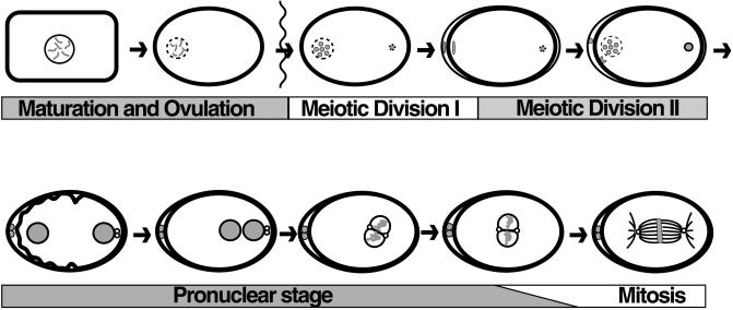
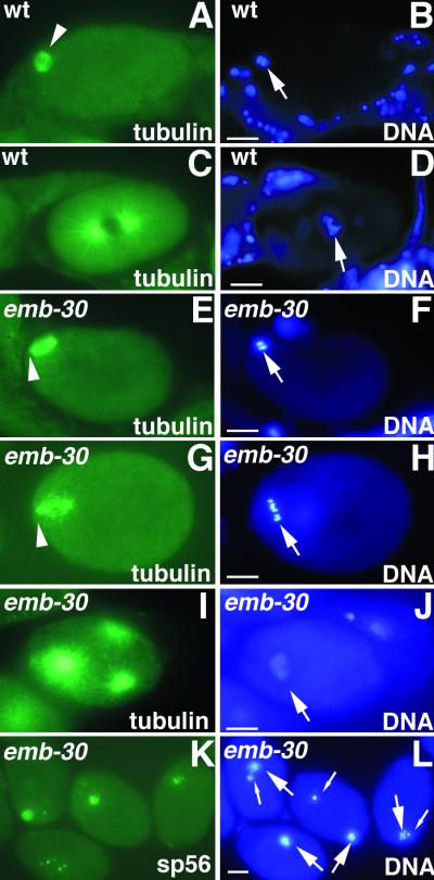
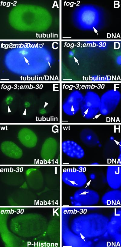
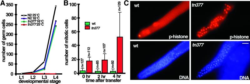
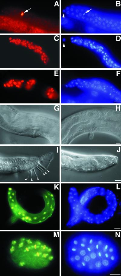
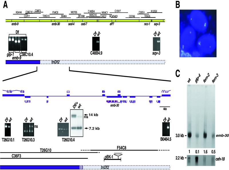
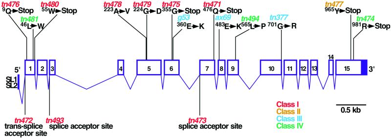
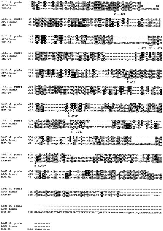
Here we show that emb-30 is required for metaphase-to-anaphase transitions during meiosis and mitosis in Caenorhabditis elegans. Germline-specific emb-30 mutant alleles block the meiotic divisions. Mutant oocytes, fertilized by wild-type sperm, set up a meiotic spindle but do not progress to anaphase I. As a result, polar bodies are not produced, pronuclei fail to form, and cytokinesis does not occur. Severe-reduction-of-function emb-30 alleles (class I alleles) result in zygotic sterility and lead to germline and somatic defects that are consistent with an essential role in promoting the metaphase-to-anaphase transition during mitosis. Analysis of the vulval cell lineages in these emb-30(class I) mutant animals suggests that mitosis is lengthened and eventually arrested when maternally contributed emb-30 becomes limiting. By further reducing maternal emb-30 function contributed to class I mutant animals, we show that emb-30 is required for the metaphase-to-anaphase transition in many, if not all, cells. Metaphase arrest in emb-30 mutants is not due to activation of the spindle assembly checkpoint but rather reflects an essential emb-30 requirement for M-phase progression. A reduction in emb-30 activity can suppress the lethality and sterility caused by a null mutation in mdf-1, a component of the spindle assembly checkpoint machinery. This result suggests that delaying anaphase onset can bypass the spindle checkpoint requirement for normal development. Positional cloning established that emb-30 encodes the likely C. elegans orthologue of APC4/Lid1, a component of the anaphase-promoting complex/cyclosome, required for the metaphase-to-anaphase transition. Thus, the anaphase-promoting complex/cyclosome is likely to be required for all metaphase-to-anaphase transitions in a multicellular organism.


Figures

Figure 1
Figure 1
The oocyte meiotic divisions and early embryogenesis. Oocyte meiotic maturation involves nuclear envelope breakdown (meiotic M-phase entry) and cortical rearrangement. The oocyte is fertilized as it enters the spermatheca. The condensed sperm chromatin is shown in the posterior (at right) of the embryo. The MI and MII divisions are completed in the uterus, forming two polar bodies at the anterior. Pronuclei form (bottom left), meet in the posterior, and fuse as they enter mitotic M phase (the small unfilled circles are centrosomes). The time from nuclear envelope breakdown to pronuclear meeting is ∼45 min.
Figure 2
Figure 2
emb-30 is required for completion of the oocyte meiotic divisions. Cytological analysis of embryos stained for tubulin, a sperm membrane protein (sp56), or DNA, as indicated. Wild-type (A and B) and emb-30(tn377ts) (E and F) embryos in MI metaphase. The MI spindle (arrowheads) is associated with the anterior cortex. (C and D) Wild-type pronuclear-stage embryo in prometaphase of the first mitotic division. (G and H) emb-30(tn377ts) one-cell embryo with a disorganized meiotic spindle (arrowhead). Six bivalents are visible (arrow), but there is no evidence of chromosome segregation. The spindle and chromatin are not closely associated with the anterior cortex. This particular spindle and chromatin morphology is not observed in the wild type. (I and J) emb-30(tn377ts) one-cell embryo with multipolar spindles. Three microtubule-organizing centers are seen. (K and L) emb-30(tn377ts) one-cell embryos stain for a sperm membrane protein and contain both maternal DNA (large arrows) and paternal DNA (small arrows). It is unclear how the female and male chromatin can on occasion come to be adjacent in the arrested embryos. This does not reflect nuclear migration processes because pronuclei do not form. Bars, 10 μm.
Figure 3
Figure 3
Cytological analysis of embryos or oocytes stained for tubulin, nuclear pore complex (Mab414), phosphohistone H3 (P-Histone), or DNA, as indicated. (A and B) Unfertilized oocyte from a fog-2 female showing intense DAPI staining (arrow) and diffuse tubulin staining. (C) One-cell embryo after fertilization of a fog-2; emb-30(tn377ts) oocyte with wild-type sperm. The MI spindle (large arrow) is in metaphase but is not associated with the anterior cortex. The paternal DNA is in the posterior (small arrow) but slightly out of the plane of focus. (D) Unfertilized oocyte from a fog-3; emb-30(tn377ts) female. An MI spindle can assemble in the absence of fertilization (arrow) and associate with the anterior cortex, but the oocyte does not become endomitotic. (E and F) Unfertilized oocytes from fog-3; emb-30(tn377ts) females. Disorganized meiotic spindle–like structures (arrowheads) associate with the maternal chromatin (arrows). Note that there are three separate oocytes in panels E and F. (G and H) Wild-type embryos form nuclear envelopes. Gastrula-stage embryo (left), pronuclear-stage embryo (middle), and meiotic embryo (right). (I and J) In emb-30(tn377ts), there is no indication of nuclear envelope formation. Instead, an amorphous aggregate of nuclear pore complex material is found in 8% of the arrested embryos. Maternal DNA is indicated by arrows. (K and L) Less condensed chromatin (arrow) contains phosphohistone H3, indicating that the embryo arrests in M phase. Bars, 10 μm.
Figure 4
Figure 4
emb-30 is required for germline proliferation. (A) Time course and temperature sensitivity of germline proliferation in emb-30(tn377ts) compared with the wild type. Larvae were synchronized by collecting L1-stage animals that hatched during a 1-h period. Germ cell number per gonad (L1 and L2) or per gonad arm (L3 and L4) was determined at 16 and 25°C. After transfer of gastrula-stage embryos to 25°C, Z2 and Z3 block in mitosis of their first division during the L1 stage; thus, at most, two germ cells are observed. At the L4 stage, germ cells were often absent (0.2 ± 0.5 germ cells per gonad arm; n = 22 gonad arms scored). For the L3 time point, the larvae in the wild-type 25°C sample were at a developmentally later stage (late L3), accounting for the slight increase in germ cell number compared with the 16°C samples. (B and C) Mitotic germ cell accumulation in the distal zone of the emb-30(tn377ts) adult hermaphrodite gonad. emb-30(tn377ts) and wild-type hermaphrodites were grown at 16°C and shifted to 25°C as young adults (92 h after hatch at 16°C). Quantitation of mitotic nuclei (mean ± SD; n = number of gonad arms scored at each time point) is shown in B, and representative phosphohistone H3 and DAPI immunofluorescence images from the 4-h time point are shown in C. Bar, 10 μm.
Figure 5
Figure 5
Germline and somatic phenotypes in emb-30 mutants. L4-stage gonads stained for phosphohistone H3 (A, C, and E) and DNA (B, D, and F). (A and B) Wild-type gonad. A single mitotic germ cell is observed in the distal arm (arrow; arrowhead indicates a distal tip cell). Mitotic germ cells accumulate in unc-32(e189)emb-30(tn475) (C and D) and emb-30(tn475); unc-46(e177)mdf-1(gk2) (E and F) gonads [mdf-1(gk2) homozygotes do not accumulate mitotic cells (Kitagawa and Rose, 1999)]. (G–I) Somatic defects in emb-30(tn475) homozygotes. (G) Normal vulva of a rescued unc-32(e189)emb-30(tn475); tnEx12(F54C8) adult. (H) Abnormal vulva of an unc-32(e189)emb-30(tn475) adult. (I) Wild-type male tail with sensory rays (arrowheads). (J) unc-32(e189)emb-30(tn475) male tail; sensory rays are absent. (K–N) Metaphase arrest in emb-30(mts, z-) animals. (K and M) Merged images of tubulin (green) and phosphohistone H3 (red) staining. (L and N) DAPI staining. (K and L) Arrested emb-30(mts, z-) larva with many metaphase cells 24 h after transfer to 25°C. Note the absence of anaphase figures. (M and N) Forty-cell emb-30(mts, z-) embryo containing 23 metaphase cells 2 h after transfer to 25°C from 20°C. Note the absence of anaphase figures. Panels A–F, G and H, I–L, and M and N are at the same scales. Bars, 10 μm.
Figure 6
Figure 6
Molecular identification of emb-30. (A) Genetic and physical map of the emb-30III region. (Top) Sequenced cosmids. Sequence tag site analysis (panels with black background) positioned the tnDf2 left breakpoint between C38C10.4 and C48B4.9. (Bottom) Enlarged physical map. ORFs are shown above and below the line, with their directions of transcription being left to right and right to left, respectively. The sod-4 ORF is not shown but would be to the right. Predicted genes from cosmid T26G10 are not deleted, whereas B0464.5 is deleted. The tnDf2 left breakpoint was detected by Southern hybridization with the use of a T26G10.4 probe. A novel 14-kb band is detected in tnDf2/+ heterozygotes (wild-type band, 7.2 kb; ns, nonspecific band). (B) Identification of emb-30 with the use of RNAi. DAPI-stained embryos resulting from injection of F54C8.3 double-stranded RNA (exons 2–15) arrest at the one-cell stage and do not form polar bodies (emb-30 mutant phenocopy). (C) emb-30 is expressed in the germ line and the soma. Northern hybridization to young-adult-stage poly(A)+ RNA from germline developmental mutants: glp-4(bn2ts), no germ line; fem-2(b245ts), oocytes only; and fem-3(q20 gf, ts), sperm only.
Figure 7
Figure 7
Molecular and genomic structure of emb-30. Exons are represented by numbered boxes, with filled regions indicating noncoding sequences. emb-30 mutant alleles are indicated.
Figure 8
Figure 8
Amino acid sequence alignment of EMB-30 with S. pombe Lid1 and human APC4. Amino acid identities and similarities are represented by black and gray boxes, respectively. The positions of emb-30 missense mutations are indicated. GenBank accession numbers: EMB-30, AF192400; human APC4, AF191338; and Lid1, AB025243.
Figure 9
Figure 9
Amino acid sequence alignment of EMB-30 WD1–WD5 with WD3–WD7 from Cdc20 family members. Conserved residues are shown in colors: red, identical in all; blue, conserved in at least three; and green, conserved in two. Cyan represents the chemical similarity of arginine to lysine and threonine to serine. EMB-30 WD2 is the most conserved repeat. The positions of emb-30 mutant alleles in the WD domain are indicated with arrows. GenBank accession numbers: S. cerevisiae (Sc) Hct1/Cdh1, Z72525; Drosophila melanogaster (Dm) fzy, AAA83150; Homo sapiens (Hs) Cdc20 (p55CDC), U05340; S. pombe (Sp) Cdc20, AAC49621; S. cerevisiae (Sc) Cdc20, P26309; and putative C. elegans (Ce) Cdc20 (predicted gene ZK1307.6), CAA87433.

Similar articles

Cited by

References

    1. Albertson DG. Formation of the first cleavage spindle in nematode embryos. Dev Biol. 1984;101:61–72. - PubMed
    1. Albertson DG, Sulston JE, White JG. Cell cycling and DNA replication in a mutant blocked in cell division in the nematode C. elegans. Dev Biol. 1978;63:165–178. - PubMed
    1. Albertson DG, Thomson JN. Segregation of holocentric chromosomes at meiosis in the nematode, Caenorhabditis elegans. Chromosome Res. 1993;1:15–26. - PubMed
    1. Altschul SF, Madden TL, Schäffer AA, Zhang J, Zhang Z, Miller W, Lipman DJ. Gapped BLAST and PSI-BLAST: a new generation of protein database search programs. Nucleic Acids Res. 1997;25:3389–3402. - PMC - PubMed
    1. Amon A. The spindle checkpoint. Curr Opin Genet Dev. 1999;9:69–75. - PubMed

Publication types

MeSH terms

Substances

Associated data