. 2000 Apr;11(4):1487-98.
doi: 10.1091/mbc.11.4.1487.

A trypanosomal protein synergizes with the cytokines ciliary neurotrophic factor and leukemia inhibitory factor to prevent apoptosis of neuronal cells

Affiliations
Free PMC article

A trypanosomal protein synergizes with the cytokines ciliary neurotrophic factor and leukemia inhibitory factor to prevent apoptosis of neuronal cells

M V Chuenkova et al. Mol Biol Cell. 2000 Apr.
Free PMC article

Abstract

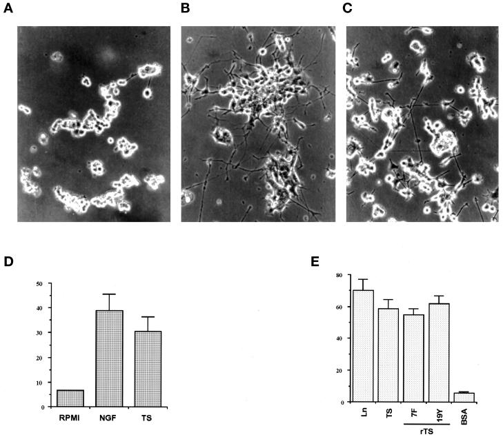
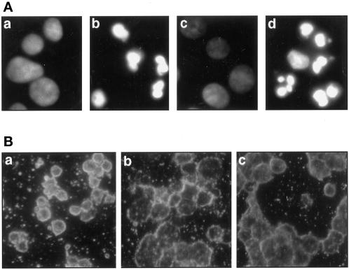
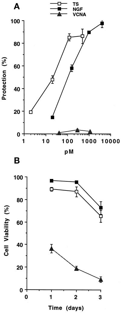
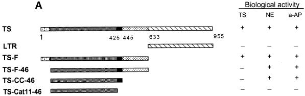
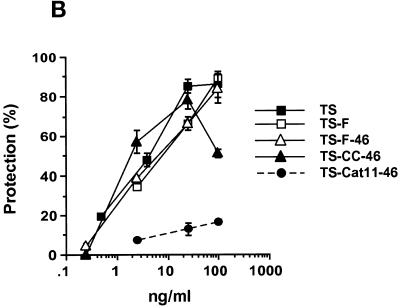
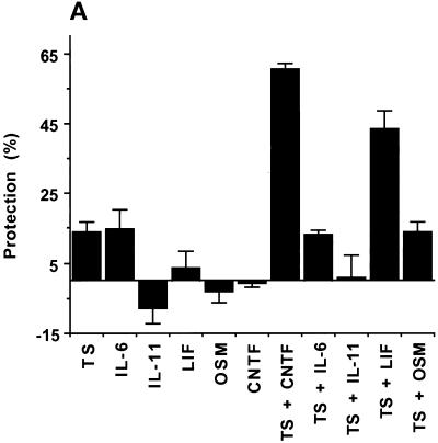
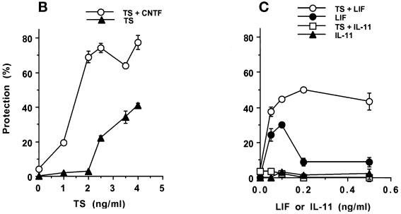
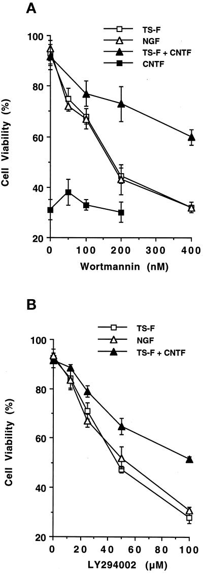
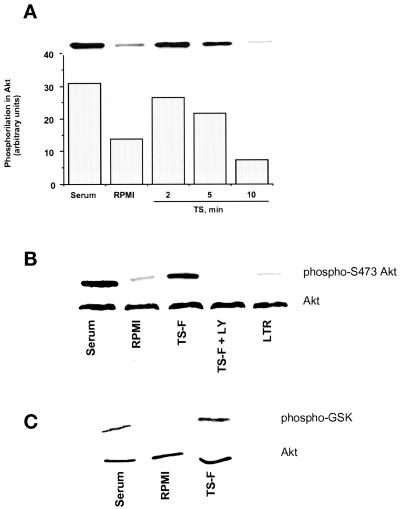
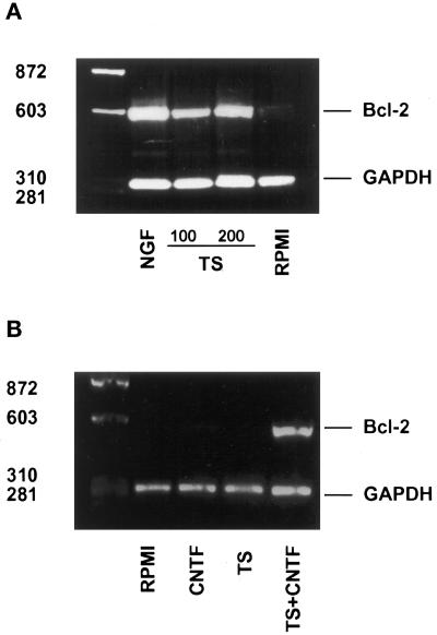
Despite the neuronal degeneration in the chronic stage of Chagas' disease, neuron counts actually increase in the preceding, asymptomatic stage, in contrast to the age-related decrease in neuron counts in age-matched normal individuals. Relevant to this observation, we found that the trans-sialidase (TS) of Trypanosoma cruzi, the etiologic agent of Chagas' disease, induces neurite outgrowth and rescues PC12 cells from apoptotic death caused by growth factor deprivation. These properties, novel for a parasite protein, were independent of catalytic activity and were mapped to the C terminus of the catalytic domain of TS. TS activated protein kinase Akt in a phosphoinositide-3 kinase-inhibitable manner, suggesting a molecular mechanism for the TS-induced neuroprotection. TS also triggered bcl-2 gene expression in growth factor-deprived cells, an effect consistent with TS protecting against apoptosis. Ciliary neurotrophic factor and leukemia inhibitory factor, two cytokines critical to the repair of injured motor neurons, specifically potentiated the TS action. The results suggest that TS acts in synergy with host ciliary neurotrophic factor or leukemia inhibitory factor to promote neuronal survival in T. cruzi-infected individuals.


Figures

Figure 1
Figure 1
Neurite outgrowth in PC12 and N18 cells in response to TS. (A–D) PC12 cells, mechanically stripped of their neurites, were seeded in serum-free RPMI medium on collagen-coated plastic dishes with no addition (A), 50 ng/ml NGF (B), or 100 ng/ml TS (C). Pictures were taken 24 h after addition of the reagents. Magnification, 400×. (D) Quantitation of PC12 cells exhibiting neurite outgrowth (triplicate samples) from an experiment similar to that shown in A–C. (E) Neurite outgrowth of N18 neuroblastoma cells by TS and rTS-F. Microtiter wells were coated overnight in triplicate with laminin (Ln), TS, rTS clones 7F and 19Y, and BSA and used as substratum for attachment of N18 cells. Cells exhibiting neurite extension were counted 17 h later. The values are the mean ± SEM of three separate experiments.
Figure 2
Figure 2
Inhibition of apoptosis in PC12 cells by TS. PC12 cells in serum-free RPMI were plated in collagen-coated plastic dishes in the presence of various reagents for 17 h and stained with DAPI (A) or TUNEL (B). The additions in A were (a) 0.1 μg/ml NGF; (b) no addition; (c) 0.1 μg/ml TS; and (d) 0.2 μg/ml VCNA. Magnification, 1000×. Additions in B were (a) no addition; (b) 0.1 μg/ml NGF; and (c) 0.1 μg/ml TS. Magnification, 400×.
Figure 3
Figure 3
Dose response and kinetics of TS inhibition of apoptosis in PC12 cells. (A) Dose response. PC12 cells were grown in triplicate in serum-free RPMI in the presence of the indicated concentrations of TS, NGF, and VCNA for 17 h. Apoptosis was measured by counting cells with nuclear fragmentation after DAPI staining. Protection against apoptosis was measured by the formula described in MATERIALS AND METHODS. Data are representative of an experiment repeated three times with similar results. The molar concentration was based on a molecular mass of 200 kDa for TS, 26 kDa for NGF, and 90 kDa for VCNA. (B) Kinetics. PC12 cells in serum-free RPMI medium were grown in the absence (RPMI) and in the presence of 4.0 nM NGF and 0.5 nM TS for the times indicated. Cell viability was measured by determining the number of cells without nuclear fragmentation relative to those with nuclear fragmentation (DAPI staining) in a sample of 300–400 cells. The average of two experiments is shown, each point in triplicate.
Figure 4
Figure 4
Identification of a TS domain that promotes neuroprotection. (A) Diagram of the linear structure of rTS and truncated derivatives obtained by PCR technology (see MATERIALS AND METHODS). Numbers under the TS diagram refer to amino acid positions in clone 19Y (GenBank accession number AJ002174). The biological activities indicated on the right are TS activity, neurite extension (NE), and antiapoptose (a-AP). These activities were measured using purified polypeptides (see MATERIALS AND METHODS). + indicates that the polypeptide promoted significant NE or a-AP in a dose-dependent manner, as in B below. Otherwise, the polypeptide was considered inactive (−). (B) Inhibition of apoptosis by endogenous and recombinant fragments of TS. PC12 cells were grown in serum-free RPMI in the presence of the indicated concentrations of TS and rTS-F, isolated from trypomastigotes and E. coli, respectively.
Figure 4
Figure 4
Identification of a TS domain that promotes neuroprotection. (A) Diagram of the linear structure of rTS and truncated derivatives obtained by PCR technology (see MATERIALS AND METHODS). Numbers under the TS diagram refer to amino acid positions in clone 19Y (GenBank accession number AJ002174). The biological activities indicated on the right are TS activity, neurite extension (NE), and antiapoptose (a-AP). These activities were measured using purified polypeptides (see MATERIALS AND METHODS). + indicates that the polypeptide promoted significant NE or a-AP in a dose-dependent manner, as in B below. Otherwise, the polypeptide was considered inactive (−). (B) Inhibition of apoptosis by endogenous and recombinant fragments of TS. PC12 cells were grown in serum-free RPMI in the presence of the indicated concentrations of TS and rTS-F, isolated from trypomastigotes and E. coli, respectively.
Figure 5
Figure 5
Synergy between TS and CNTF or LIF to rescue PC12 cells from apoptosis. (A) PC12 cells were cultured in serum-free RPMI for 17 h without TS and with TS at 2.5 ng/ml plus IL-6, 10 ng/ml, IL-11, 1 ng/ml, LIF, 1 ng/ml, OSM, 1 ng/ml, and CNTF, 50 ng/ml. Apoptosis was measured by DAPI staining 48 h after addition of reagents. (B) Dose response of the synergistic antiapoptotic action of TS with CNTF. PC12 cells were cultured for 17 h in serum-free RPMI medium without additions, with the indicated concentrations of TS, and with 50 ng/ml CNTF without and with the indicated concentrations of TS (TS + CNTF). (C) Dose response of the synergistic antiapoptotic action of TS with LIF. PC12 cells were cultured for 17 h in serum-free RPMI medium without additions and with the indicated concentrations of LIF or IL-11 without (LIF or IL-11) or with 2.5 ng/ml TS (TS + LIF and TS + IL-11, respectively).
Figure 5
Figure 5
Synergy between TS and CNTF or LIF to rescue PC12 cells from apoptosis. (A) PC12 cells were cultured in serum-free RPMI for 17 h without TS and with TS at 2.5 ng/ml plus IL-6, 10 ng/ml, IL-11, 1 ng/ml, LIF, 1 ng/ml, OSM, 1 ng/ml, and CNTF, 50 ng/ml. Apoptosis was measured by DAPI staining 48 h after addition of reagents. (B) Dose response of the synergistic antiapoptotic action of TS with CNTF. PC12 cells were cultured for 17 h in serum-free RPMI medium without additions, with the indicated concentrations of TS, and with 50 ng/ml CNTF without and with the indicated concentrations of TS (TS + CNTF). (C) Dose response of the synergistic antiapoptotic action of TS with LIF. PC12 cells were cultured for 17 h in serum-free RPMI medium without additions and with the indicated concentrations of LIF or IL-11 without (LIF or IL-11) or with 2.5 ng/ml TS (TS + LIF and TS + IL-11, respectively).
Figure 6
Figure 6
Reversal of the survival-promoting activity of TS by the PI-3 kinase inhibitors. PC12 cells were switched to serum-free RPMI without and with TS-F (100 ng/ml), NGF (100 ng/ml), CNTF (50 ng/ml), and TS-F (2.5 ng/ml) plus CNTF (50 ng/ml). After 24 h, wortmannin (A) or LY294002 (B) was added to the cultures at the indicated concentrations. Neuronal viability was measured by DAPI staining 24 h after addition of wortmannin or LY294002. Viability of PC12 cells in RPMI alone (33%) was similar to the viability of the cells in RPMI 1640 plus CNTF. Values are from a representative experiment (each point in triplicate) repeated three times, with similar results.
Figure 7
Figure 7
Activation of signaling pathways in PC12 cells by TS. (A) TS induces serine phosphorylation of Akt kinase. PC12 cells were kept in RPMI containing 0.1% FCS for 2 d. Then some monolayers were switched to 20% FCS (Serum) and others to RPMI without (RPMI) and with 100 ng/ml TS (TS) for the indicated times. Cells were lysed in 2% SDS, and proteins in the lysate were resolved by SDS-PAGE on 10% gels and stained with anti-phospho-Akt antibody. The bar graph represents the quantitation by scanning densitometry of the corresponding bands in the immunoblot (inset). (B) TS-induced phosphorylation of Akt kinase is inhibited by the PI-3 kinase inhibitor LY294002. The protocol was similar to the one in A, except that, where indicated, cells were preincubated with 1 μM LY294002 (TS-F + LY) before the addition of 100 ng/ml catalytic domain of TS (TS-F). Note that LY294002 completely blocked TS-F-induced phosphorylation of Akt kinase. LY294002 was similarly effective in inhibiting Akt phosphorylation induced by full-length TS (our unpublished data). (C) TS-F induces PKB/Akt kinase activity. PC12 cells were treated as described in A. Then some were switched to 20% FCS (Serum) and others to RPMI without (RPMI) and with 100 ng/ml TS-F (TS-F) for 2 min. Cell lysates were immunoprecipitated with Akt antibodies coupled to agarose beads The resulting immunoprecipitates were incubated with GSK-3 fusion protein as an Akt kinase substrate. Phosphorylation of GSK-3 was measured by Western blotting using a phospho-GSK-3α/β (Ser-21/9) antibody.
Figure 8
Figure 8
Induction of Bcl-2 Expression in PC12 cells. Bcl-2 expression in total RNA was isolated 17 h after addition of the growth factors and was assessed by RT-PCR using primers specific for Bcl-2 and for control GAPDH. Molecular weight markers (in base pairs) are indicated. (A) Bcl-2 expression in PC12 cells cultured in serum-free medium without (RPMI) and with NGF (100 ng/ml) or TS (100 and 200 ng). (B) Bcl-2 expression induced by TS (2.5 ng/ml) plus CNTF (50 ng/ml). Bcl-2 transcripts were not detected when TS (2.5 ng/ml) and CNTF (50 ng/ml) were added separately to PC12 cells in serum-free medium (RPMI).

References

    1. Adad SJ, Andrade DC, Lopes ER, Chapadeiro E. Pathological anatomy of chagasic megaesophagus. Rev Inst Med Trop São Paulo. 1991;33:443–450. - PubMed
    1. Ahmed NN, Grimes HL, Bellacosa A, Chan TO, Tsichlis PN. Transduction of interleukin-2 antiapoptotic and proliferative signals via Akt protein kinase. Proc Natl Acad Sci USA. 1997;94:3627–3632. - PMC - PubMed
    1. Andrade ZA. Mechanisms of myocardial damage in Trypanosoma cruzi infection. Ciba Found Symp. 1983;99:214–233. - PubMed
    1. Arakawa Y, Sendtner M, Thoenen H. Survival effect of ciliary neurotrophic factor (CNTF) on chick embryonic motorneurons in culture: comparison with other neurotrophic factors and cytokines. J Neurosci. 1990;10:3507–3515. - PMC - PubMed
    1. Bear M, Connors BW, Paradiso MA. Neurons and glia. In: Satterfield TS, editor. Neuroscience: Exploring the Brain. Baltimore: Williams & Wilkins; 1996. pp. 22–45.

Publication types

MeSH terms

LinkOut - more resources