Review
. 2000 Apr;156(4):1117-32.
doi: 10.1016/s0002-9440(10)64980-2.

Rous-Whipple Award Lecture. Viruses, immunity, and cancer: lessons from hepatitis B

Affiliations
Review

Rous-Whipple Award Lecture. Viruses, immunity, and cancer: lessons from hepatitis B

F V Chisari. Am J Pathol. 2000 Apr.
No abstract available


Figures

Figure 1.
Figure 1.
The infection.
Figure 2.
Figure 2.
HBV map. The partially double-stranded 3.2-kb open circular genome present in circulating virions is shown at the center. The genome contains the core (C), pre-S (PS), HBs (S), and HBx (X) promoters shown inside round icons as indicated, two enhancers (En I and En II) shown as shaded regions and a single polyadenylation signal (Poly A) resulting in the production of four extensively overlapping transcripts that are 3.5 kb, 2.4 kb, 2.1 kb, and 0.7 kb in length. The 3.5-kb RNA is translated to produce the viral capsid (core) and secreted precore proteins and the polymerase (pol) protein which has reverse transcriptase, RNase H, and DNA polymerase activity. The 3.5-kb RNA is also the viral pregenomic RNA that is packaged with the polymerase protein inside of capsid particles in the cytoplasm where viral replication occurs. The 2.4-kb transcript is translated to produce the large envelope protein, whereas the 2.1-kb transcript is heterogeneous at its 5′ end with species that flank the translational start site of the middle envelope protein, such that the shorter species produce the major (most abundant) envelope protein. The 0.7-kb transcript is translated to produce the X protein, which transcriptionally transactivates the viral promoters and several cellular RNAs as well.
Figure 3.
Figure 3.
The HBV life cycle. Entry of the HBV virion into the cell is a poorly defined process that is presumably receptor-mediated and leads to uncoating and transport of the capsid to the nucleus, where evidence suggests disassembly occurs, releasing the open circular viral genome into the nucleus where the second strand is completed and the ends of each strand are ligated. This leads to the production of a covalently closed circular DNA (cccDNA) molecule, which is the transcriptional template of the virus. Pol II-driven transcription results in production of the 4 viral RNAs that are thought to be actively transported out of the nucleus via shared sequences at the 3′ end of the transcripts that apparently interact with cellular RNA export proteins. Once in the cytoplasm, the transcripts are translated into the corresponding proteins as shown. The precore protein contains a leader sequence that transports it into the endoplasmic reticulum (ER) where it is further processed and eventually secreted as HBeAg. The envelope proteins traverse the ER membrane as integral membrane proteins as shown. The core and polymerase proteins assemble around the pregenomic RNA (pRNA) to form HBV RNA-containing capsids within which the RNA is reverse transcribed to produce the first strand viral DNA that serves as the template for second strand DNA synthesis. While the RNA-containing capsid is maturing into a DNA-containing capsid it migrates bidirectionally within the cytoplasm. One pathway terminates at the ER membrane where it interacts with the envelope proteins which trigger an internal budding reaction resulting in the formation of virions that are transported out of the cell by the default secretory pathway. The second pathway transports the maturing capsid to the the nucleus to amplify the pool of cccDNA.
Figure 4.
Figure 4.
Overview.
Figure 5.
Figure 5.
HBV-specific CTL response during acute and chronic infection. The CTL response to 5 HLA A2-restricted epitopes derived from the viral core, envelope, and polymerase proteins is indicated by vertical bars. Each set of bars represents the cytolytic activity of 8 replicate assays for each peptide in each patient. Acutely infected patients typically respond vigorously to multiple epitopes, as shown, and the response persists for many decades in patients who are convalescent from acute infection. In contrast, the CTL response is characteristically weak or undetectable in chronically infected patients. However, it is detectable in previously infected patients who clear the virus in response to interferon therapy, indicating CTLs are present in chronically infected patients but either too infrequent to be detected or functionally suppressed.
Figure 6.
Figure 6.
CTL-induced apoptosis and inflammation in HBV transgenic mice. A: Within 1 hour after intravenous injection of murine HBsAg-specific CTLs into HBV transgenic mice that express all viral gene products, replicate the viral genome and produce infectious virions, the CTLs recognize processed antigenic peptides presented by class I molecules on the surface of hepatocytes and stimulate them to undergo apoptosis. In this experiment, bromodeoxyuridine (BrdU)-labeled CTLs (arrow) were injected and the liver was stained with an anti-BrdU specific antibody imparting a brown stain to the CTL. Note the condensation and fragmentation of the cytoplasm and nucleus of the hepatocyte (asterisk), indicating apoptosis. B: Between 24 and 48 hours later, the CTL-induced necroinflammatory disease is maximal and the CTLs (arrow) have recruited a mixed population of host-derived, HBV-nonspecific inflammatory cells (arrowheads), many of which are associated with necrotic and apoptotic hepatocytes at a distance from the CTL. Under the conditions of this experiment, the CTLs and associated foci are widely scattered such that fewer than 10% of the hepatocytes are killed. Note that the hepatocytes surrounding the inflammatory focus are histologically normal.
Figure 7.
Figure 7.
Noncytopathic antiviral effect of the CTLs. A displays the presence of HBcAg (red stain) in the nucleus and the cytoplasm of hepatocytes in an HBV transgenic mouse before the injection of HBV-specific CTLs. B displays the absence of HBcAg from the liver of an HBV transgenic mouse 5 days after the injection of HBV-specific CTLs. The upper and lower insets represent Northern and Southern blots, respectively, of liver RNA and DNA from the same control (left side) and CTL-injected (right side) mouse livers stained in A and B. GAPDH RNA and integrated transgene DNA serve as loading controls for the Northern and Southern blots, respectively. Note that virtually 100% of the viral RNA, replicative DNA intermediates, and core protein disappear from the liver after CTL injection. Under the conditions of this experiment, less than 10% of the hepatocytes were destroyed. Thus, viral clearance from the rest of the hepatocytes was not due to their destruction.
Figure 8.
Figure 8.
IFN-γ mediates the antiviral effect of the CTLs. Mice were injected with equal numbers of HBV-specific CTL clones that recognize the same viral epitope with equal affinity. CTL clones were produced in wild-type mice and in mice that lack the genes for either Fas ligand, perforin, or IFN-γ. As shown in A, the wild-type (wt) and IFN-γ-deficient (IFN-γ) CTL clones cause a transient episode of hepatitis, indicated by elevated serum alanine aminotransferase (ALT) activity. In contrast, the Fas ligand-deficient (FasL) and perforin-deficient (Perf.) CTL clones failed to cause hepatitis. As shown in B, the wt clone abolished HBV replicative intermediates from the liver while leaving the integrated transgene unaffected, since most cells were not destroyed. Similarly, the Perf. and FasL CTL clones also inhibited HBV replication, even though they didn’t kill any hepatocytes, as shown in A. Importantly, the IFNγ clone did not inhibit HBV replication, even though it caused hepatitis. Similar results were obtained in separate experiments in which the antiviral effects of the wt CTLs were completely blocked by a cocktail of antibodies to IFN-γ and TNF-α, which had no effect on the cytopathic effect of the CTLs in vivo. These results demonstrate at the genetic and functional levels that the cytopathic and antiviral functions of the CTLs are independent of one another, although both functions require antigen recognition by the CTLs.
Figure 10.
Figure 10.
Noncytolytic clearance of HBV in an acutely infected chimpanzee. In this experiment, serum and needle liver biopsies were obtained on a weekly basis after inoculation of a chimpanzee with HBV-positive plasma from an HBV transgenic mouse. The chimpanzee became transiently infected as indicated by the appearance and eventual clearance of HBV DNA from the liver. The chimpanzee also developed an episode of acute hepatitis as seen by the transient elevation of serum ALT activity. Note that the kinetics of viral clearance (determined by competitive polymerase chain reaction in the top panel and by Southern blot in the middle panel) preceded the kinetics of disease activity by several weeks, indicating that the two events are independent during acute HBV infection in this model, similar to the observations in the HBV transgenic mice. Note also, that the cccDNA species as well as the viral replicative intermediates disappeared from the liver with similar kinetics, suggesting that the cccDNA is susceptible to noncytolytic clearance mechanisms as well as the replicative intermediates. Finally, note that the decrease in viral DNA coincides with the appearance of IFN-γ mRNA in the liver, whereas the liver disease correlates primarily with the appearance of CD3 mRNA, a T cell marker. This suggests that antiviral inflammatory cytokines produced by non-T cells may play an important role in the early viral clearance process, while the disease is more closely related to the influx of T cells into the liver.
Figure 11.
Figure 11.
Mechanisms of viral persistence.
Figure 9.
Figure 9.
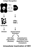
Noncytolytic clearance of HBV from the hepatocyte by T cell-derived cytokines. On antigen recognition, CD8-positive CTL deliver an apoptotic signal to their target cells, killing them. They also secrete IFN-γ and TNF-α, cytokines that abolish HBV gene expression and viral replication in vivo, curing them. The curative effect of the CTL response is more efficient than its destructive effect. The outcome of an infection may depend on the relative balance of these two effects, with a predominantly curative response leading to viral clearance and a predominantly destructive response leading to viral persistence and chronic liver disease. Importantly, if the curative process only partially inhibits viral gene expression and replication, it could paradoxically lead to viral persistence by reducing the immunological visibility of the virus without removing it. Note that antiviral cytokines produced by non-T cells during acute infection or by HBV-nonspecific T cells in the event of a superinfection could also inhibit HBV replication by a bystander mechanism. This is common during hepatitis delta virus superinfection of patients chronically infected by HBV, and it has been described during hepatitis A virus and HCV superinfection as well.
Figure 12.
Figure 12.
The chronic injury → HCC hypothesis. According to this hypothesis, a vigorous (+++) immune response to HBV leads to viral clearance while an absent (−) immune response leads to the “healthy” carrier state, and an intermediate (+) immune response produces chronic hepatitis. This indolent necroinflammatory liver disease is characterized by chronic liver cell necrosis which stimulates a sustained regenerative response. The inflammatory component includes activated macrophages that are a rich source of free radicals. The collaboration of these mitogenic and mutagenic stimuli has the potential to cause cellular and viral DNA damage, chromosomal abnormalities, genetic mutations, etc, that deregulate cellular growth control in a multistep process that eventually leads to hepatocellular carcinoma.

References

    1. Barker LF, Chisari FV, McGrath PP, Dalgard DW, Kirschstein RL, Almeida JD, Edington TS, Sharp DG, Peterson MR: Transmission of type B viral hepatitis to chimpanzees. J Infect Dis 1973, 127:648-662 - PubMed
    1. Chisari FV, Ferrari C: Hepatitis B virus immunopathogenesis. Annu Rev Immunol 1995, 13:29-60 - PubMed
    1. Beasley RP, Lin C-C, Hwang LY, Chen C-S: Hepatocellular carcinoma and hepatitis B virus: a prospective study of 22,707 men in Taiwan. Lancet 1981, 2:1129-1133 - PubMed
    1. Chisari FV: Hepatitis B virus biology and pathogenesis. Friedmann T eds. Molecular Genetic Medicine. 1992, :pp 67-104 Academic Press, San Diego - PubMed
    1. Chisari FV, Ferrari C, Mondelli MU: Hepatitis B virus structure and biology. Microb Pathog 1989, 6:311-325 - PubMed