. 2000 Apr;156(4):1157-63.
doi: 10.1016/S0002-9440(10)64984-X.

Gastrointestinal stromal tumors may originate from a subset of CD34-positive interstitial cells of Cajal

Affiliations

Gastrointestinal stromal tumors may originate from a subset of CD34-positive interstitial cells of Cajal

T L Robinson et al. Am J Pathol. 2000 Apr.

Abstract

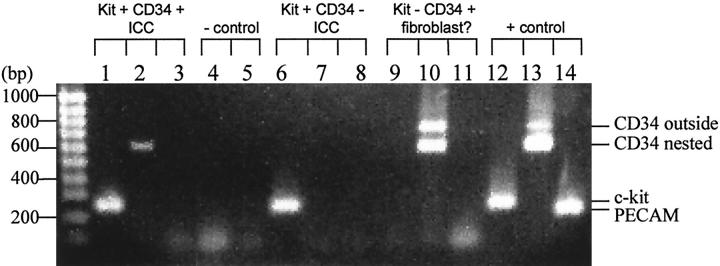
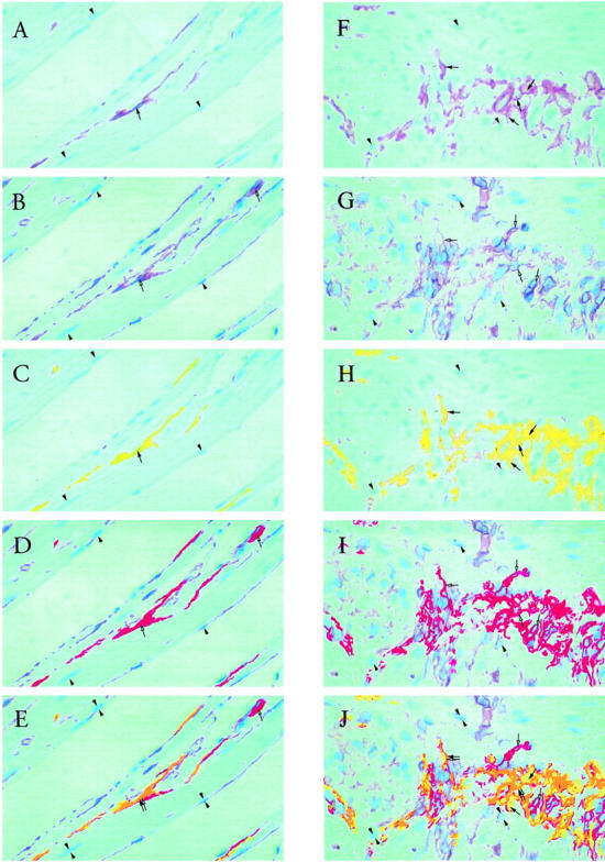
Most gastrointestinal stromal tumors (GISTs), a subgroup of mesenchymal neoplasms of the gut wall, express both Kit (CD117) and CD34 proteins. It has been suggested that GISTs originate from or differentiate into interstitial cells of Cajal (ICC), after several reports indicated that ICC are likely the only cells in the gut which express both Kit and CD34. ICC are among the few cell types resident in the gut which express Kit, together with mast cells. However, the question whether or not ICC express CD34 is currently disputed. Using single-cell reverse transcriptase-polymerase chain reaction (RT-PCR) on cultured murine intestinal cells, single ICC were selected by morphology and tested for the expression of c-kit and CD34 mRNA. Most ICC were only c-kit-positive, however a subset (7 out of 43) were double positive for both c-kit and CD34. In the human small intestine, sequential immunohistochemical staining for Kit and CD34 proteins on the same 3-microm sections showed that some of the ICC surrounding Auerbach's plexus and ICC within the circular muscle layer of the small intestine were positive for both Kit and CD34. In addition, CD34(+)Kit(-) cells were seen adjacent to ICC. These data from two different techniques indicate that ICC can be double positive for Kit and CD34. Thus, GISTs with the Kit(+)CD34(+) phenotype may arise from a subpopulation of CD34(+) Kit(+) ICC.


Figures

Figure 1.
Figure 1.
Two phenotypes were discovered in the morphologically identified ICC from the murine myenteric plexus: c-kit+CD34+ PECAM (lanes 1–3) and c-kit+CD34 PECAM (lanes 6–8). One cell that was not identified morphologically as an ICC was c-kit CD34+, and did not express detectable levels of PECAM (lanes 9–11). Two negative controls were performed, one for c-kit (lane 4) the other for CD34 (lane 5), in which water was substituted for cell contents in the RT-PCR reactions. As a positive control, an mRNA extraction from a portion of the myenteric plexus and longitudinal muscle of the murine intestine was tested for c-kit, CD34, and PECAM (lanes 12–14). The c-kit band from the nested primers (second PCR reaction) appears at 258 bp in the morphologically identified ICC. The CD34 band from nested primers appears at 608 bp in a subset of morphologically identified ICC. Another CD34 band appears at 772 bp in the one cell tentatively labeled as a fibroblast, which is the product from the first PCR reaction (outside primers). The PECAM band from the nested primers appears at 218 bp only in the positive control.
Figure 2.
Figure 2.
Left column: sequential immunohistochemical staining for CD117 (Kit) and CD34, using AEC chromagen, counterstained with hematoxylin, on single 3-μm sections of the circular smooth muscle region of the human small intestine. A: Kit staining of ICC in the muscle layer (brown, selected area marked by filled arrow). Arrowheads mark the tops of selected nuclei for location reference. B: CD34 staining (brown, selected areas marked by open arrows) of the same area of the section as in A. Arrowheads mark the bottoms of the same nuclei marked in A. Far more CD34 positivity is present than Kit positivity (compare B to A). C: Digital recoloring (to yellow) of selected Kit-stained areas in A. D: Digital recoloring (to red) of selected CD34-stained areas in B. Note that the recoloring produces one color intensity, compared to several color intensities in the original stain. Very low intensities of the original stain are not recolored. E: Merged fields C and D. Kit+CD34+ ICC are present in the muscle layer (orange, marked by double arrows). Also present in the muscle layer are Kit CD34+ cells (red, single open arrow). An example of a Kit +CD34 ICC adjacent to a Kit CD34+ fibroblast is found in the right bottom corner, confirming the recent observation from Vanderwinden et al Magnification, ×200. Right column: sequential immunohistochemical staining for CD117 (Kit) and CD34, using AEC chromagen, counterstained with hematoxylin, on single 3-μm sections of the Auerbach’s plexus region of the human small intestine. F: Kit staining of ICC in the plexus (brown, selected areas marked by filled arrows). Arrowheads mark the tops of selected nuclei for location reference. G: CD34 staining (brown, selected areas marked by open arrows) of the same area of the section as in F. Arrowheads mark the bottoms of the same nuclei marked in F. Far more CD34 positivity is present than Kit positivity (compare G to F). H: Digital recoloring (to yellow) of selected Kit-stained areas in F. I: Digital recoloring (to red) of selected CD34-stained areas in G. Note that the recoloring produces one color intensity, compared to the graded color intensities in the original stain. Very low intensities of the original stain were not recolored. Hence the total staining in H and I is less than in, respectively, F and G. The staining is only much brighter for purpose of overlap identification. J: Merged fields H and I. In many instances, co-localization of Kit and CD34 was observed (orange, selected areas marked by double arrows), representing Kit+CD34+ ICC. Close to the double-positive ICC, are Kit CD34+ cells, which may be fibroblasts (red, single open arrows). The merged fields also revealed the presence of Kit+CD34 ICC (yellow, single filled arrows). The Kit CD34+ cells are far more numerous than Kit+CD34 or Kit+CD34+ cells. Both Kit and CD34 preferentially stained cells located around the ganglia and into the septa. Magnification, ×200.

References

    1. Vanderwinden JM, Rumessen JJ, De Laet MH, Vanderhaeghen JJ, Schiffmann SN: CD34+ cells in human intestine are fibroblasts adjacent to, but distinct from, interstitial cells of Cajal. Lab Invest 1999, 79:59-65 - PubMed
    1. Editorial: Setting the pace for gastrointestinal stromal tumors. Lab Invest 1999, 79:1
    1. Saul SH, Rast ML, Brooks JJ: The immunohistochemistry of gastrointestinal stromal tumors: evidence supporting an origin from smooth muscle. Am J Surg Pathol 1987, 11:464-473 - PubMed
    1. Van de Rijn M, Hendrickson MR, Rouse RV: CD34 expression by gastrointestinal tract stromal tumors. Hum Pathol 1994, 25:766-771 - PubMed
    1. Kindblom LG, Remotti HE, Aldenborg F, Meis-Kindblom JM: Gastrointestinal pacemaker cell tumor (GIPACT): gastrointestinal stromal tumors show phenotypic characteristics of the interstitial cells of Cajal. Am J Pathol 1998, 152:1259-1269 - PMC - PubMed

Publication types

MeSH terms

LinkOut - more resources