. 2000 Apr;156(4):1253-61.
doi: 10.1016/S0002-9440(10)64996-6.

The Fn14 immediate-early response gene is induced during liver regeneration and highly expressed in both human and murine hepatocellular carcinomas

Affiliations

The Fn14 immediate-early response gene is induced during liver regeneration and highly expressed in both human and murine hepatocellular carcinomas

S L Feng et al. Am J Pathol. 2000 Apr.

Abstract

Polypeptide growth factors stimulate mammalian cell proliferation by binding to specific cell surface receptors. This interaction triggers numerous biochemical responses including the activation of protein phosphorylation cascades and the enhanced expression of specific genes. We have identified several fibroblast growth factor (FGF)-inducible genes in murine NIH 3T3 cells and recently reported that one of them, the FGF-inducible 14 (Fn14) immediate-early response gene, is predicted to encode a novel, cell surface-localized type Ia transmembrane protein. Here, we report that the human Fn14 homolog is located on chromosome 16p13.3 and encodes a 129-amino acid protein with approximately 82% sequence identity to the murine protein. The human Fn14 gene, like the murine Fn14 gene, is expressed at elevated levels after FGF, calf serum or phorbol ester treatment of fibroblasts in vitro and is expressed at relatively high levels in heart and kidney in vivo. We also report that the human Fn14 gene is expressed at relatively low levels in normal liver tissue but at high levels in liver cancer cell lines and in hepatocellular carcinoma specimens. Furthermore, the murine Fn14 gene is rapidly induced during liver regeneration in vivo and is expressed at high levels in the hepatocellular carcinoma nodules that develop in the c-myc/transforming growth factor-alpha-driven and the hepatitis B virus X protein-driven transgenic mouse models of hepatocarcinogenesis. These results indicate that Fn14 may play a role in hepatocyte growth control and liver neoplasia.


Figures

Figure 1.
Figure 1.
Comparison of the mouse and human Fn14 deduced amino acid sequences. Identical residues are boxed and the numbers to the right refer to the last amino acids on the lines. The solid line indicates the predicted signal peptide sequence and the dotted line indicates the predicted transmembrane domain.
Figure 2.
Figure 2.
Chromosomal localization of the human Fn14 gene by FISH. Two partial human metaphase spreads demonstrating specific hybridization signals at chromosome 16p13.3 are shown. Inset in right panel shows specific hybridization to individual chromosome 16 homologues from other metaphase spreads. The photographs represent computer-generated, merged images of fluorescein signals (arrows) and DAPI-stained chromosomes.
Figure 3.
Figure 3.
Regulation of Fn14 mRNA expression in human M426 cells. A: RNA was isolated from human M426 fibroblasts and murine NIH3T3 fibroblasts and equivalent amounts of each sample were analyzed by Northern blot hybridization. The positions of 28S and 18S rRNA are noted on the left. In the bottom section of this panel and the subsequent panels, a photograph of that portion of the RNA gel containing the 18S rRNA band is shown to demonstrate that similar amounts of RNA were present in each gel lane. B−D: Serum-starved M426 cells were either left untreated or treated with FGF-1 (B), FBS (C), or PMA (D) for the indicated time periods. RNA was isolated and equivalent amounts of each sample were analyzed by Northern blot hybridization.
Figure 4.
Figure 4.
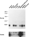
Fn14 mRNA expression in various human tissues. A Northern blot containing equivalent amounts of poly(A)+ RNA isolated from eight human tissues was obtained and hybridization analysis was performed using the two cDNA probes indicated. RNA size markers (in kb) are shown on the left.
Figure 5.
Figure 5.
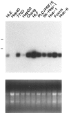
Fn14 mRNA expression in human liver cell lines. RNA was isolated from the indicated liver cell lines and equivalent amounts of each sample were analyzed by Northern blot hybridization. The positions of 28S and 18S rRNA are noted on the left. In the bottom panel, a photograph of the RNA gel is shown to demonstrate that similar amounts of RNA were present in each gel lane.
Figure 6.
Figure 6.
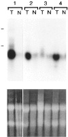
Fn14 mRNA expression in human HCC specimens. Northern blots containing equivalent amounts of total RNA isolated from HCC tumor tissue (T) and adjacent nontumoral liver tissue (N) from four individuals were obtained and hybridization analysis was performed. The RNA samples from individual no. 1 were on one blot, whereas the other samples were on a second blot, and the hybridization results were combined into this panel. The positions of 28S and 18S rRNA are noted on the left. In the bottom panel, a photograph of the RNA gel is shown to demonstrate that similar amounts of RNA were present in each gel lane.
Figure 7.
Figure 7.
Regulation of Fn14 mRNA expression during liver regeneration in mice. RNA was isolated from regenerating mouse liver harvested at various times after partial hepatectomy, and equivalent amounts were used for Northern blot hybridization analysis using the two cDNA probes indicated. The positions of 28S and 18S rRNA are noted on the left.
Figure 8.
Figure 8.
Fn14 mRNA expression in HCC specimens from c-myc/TGF-α double transgenic mice. RNA was isolated from HCC tumor tissue (T) and adjacent nontumoral liver tissue (N) from three transgenic animals and equivalent amounts of each sample were analyzed by Northern blot hybridization using the two cDNA probes indicated. The positions of 28S and 18S rRNA are noted on the left.
Figure 9.
Figure 9.
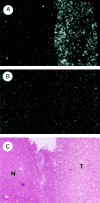
Fn14 mRNA expression in HCC nodules from HBx transgenic mice. Serial sections of liver harvested from a transgenic animal were used for in situ hybridization analysis using Fn14 antisense (A) or sense (B) riboprobes. These two dark-field photographs, which reveal the hybridization signal grains in white, were taken at the same exposure level. A bright-field view showing another serial section stained with hematoxylin and eosin is shown in C. The HCC tumor nodule (T) and the adjacent nontumoral region of the liver (N) are indicated.

References

    1. Van der Geer P, Hunter T, Lindberg RA: Receptor protein-tyrosine kinases and their signal transduction pathways. Annu Rev Cell Biol 1994, 10:251-337 - PubMed
    1. Winkles JA: Serum-and polypeptide growth factor-inducible gene expression in mouse fibroblasts. Prog Nucleic Acid Res Mol Biol 1998, 58:41-78 - PubMed
    1. Iyer VR, Eisen MB, Ross DT, Schuler G, Moore T, Lee JCF, Trent JM, Staudt LM, Hudson J, Boguski MS, Lashkari D, Shalon D, Botstein D, Brown PO: The transcriptional program in the response of human fibroblasts to serum. Science 1999, 283:83-87 - PubMed
    1. Fambrough D, McClure K, Kazlauskas A, Lander ES: Diverse signaling pathways activated by growth factor receptors induce broadly overlapping, rather than independent, sets of genes. Cell 1999, 97:727-741 - PubMed
    1. Winkles JA, Donohue PJ, Hsu DKW, Guo Y, Alberts GF, Peifley KA: Identification of FGF-1-inducible genes by differential display. Gallo L eds. Cardiovascular Disease 2. 1995, :pp 109-120 Plenum Press, New York

Publication types

MeSH terms