. 2000 Apr;156(4):1299-306.
doi: 10.1016/S0002-9440(10)65000-6.

Dual-hit hypothesis explains pulmonary hypoplasia in the nitrofen model of congenital diaphragmatic hernia

Affiliations

Dual-hit hypothesis explains pulmonary hypoplasia in the nitrofen model of congenital diaphragmatic hernia

R Keijzer et al. Am J Pathol. 2000 Apr.

Abstract

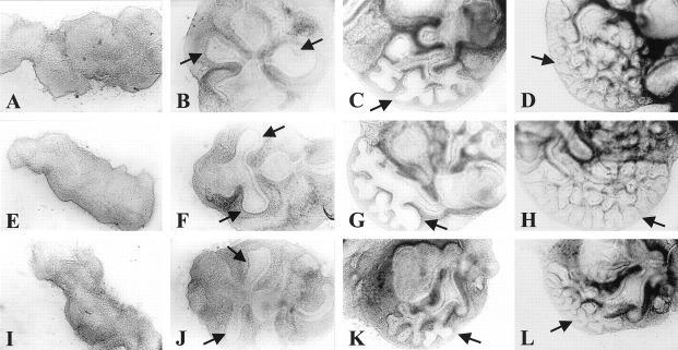
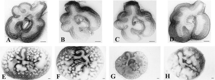
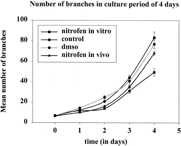
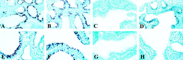
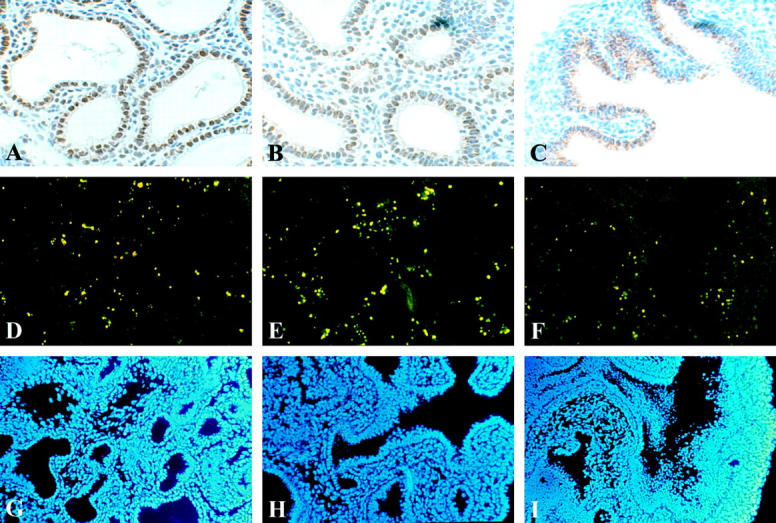
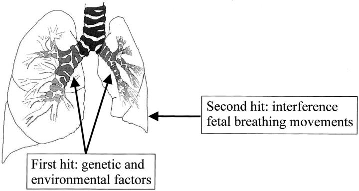
Pulmonary hypoplasia associated with congenital diaphragmatic hernia (CDH) remains a major therapeutic problem. Moreover, the pathogenesis of pulmonary hypoplasia in case of CDH is controversial. In particular, little is known about early lung development in this anomaly. To investigate lung development separate from diaphragm development we used an in vitro modification of the 2, 4-dichlorophenyl-p-nitrophenylether (Nitrofen) animal model for CDH. This enabled us to investigate the direct effects of Nitrofen on early lung development and branching morphogenesis in an organotypic explant system without the influence of impaired diaphragm development. Epithelial cell differentiation of the lung explants was assessed using surfactant protein-C and Clara cell secretory protein-10 mRNA expression as markers. Furthermore, cell proliferation and apoptosis were investigated. Our results indicate that Nitrofen negatively influences branching morphogenesis of the lung. Initial lung anlage formation is not affected. In addition, epithelial cell differentiation and cell proliferation are attenuated in lungs exposed to Nitrofen. These data indicate that Nitrofen interferes with early lung development before and separate from (aberrant) diaphragm development. Therefore, we postulate the dual-hit hypothesis, which explains pulmonary hypoplasia in CDH by two insults, one affecting both lungs before diaphragm development and one affecting the ipsilateral lung after defective diaphragm development.


Figures

Figure 1.
Figure 1.
Lung formation and branching morphogenesis of the lung in an explant system of rat foregut. Foreguts of rats were harvested at 11 days of gestation and cultured in a semidry system. Pictures representative of a series of experiments of untreated (A−D), vehicle DMSO-exposed (E−H) and Nitrofen-exposed (I−L) foreguts developing into branching lungs are shown at culture day 0 (A, E, I), 2 (B, F, J), 6 (C, G, K), and 8 (D, H, L), respectively. The Nitrofen-exposed group was exposed to Nitrofen in a concentration similar to that used in vivo: 0.25 mg.200 μl−1 medium on the first culture day, and to half of this on the second culture day. Lung formation in this group occurred at the same time point as in the control groups (J), which were cultured in medium with vehicle DMSO (F) or in medium alone (B). Branching morphogenesis was clearly reduced in the explants exposed to Nitrofen during the remaining culture period (L). Arrow indicates lung. All images at same magnification.
Figure 2.
Figure 2.
Branching morphogenesis in a rat lung explant system. At 13 days of gestation, lungs were removed from the fetuses and cultured in a semidry system. The upper row of pictures are representative for lungs at 13 days of gestation at culture day 0 just after isolation of the lungs from the fetus. The bottom row of pictures represents lungs after a culture period of 4 days. One group was treated with Nitrofen in vivo by administration of the Nitrofen to the mother at 9 days of gestation (D and H) Another group was treated with Nitrofen in vitro as described before (G). Branching morphogenesis was compared to groups cultured in medium alone (E), and groups cultured in medium plus vehicle DMSO (F). In all Nitrofen-exposed explants a statistically significant (P < 0.05) reduced number of branches was observed after 4 days of culture (G and H), whereas no effects of exposure to DMSO were observed (F; see also Figure 3 ▶ and Table 2 ▶ ). In addition, the size of the explants exposed to Nitrofen was clearly reduced (G and H). The effects of Nitrofen exposure in vitro were more severe than the effects after Nitrofen exposure in vivo. All pictures are representative of a series of experiments. Scale bar represents magnification level.
Figure 3.
Figure 3.
Branching morphogenesis plotted as a graph. Number of branches, mean ± SE, is plotted against days of culture. Explants exposed to Nitrofen in vitro showed an arrest in branching during the first 2 days of culture, the period of exposure to Nitrofen. In addition, the rate of branching (slope) was also reduced after the first 2 days of culture during the remaining culture-period. After exposure to Nitrofen in vivo explants showed also a reduced number of branches after 3 days of culture and a reduced rate of branching (slope) although to a lesser extent than the ones exposed to Nitrofen in vitro.
Figure 4.
Figure 4.
Expression of markers for distal and proximal epithelial cell differentiation, SP-C (upper row) and CC-10 (bottom row) mRNA respectively, in lung explants after 4 days of culture. Expression of SP-C mRNA was observed in control, DMSO, and Nitrofen in vivo treated explants in the epithelial lining of the terminal lung buds after 4 days of culture (A, B, and D). No expression was observed in the explants exposed to Nitrofen in vitro (C). Expression of CC-10 mRNA was observed in control and DMSO explants in the epithelial lining of the major proximal airways. (E and F). No expression was observed in the explants exposed to Nitrofen both in vitro (G) and in vivo (H). All pictures are representative of a series of experiments. All images at same magnification.
Figure 5.
Figure 5.
Following a culture period of 4 days, proliferation (A−C) and apoptosis (D−F) were investigated. Immunolocalization of PCNA indicates proliferating cells. Strong PCNA immunoreactivity was observed in the epithelial lining of the terminal buds of the control and DMSO explants, and also, but lower in the mesenchyme (A and B). Surprisingly immunoreactivity was not observed in the mesenchyme and only perinuclear in the explants exposed to Nitrofen (C). Apoptosis in the explants was assessed using TUNEL assay. TUNEL positive cells were observed in all explants in the mesenchyme only, and the number of TUNEL positive cells appeared similar in all explants (D−F). All nuclei were stained using DAPI mounting solution (G−I). All pictures are representative of a series of experiments. All images at same magnification.
Figure 6.
Figure 6.
The dual-hit hypothesis: pulmonary hypoplasia in case of CDH is explained by two developmental insults. The first hit affects both lungs before and separate from diaphragm development in a background (unidentified until now) of genetic and environmental factors. The second hit affects only the ipsilateral lung after defective development of the diaphragm because of interference of the herniated abdominal organs with fetal breathing movements of this lung.

References

    1. Torfs CP, Curry CJ, Bateson TF, Honore LH: A population-based study of congenital diaphragmatic hernia. Teratology 1992, 46:555-565 - PubMed
    1. Katz AL, Wiswell TE, Baumgart S: Contemporary controversies in the management of congenital diaphragmatic hernia. Clin Perinatol 1998, 25:219-248 - PubMed
    1. Reickert CA, Hirschl RB, Atkinson JB, Dudell G, Georgeson K, Glick P, Greenspan J, Kays D, Klein M, Lally KP, Mahaffey S, Ryckman F, Sawin R, Short BL, Stolar CJ, Thompson A, Wilson JM: Congenital diaphragmatic hernia survival and use of extracorporeal life support at selected level III nurseries with multimodality support. Surgery 1998, 123:305-310 - PubMed
    1. Wung JT, Sahni R, Moffitt ST, Lipsitz E, Stolar CJ: Congenital diaphragmatic hernia: survival treated with very delayed surgery, spontaneous respiration, and no chest tube. J Pediatr Surg 1995, 30:406-409 - PubMed
    1. Thebaud B, Mercier JC, Dinh-Xuan AT: Congenital diaphragmatic hernia: a cause of persistent pulmonary hypertension of the newborn which lacks an effective therapy. Biol Neonate 1998, 74:323-336 - PubMed

Publication types

LinkOut - more resources