

Vascular endothelial growth factor and hepatocyte growth factor levels are differentially elevated in patients with advanced retinopathy of prematurity
- PMID: 10751359
- PMCID: PMC1876877
- DOI: 10.1016/S0002-9440(10)65004-3
Vascular endothelial growth factor and hepatocyte growth factor levels are differentially elevated in patients with advanced retinopathy of prematurity
Abstract
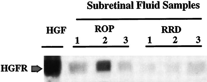
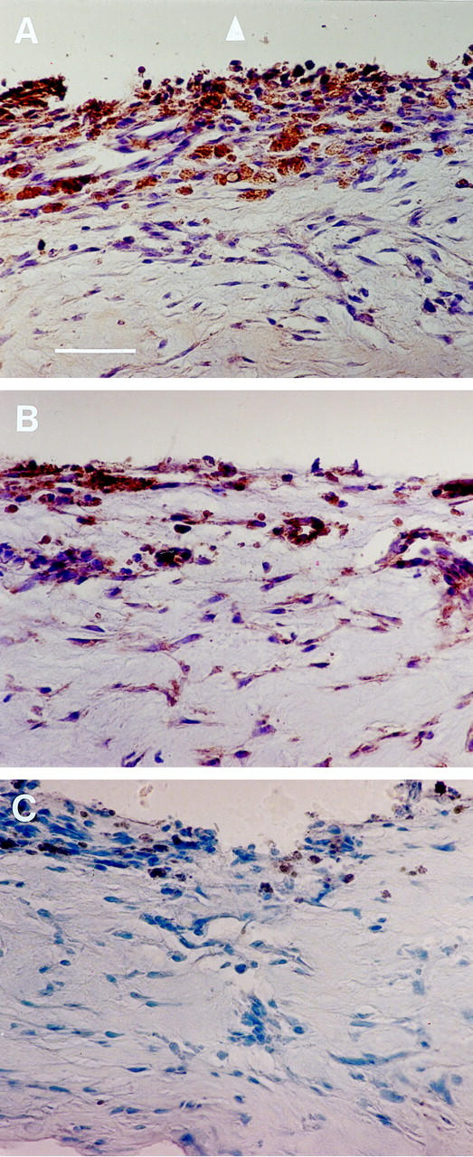
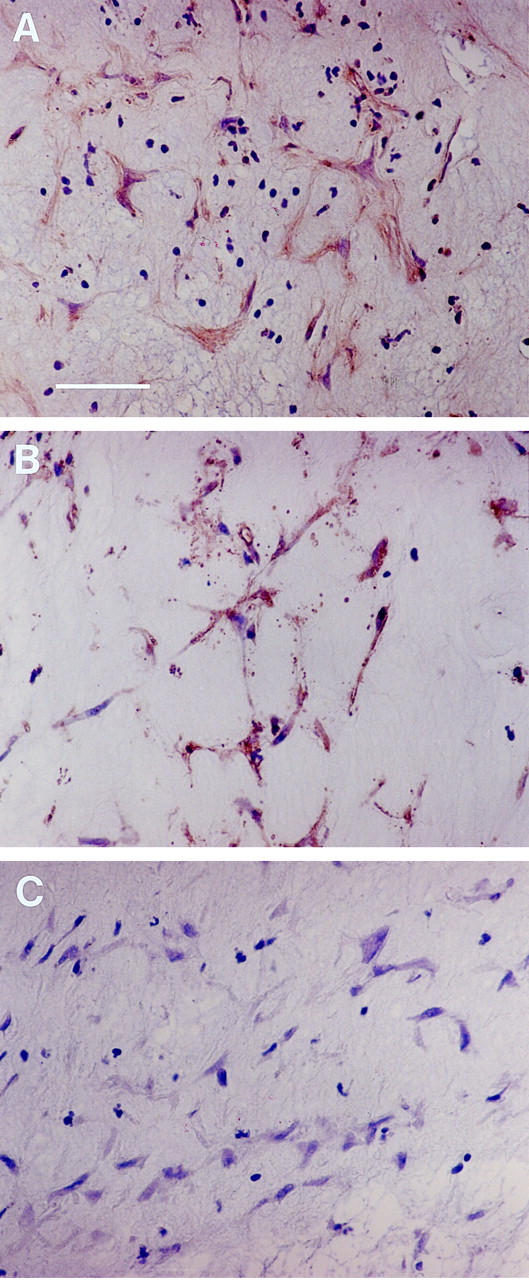
Although the roles of vascular endothelial growth factor (VEGF) and hepatocyte growth factor (HGF) in angiogenesis are well described, the putative roles of these factors in retinopathy of prematurity (ROP) remain unknown. We evaluated VEGF and HGF protein levels in subretinal fluid of eyes with ROP, and expression of their corresponding receptors in retrolental membranes associated with stage 5 ROP. We examined subretinal fluid samples from eyes using rhegmatogenous retinal detachment as a control. VEGF and HGF were differentially elevated in eyes with ROP. In Stage 5 ROP (n = 22), the mean VEGF and HGF levels were 14.77 +/- 14.01 ng/ml and 16.56 +/- 9.62 ng/ml, respectively. Interestingly, in patients with active stage 4 ROP, mean VEGF levels were highly elevated (44.16 +/- 18.72 ng/ml), whereas mean HGF levels remained very low (4.77 +/- 2.50 ng/ml). Next, we investigated in vivo expression of VEGF receptor-2 and HGF receptor in retrolental membranes from 16 patients with stage 5 ROP. Both VEGF receptor-2 and HGF receptor proteins were detected mainly in posterior portions of the membrane as well as in vessel walls and along the retinal interface where angiogenesis was active. These findings together suggest that VEGF and HGF play important roles in the pathogenesis of ROP.
Figures
References
-
- Wiedemann P: Growth factors in retinal diseases: proliferative vitreoretinopathy, proliferative diabetic retinopathy, and retinal degeneration. Surv Ophthalmol 1992, 36:373-384 - PubMed
-
- Forrester JV, Shafiee A, Schroder S, Knott R, McIntosh L: The role of growth factors in proliferative diabetic retinopathy. Eye 1993, 7:276-287 - PubMed
-
- Zarbin MA: Age-related macular degeneration: review of pathogenesis. Eur J Ophthalmol 1998, 8:199-206 - PubMed
-
- Risau W: Mechanisms of angiogenesis. Nature 1997, 386:671-674 - PubMed
-
- Neufeld G, Cohen T, Gengrinovitch S, Poltorak Z: Vascular endothelial growth factor (VEGF) and its receptors. FASEB J 1999, 13:9-22 - PubMed
Publication types
MeSH terms
Substances
LinkOut - more resources
Full Text Sources
Other Literature Sources
Medical
