. 2000 Apr;156(4):1345-62.
doi: 10.1016/s0002-9440(10)65005-5.

Regulation of angiogenesis in vivo by ligation of integrin alpha5beta1 with the central cell-binding domain of fibronectin

Affiliations

Regulation of angiogenesis in vivo by ligation of integrin alpha5beta1 with the central cell-binding domain of fibronectin

S Kim et al. Am J Pathol. 2000 Apr.

Abstract

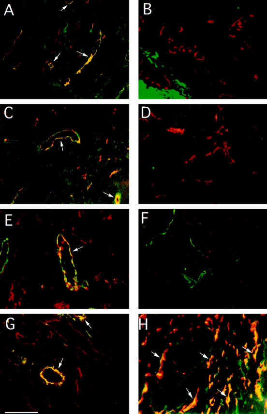
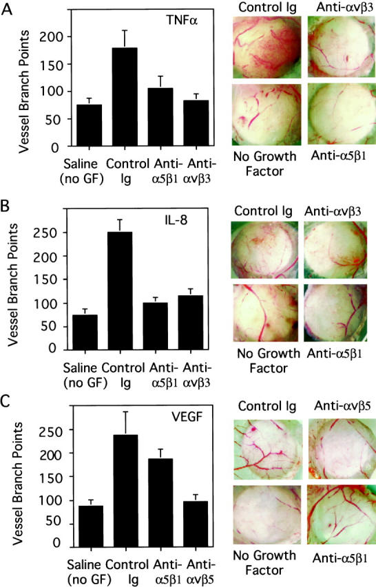
Angiogenesis depends on the cooperation of growth factors and cell adhesion events. Although alphav integrins have been shown to play critical roles in angiogenesis, recent studies in alphav-null mice suggest that other adhesion receptors and their ligands also regulate this process. Evidence is now provided that the integrin alpha5beta1 and its ligand fibronectin are coordinately up-regulated on blood vessels in human tumor biopsies and play critical roles in angiogenesis, resulting in tumor growth in vivo. Angiogenesis induced by multiple growth factors in chick embryos was blocked by monoclonal antibodies to the cell-binding domain of fibronectin. Furthermore, application of fibronectin or a proteolytic fragment of fibronectin containing the central cell-binding domain to the chick chorioallantoic membrane enhanced angiogenesis in an integrin alpha5beta1-dependent manner. Importantly, antibody, peptide, and novel nonpeptide antagonists of integrin alpha5beta1 blocked angiogenesis induced by several growth factors but had little effect on angiogenesis induced by vascular endothelial growth factor (VEGF) in both chick embryo and murine models. In fact, these alpha5beta1 antagonists inhibited tumor angiogenesis, thereby causing regression of human tumors in animal models. Thus, fibronectin and integrin alpha5beta1, like integrin alphavbeta3, contribute to an angiogenesis pathway that is distinct from VEGF-mediated angiogenesis, yet important for the growth of tumors.


Figures

Figure 1.
Figure 1.
Expression of integrin α5β1 and fibronectin on human and murine tumor blood vessels. Cryostat sections 5 μm in width of human colon carcinoma (A), normal colon (B), breast carcinoma (C, E, and G), normal breast (D and F), and subcutaneous human tumor xenotransplants in SCID mice (H) were analyzed by fluorescence microscopy at 200× magnification for expression of integrin α5β1, fibronectin, CD31, or vWF, as described in Materials and Methods. A−D, H: Tissue sections stained for integrin α5β1 (FITC) and CD31 (rhodamine) expression. E and F: Tissue sections stained for fibronectin (rhodamine) and vWF (FITC) expression. G: Tissue sections stained for integrin α5β1 (FITC) and fibronectin (rhodamine) expression. Merged images of these tissues stained with both antibodies indicate where colocalization (yellow) occurs. Representative costaining vessels are indicated by arrows. Scale bar, 10 μm.
Figure 2.
Figure 2.
Enhanced expression of integrin α5β1 and fibronectin on blood vessels after growth factor stimulation. Cryostat sections of normal unstimulated (A and B), bFGF-stimulated (C and D), or VEGF-stimulated CAMs (E and F) were stained with anti-integrin α5β1 (rhodamine) and anti-vWF (FITC) antibodies (A, C, and E), anti-fibronectin (rhodamine) and anti-vWF (FITC) antibodies (B, D, and F). Merged images of these tissues stained with both antibodies indicate where colocalization (yellow) occurs. Scale bar, 10 μm.
Figure 3.
Figure 3.
Role of fibronectin in angiogenesis. A: Adhesion of HUVECs to fibronectin in the presence of adhesion medium (medium) or 25 μg/ml antibodies directed to the cell-binding peptide region of fibronectin (Anti-CBP) or to the N-terminal heparin binding region of fibronectin (Anti-NT). B: Angiogenesis induced on the CAM by bFGF or VEGF in the presence of saline, 25 μg of an antibody directed against the cell-binding peptide of fibronectin (Anti-CBP) or 25 μg of an antibody directed against the fibronectin N-terminus (Anti-NT). The number of blood vessel branch points within a standard 5-mm area are shown. C: bFGF-induced angiogenesis on the CAM in the presence of saline or equimolar amounts of full length fibronectin, the 120-kd cell-binding fibronectin fragment, the 40-kd fibronectin fragment, or full length fibronectin plus 10 μg anti-integrin α5β1. The data are presented as blood vessel branch points above background; * indicates treatments that resulted in significantly different numbers of vessel branch points than bFGF treatment alone, fibronectin (P = 0.05), and 120-kd fibronectin fragments (P = 0.05).
Figure 4.
Figure 4.
Inhibition of cell adhesion and migration by integrin α5β1 antagonists. A: The adhesion of HT29 colon carcinoma cells transfected with the integrin α5 cDNA (HT29 α5-positive), chick embryo fibroblasts (CEF) and human umbilical vein endothelial cells (HUVEC) to fibronectin in the presence of 25 μg/ml function-blocking (▪) or non-function-blocking anti-integrin α5β1 antibodies (▨) expressed as percent of cells adhering in adhesion buffer alone. The adhesion of HUVECs to vitronectin in the presence of 25 μg/ml function-blocking anti-α5β1 antibodies (▪) or LM609, a function-blocking anti-integrin αvβ3 antibody (▩) B: The adhesion of HT29 α5-positive cells, CEFs, and HUVECs to fibronectin in the presence of 10 μmol/L cyclic peptide CRRETAWAC (▪) or the control scrambled peptide, CATAERWRC (▨). C: Adhesion of HT29 α5-positive cells to fibronectin in the presence of dilutions of the small molecule integrin α5β1 antagonist SJ749. D: Migration of HUVECs on fibronectin or collagen in the presence of migration medium (▪), 25 μg/ml function-blocking (▨), and 25 μg/ml non-function-blocking (▩) antibodies to integrin α5β1 or 25 μg/ml antibodies to integrin β1 (formula image).
Figure 5.
Figure 5.
Inhibition of angiogenesis by anti-integrin α5β1 antibody, peptide, and nonpeptide small molecule antagonists. A: Chick chorioallantoic membranes stimulated by bFGF were treated with either saline, 10 μg anti-α5β1 monoclonal (Anti-α5β1), 10 μg non-function-blocking anti-α5β1 antibodies (control IgG), or 10 μg of anti-αvβ3 antibodies. Forty-eight hours after administering the antagonists, CAMs were excised. Selected representative CAMS were photographed at 10× magnification. B: Blood vessel branch points within the 5-mm treatment area were counted under 30× magnification using a stereo dissecting microscope for CAMs treated as in A. C: Blood vessel branch points within the 5-mm treatment area of CAMs stimulated by saline (PBS) or by bFGF and treated with saline (bFGF), 750 pmoles cyclic peptide CRRETAWAC, or 750 pmoles control peptide CATAERWRC. were counted at 30× magnification. D: Blood vessel branch points within the 5-mm treatment area of CAMs stimulated by bFGF and treated with dilutions of the nonpeptide integrin α5β1 antagonist SJ749 were counted at 30× magnification and are expressed as a percentage of bFGF-induced branch points. E and F: Blood vessel branch points on bFGF-stimulated CAMs treated by intravenous injections of saline, anti-α5β1 or control antibody (P1F6), cyclic peptide CRRETAWAC, or control peptide CATAERWRC (25 μmol/L, final serum concentration), nonpeptide integrin α5β1 antagonist SJ749 or control nonpeptide XU065 were counted at 30× magnification. At least 10 embryos were used per treatment group. Each experiment was performed a minimum of three times. Data were evaluated in terms of average number of blood vessel branch points per treatment group ± SEM. Statistical analyses were performed using Student’s t-test.
Figure 6.
Figure 6.
Inhibition of angiogenesis by anti-integrin α5β1 in the SCID mouse/human skin chimera. Angiogenesis was induced by intradermal injection of growth factor depleted matrigel supplemented with 1 μg/ml bFGF and 25 μg/ml function-blocking or control anti-integrin α5β1 antibodies into human skin transplanted onto SCID mice. A: Anti-human CD31 immunohistochemical analysis of frozen sections of function-blocking or control treated human skin at 100× magnification. Arrows indicate human CD31-positive blood vessels. Scale bar, 10 μm. B: Quantification of CD31-positive blood vessels per 100× microscopic field in bFGF-stimulated human skin treated with function-blocking or control antibodies. The data are presented as mean CD31-positive blood vessel numbers per 100× microscopic field, ± SEM. Statistical analyses were performed using Student’s t-test.
Figure 7.
Figure 7.
Inhibition of TNF-α and IL-8 but not VEGF angiogenesis by integrin α5β1 antagonists. Chick chorioallantoic membranes stimulated by TNFα (A), IL-8 (B), or VEGF (C) were treated with either saline, 25 μg anti-α5β1 monoclonal (Anti-α5β1), 25μg non-function-blocking anti-α5β1 antibodies (control IgG), or 25 μg of anti-αvβ3 antibodies (TNF-α- or IL-8-stimulated CAMs) or 25 μg of anti-αvβ5 antibodies (VEGF-stimulated CAMs). Forty-eight hours after administering the antagonists, CAMs were excised. Blood vessel branch points within the 5-mm treatment area were counted under 30× magnification using a stereo dissecting microscope (Left panels). At least 10 embryos were used per treatment group. Each experiment was performed a minimum of three times. Data were evaluated in terms of average number of blood vessel branch points per treatment group ± SEM. Statistical analyses were performed using Student’s t-test. Right panels: Selected representative CAMS were photographed at 10× magnification.
Figure 8.
Figure 8.
Inhibition of tumor angiogenesis by antagonists of integrin α5β1. Tumor fragments (50 mg) cultured on CAMs were treated topically with function-blocking or control anti-α5β1 or systemically with active (CRRETAWAC) and control (CATAERWRC) peptides and active (SJ749) and control small molecule inhibitors of integrin α5β1. Forty-eight hours later, CAMs were excised from the egg and representative tumors from antibody treatment groups were photographed under 10× magnification (A). Tumor-associated blood vessels were quantified by counting blood vessel branch points. The data are presented as mean blood vessel number per treatment group (± SEM). Each treatment group incorporated at least 10 tumors per experiment (B). Tumors excised from the egg and tumor weights were determined for the antibody (C), peptide (D), and nonpeptide antagonist (E) treatment groups. The data are presented as mean tumor weight per treatment group (± SEM). Each treatment group incorporated at least 10 tumors per experiment. Immunohistochemical analysis of frozen sections from representative tumors for expression of vWF, a marker of blood vessels. Representative 200× fields were photographed (F). Arrows indicate individual vWF-positive vessels. vWF-positive blood vessels were counted in random 200× fields from each of 6 tumors per treatment group (G). The data are presented as mean blood vessel number per treatment group (± SEM). Hematoxylin-and-eosin-stained, paraffin-embedded sections of control and anti-α5β1-treated tumors were photographed at 100× magnification (H) and at 400× magnification (I). Arrows indicate blood vessels at the tumor periphery. All statistical analyses were performed using Student’s t-test.
Figure 8.
Figure 8.
Inhibition of tumor angiogenesis by antagonists of integrin α5β1. Tumor fragments (50 mg) cultured on CAMs were treated topically with function-blocking or control anti-α5β1 or systemically with active (CRRETAWAC) and control (CATAERWRC) peptides and active (SJ749) and control small molecule inhibitors of integrin α5β1. Forty-eight hours later, CAMs were excised from the egg and representative tumors from antibody treatment groups were photographed under 10× magnification (A). Tumor-associated blood vessels were quantified by counting blood vessel branch points. The data are presented as mean blood vessel number per treatment group (± SEM). Each treatment group incorporated at least 10 tumors per experiment (B). Tumors excised from the egg and tumor weights were determined for the antibody (C), peptide (D), and nonpeptide antagonist (E) treatment groups. The data are presented as mean tumor weight per treatment group (± SEM). Each treatment group incorporated at least 10 tumors per experiment. Immunohistochemical analysis of frozen sections from representative tumors for expression of vWF, a marker of blood vessels. Representative 200× fields were photographed (F). Arrows indicate individual vWF-positive vessels. vWF-positive blood vessels were counted in random 200× fields from each of 6 tumors per treatment group (G). The data are presented as mean blood vessel number per treatment group (± SEM). Hematoxylin-and-eosin-stained, paraffin-embedded sections of control and anti-α5β1-treated tumors were photographed at 100× magnification (H) and at 400× magnification (I). Arrows indicate blood vessels at the tumor periphery. All statistical analyses were performed using Student’s t-test.

References

    1. Breier G, Risau W: The role of vascular endothelial growth factor in blood vessel formation. Trends Cell Biol 1996, 6:454-456 - PubMed
    1. Breier G, Damert A, Plate KH, Risau W: Angiogenesis in embryos and ischemic diseases. Thromb Haemost 1997, 8:678-683 - PubMed
    1. Folkman J: Angiogenesis in cancer, vascular, rheumatoid and other disease. Nat Med 1995, 1:27-31 - PubMed
    1. Risau W: Mechanism of angiogenesis. Nature 1997, 386:671-674 - PubMed
    1. Stromblad S, Cheresh DA: Integrins, angiogenesis and vascular cell survival. Chem Biol 1996, 3:881-885 - PubMed

Publication types

MeSH terms