. 2000 Apr;156(4):1469-76.
doi: 10.1016/S0002-9440(10)65015-8.

Overexpression of VEGF 121 in immortalized endothelial cells causes conversion to slowly growing angiosarcoma and high level expression of the VEGF receptors VEGFR-1 and VEGFR-2 in vivo

Affiliations

Overexpression of VEGF 121 in immortalized endothelial cells causes conversion to slowly growing angiosarcoma and high level expression of the VEGF receptors VEGFR-1 and VEGFR-2 in vivo

J L Arbiser et al. Am J Pathol. 2000 Apr.

Abstract

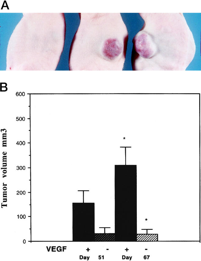
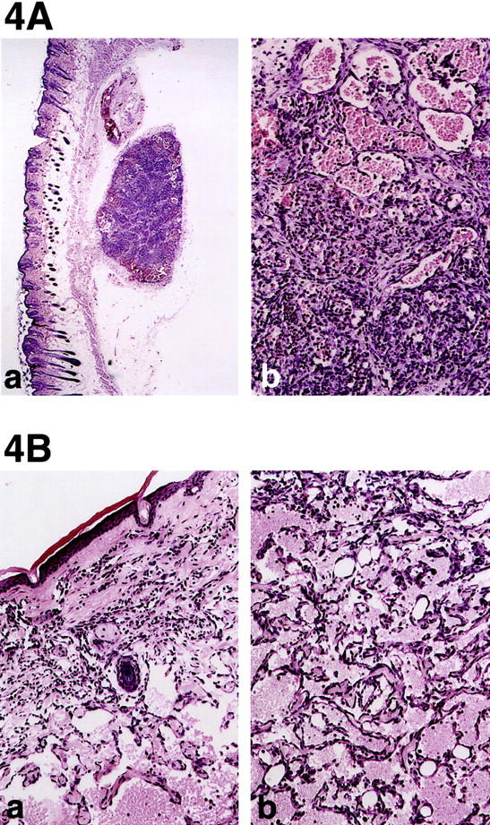
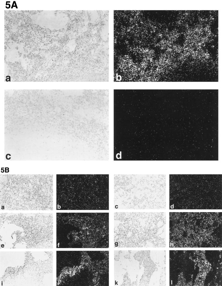
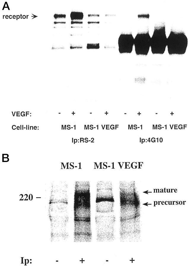
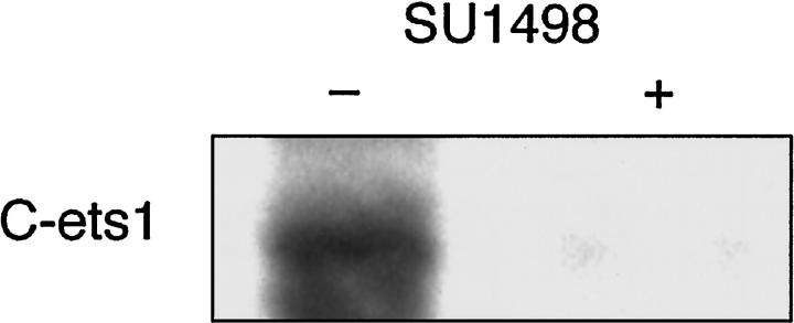
Vascular endothelial growth factor (VEGF or vascular permeability factor) is an important angiogenic factor that is up-regulated in numerous benign and malignant disorders, including angiosarcoma, hemangiomas, and solid tumors. To determine the functional role of VEGF in the development of endothelial tumors, we expressed primate VEGF 121 in an endothelial cell line, MS1, derived from primary murine cells by immortalization with a temperature-sensitive SV40 large T antigen. This cell line expresses the VEGFR-2 (Flk-1/Kdr) receptor for VEGF. Expression of VEGF 121 led to the development of slowly growing endothelial tumors, which were histologically well-differentiated angiosarcomas. The angiosarcomas generated from MS1 VEGF cells demonstrated up-regulation of the VEGF receptors VEGFR-2 and VEGFR-1 (Flt-1) in vivo compared with benign hemangiomas generated from MS1 cells. Treatment of these cells with the VEGFR-2 tyrosine kinase inhibitor SU 1498 led to decreased expression of ets-1, a transcription factor which has been shown to be stimulated by VEGF. These results suggest that high level expression of VEGF in endothelial cells may result in malignant transformation. This transformation process likely involves both autocrine and paracrine pathways.


Figures

Figure 1.
Figure 1.
Northern blot analysis of VEGFR-2 expression in MS1 and SVR cells. Expression of flk-1 is present, but is not altered by ras transformation, phorbol ester, hypoxia, or free radicals. Lanes 1 under both MS1 and SVR represent cells exposed to free radicals, lanes 2 represent cells after exposure to phorbol ester, lanes 3 represent cells exposed to hypoxia (3.5% oxygen), and lane 4 represents normoxia controls.
Figure 2.
Figure 2.
Northern blot analysis of VEGF expression in MS1puro and MS1VEGF cells using a human VEGF probe. This probe shows no hybridization with endogenous mouse VEGF
Figure 3.
Figure 3.
A: Gross appearance of tumors overexpressing VEGF 121. The mouse on the left was injected with MS1puro cells (MS1 cells with vector control), and the two mice on the right were injected with MS1 VEGF cells. This picture was taken 5 months after inoculation of cells. B: Growth of tumors in the presence of absence of VEGF overexpression. The differences in tumor volume between VEGF overexpressing and control cells is significant at P < 0.05. Four mice were injected with each cell line.
Figure 4.
Figure 4.
A. a: Hemangioma induced by MS1 cells which do not overexpress VEGF. Parental MS1 tumor cells are histologically identical to MS1puro cells. Tumor consists of two well circumscribed masses in the subcutaneous tissue (original magnification, ×40). In the larger mass the tumor is subdivided into lobules by fibrous bands similar the pattern seen in capillary hemangiomas in humans. b: Higher power of one tumor lobule shows mixture of small vessels with tiny lumina (left) to larger vessels with distended lumina filled with erythrocytes (original magnification, ×200). Note that the vascular channels do not intercommunicate with one another as they do in angiosarcoma. B: Angiosarcoma induced by MS1 VEGF cells. a: Irregular endothelial lined channels dissect through the dermal collagen isolating individual adnexal structures (center, original magnification, ×40). b: Higher power showing intercommunicating vascular channels dissecting around fat cells of subcutaneous tissue (original magnification, ×200). Solid or undifferentiated areas were not observed, however.
Figure 5.
Figure 5.
In situ hybridization of MS1, MS1 VEGF, and SVR tumors. A: Expression of VEGF is maintained in vivo. Frames a and b represent antisense human VEGF probe, whereas frames c and d represent sense VEGF probe control. Frames a and c are brightfield; frames b and d are darkfield. B: Expression of VEGFR-1 and VEGFR-2 in endothelial tumors. Frames a−d represent MS1 tumors, frames e−h represent MS1 VEGF tumors, and frames i−l represent SVR tumors. The leftmost column represents brightfield images of VEGFR-1 hybridization, the second column from the left represents darkfield images of VEGFR-1 hybridization, the second column from the right represents brightfield images of VEGFR-2 hybridization, and the rightmost column represents darkfield images of VEGFR-2 hybridization. Original magnifications, ×200.
Figure 6.
Figure 6.
A: MS1 cells and MS1 cells overexpressing VEGF were analyzed for induction of kinase activity using an in vitro kinase assay. Lanes 1–4 show the presence of the VEGFR-2 in both MS1 cells (lanes 1 and 2), and in MS1 VEGF cells which overexpress VEGF 121 (lanes 3 and 4). Cells were treated (+) or left untreated (−) withVEGF (100 ng/ml) for 7 minutes at 37°C, lysed, and immunoprecipitated with antibodies against VEGFR-2 (RS-2) or phosphotyrosine (4G10), and subjected to an in vitro kinase assay. Samples were analyzed by SDS-PAGE and autoradiography. VEGFR-2 is marked in the figure with an arrow. Demonstration of phsophorylation of VEGFR-2 is seen in lane 6. B: MS1 and MS1 VEGF cells express VEGFR-2. MS1 and MS1 VEGF cells were metabolically labeled with [35S]methionine/cysteine, lysed and immunoprecipitated with a rabbit antiserum to VEGFR-2 (+), followed by incubation with protein A Sepharose. Parallel samples were incubated with protein A Sepharose alone (−). Migration positions of marker proteins are indicated to the left; migration positions of VEGFR-2 intracellular precursor and cell surface mature forms are indicated to the right.
Figure 7.
Figure 7.
Western blot analysis of c-Ets-1 expression in the presence or absence of SU-1498. The left lane represents c-Ets-1 expression in the presence of vehicle alone. The right lane represents expression in the presence of SU-1498. Representative lanes are shown. Experiments were performed in triplicate.

References

    1. Pratt GA: Birthmarks in infants. Arch Dermatol 1953, 67:302-305 - PubMed
    1. Vin-Christian K, McCalmont TH, Frieden IJ: Kaposiform hemangioendothelioma. A locally invasive vascular tumor that can mimic hemangioma of infancy. Arch Dermatol 1997, 133:1573-1578 - PubMed
    1. Morrison WH, Byers RM, Garden AS, Evans HL, Ang KK, Peters LJ: Cutaneous angiosarcoma of the head and neck: a therapeutic dilemma. Cancer 1995, 76:319-327 - PubMed
    1. Blei F, Karp N, Rofski N, Rosen R, Greco MA: Successful multimodal therapy for kaposiform hemangioendothelioma complicated by Kasabach-Merritt phenomenon: case report and review of the literature. Pediat Hematol Oncol 1998, 15:295-305 - PubMed
    1. Schmitz-Rixen T, Horsch S, Arnold G, Peters PE: Angiosarcoma in primary lymphedema of the lower extremity-Stewart-Treves syndrome. Lymphology 1984, 17:50-53 - PubMed

Publication types

MeSH terms

Substances