. 2000 Apr 15;20(8):2875-86.
doi: 10.1523/JNEUROSCI.20-08-02875.2000.

Akt/protein kinase B prevents injury-induced motoneuron death and accelerates axonal regeneration

Affiliations

Akt/protein kinase B prevents injury-induced motoneuron death and accelerates axonal regeneration

K Namikawa et al. J Neurosci. .

Abstract

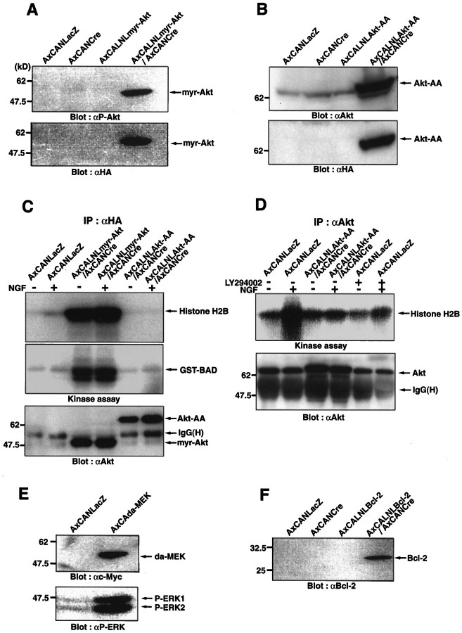
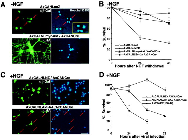
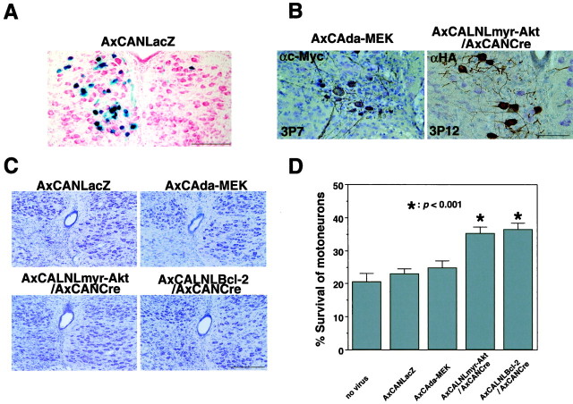
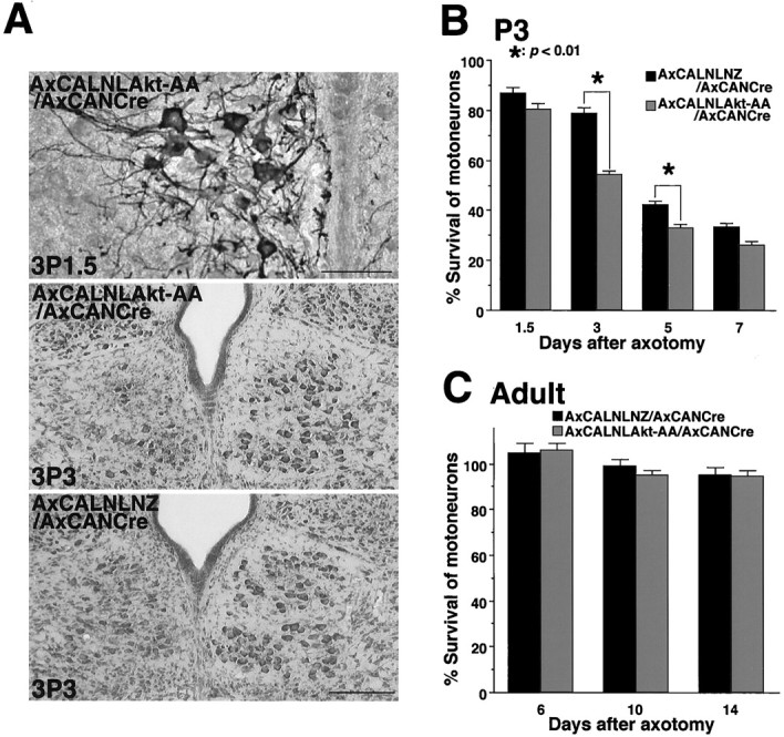
Motoneurons require neurotrophic factors for their survival and axonal projection during development, as well as nerve regeneration. By using the axotomy-induced neuronal death paradigm and adenovirus-mediated gene transfer, we attempted to gain insight into the functional significances of major growth factor receptor downstream cascades, Ras-extracellular signal-regulated kinase (Ras-ERK) pathway and phosphatidylinositol-3 kinase-Akt (PI3K-Akt) pathway. After neonatal hypoglossal nerve transection, the constitutively active Akt-overexpressing neurons could survive as well as those overexpressing Bcl-2, whereas the constitutively active ERK kinase (MEK)-overexpressing ones failed to survive. A dominant negative Akt experiment demonstrated that inhibition of Akt pathway hastened axotomy-induced neuronal death in the neonate. In addition, the dominant active Akt-overexpressing adult hypoglossal neurons showed accelerated axonal regeneration after axotomy. These results suggest that Akt plays dual roles in motoneuronal survival and nerve regeneration in vivo and that PI3K-Akt pathway is probably more vital in neuronal survival after injury than Ras-ERK pathway.


Figures

Fig. 1.
Fig. 1.
Expression and activation of Akt in injured hypoglossal motoneurons are oppositely regulated between neonate and adult. A, Expression of Akt1 mRNA demonstrated byin situ hybridization using DIG-labeled Akt1 antisense RNA probe. Photograph indicates markedly upregulated signal in injured hypoglossal motoneurons (left) of adult rat 7 d after axotomy. B, C, Immunohistochemical demonstrations of phosphorylated Akt at Ser473(P-Akt). B, In the adult rat, enhanced P-Akt expression was observed in injured hypoglossal nucleus (left) compared with the control hypoglossal nucleus (right) 7 d after axotomy. C, High-power magnification indicates that most of the injured motoneurons show intense P-Akt immunoreactivity. D, E, Decreased Akt expression and activity in the neonate injured motoneurons in response to axotomy. D, Expression of Akt1 mRNA was downregulated on the injured side (left) 3 d after axotomy.E, Immunohistochemistry also showed few P-Akt-positive neurons in the injured side (left), whereas many immunostained motoneurons are observed on control side (right) 3 d after axotomy. Scale bars:A, 600 μm; B, 500 μm;C, 50 μm; D, E, 200 μm.
Fig. 2.
Fig. 2.
Expression and activity of myr-Akt, Akt-AA, da-MEK, and Bcl-2 in PC12 cells by recombinant adenoviral gene transfer. These cells were lysed 36 hr after viral infection.A, Expression of HA-tagged myr-Akt, which is demonstrated by anti-P-Akt or anti-HA antibody, is found only in the case of the double infection with AxCALNLmyr-Akt and AxCANCre.B, Expression of HA-tagged Akt-AA was also detected by anti-Akt or anti-HA antibody only when the double infection (AxCALNLAkt-AA plus AxCANCre) was performed. C, LacZ-, myr-Akt-, or Akt-AA-expressing virus was infected into the cells and lysed (36 hr after infection) under the presence (100 ng/ml; 3 min after stimulation) or absence of NGF. Thereafter, these lysates were immunoprecipitated with anti-HA polyclonal antibody (IP: αHA), and the kinase activity of myr-Akt or Akt-AA was examined with phosphorylation of substrates such as histone H2B and recombinant BAD. The same immunoprecipitations were also used for Western blot analysis with anti-Akt antibody to confirm the expression. Note that the adenovirus-expressed myr-Akt showed strong phosphorylation activity, whereas Akt-AA completely lost the phosphorylation activity, even under the existence of NGF.D, Adenovirus-expressed Akt-AA could reduce endogenous Akt activity at a similar level to LY294002 treatment (100 μm), even after NGF stimulation. Akt kinase assay was performed after the immunoprecipitation with anti-Akt polyclonal antibody (IP: αAkt). E, Expression of c-Myc-tagged da-MEK in PC12 cells was detected by anti-c-Myc antibody. The da-MEK expression also induced phosphorylation of ERKs (ERK1 and ERK2) in the same cells, which were detected by anti-phospho ERK antibody. F, Expression of Bcl-2 in PC12 cells was observed (detected by anti-Bcl-2 antibody) only in the case of the double infection (AxCALNLBcl-2 plus AxCANCre). Migration position of prestained molecular weight protein marker is shown on the left (A–F).IgG(H) indicated by arrows inC and D shows the position of IgG heavy chain.
Fig. 3.
Fig. 3.
Effects of adenovirus-expressed myr-Akt and Akt-AA on neuronal survival in vitro. A, Photographs show PC12 cells infected with AxCANLacZ (top row) or AxCALNLmyr-Akt plus AxCANCre (bottom row) 18 hr after NGF withdrawal. Infected cells were detected by anti-β-Gal antibody or anti-HA antibody followed by FITC-conjugated secondary antibody (left column).Right column shows Hoechst 33258 staining. Thered arrows indicate cells that show condensed or fragmented chromatin. B, Viability of adenovirus-infected PC12 cells 24 and 48 hr after NGF withdrawal. Eachdot represents the average of values from four different experiments. C, Photographs show PC12 cells infected with AxCANLacZ plus AxCANCre (top row) and AxCALNL Akt-AA plus AxCANCre (bottom row) under the presence of NGF. The infected cells were detected by the same antibody described in A (left column). Hoechst staining (right column) demonstrated that Akt-AA-expressing cells show apoptotic profiles (red arrows) as seen in NGF-deprived cells (A;red arrows). D, Viability of Akt-AA-expressed and LY294002-treated PC12 cells under the presence of NGF. Viability of LacZ-expressing PC12 cells is also examined as control. LY294002-treated cells show marked decrease of viability, and Akt-AA-expressed cells also demonstrated apparent decrease of viability. Scale bars: A, C, 20 μm. Error bars indicate SEM in B and D.
Fig. 4.
Fig. 4.
Expression of myr-Akt prevents axotomy-induced neuronal death in vivo. A, Ten days after injection of AxCANLacZ into the left side of the tongue, NLS-tagged β-Gal-positive staining is detected in the ipsilateral side of hypoglossal motoneurons. This indicates a typical rate of infection efficacy in the present study. B, Immunohistochemical detection of c-Myc-tagged da-MEK (left) and HA-tagged myr-Akt (right) in injured motoneurons (3P7 or 3P12). (After the viral infection, ipsilateral nerve was transected on third day after birth and observed on postoperative day 7 or 12.) C, The survival activity by the infection of adenovirus expressing LacZ, da-MEK, myr-Akt, or Bcl-2. Sections are obtained 12 d after hypoglossal nerve transection and stained with thionin.D, The mean percentage of survival ratio of injured motoneurons 12 d after axotomy. For statistical analysis, at least 10 sections prepared from seven different animals were studied. *p < 0.001, significant differences between survival ratio of injured motoneurons in each viral infected animals and that in animals without virus infection (ANOVA). Error bars indicate SEM. Scale bars: A, C, 500 μm;B, 100 μm.
Fig. 5.
Fig. 5.
Effects of Akt pathway inhibition in injured motoneuron. A, Top, An immunohistochemical detection of adenovirus-expressed Akt-AA (detected by anti-HA antibody) in injured neonate motoneurons [3P1.5; the axotomy was done on the third day after birth, and the rats were killed 1.5 d (36 hr) after the operation]. Scale bar, 50 μm. Middle, Three days after axotomy, approximately half of motoneurons on the injured side (left) disappeared in Akt-AA-expressed animal (3P3; axotomized on the third day after birth and observed on postoperative day 3). Bottom, Many surviving neurons were observed in LacZ-expressing rat at the same time point (3P3). Scale bar, 200 μm. B,C, The mean percentage of surviving hypoglossal motoneurons after infection of either Akt-AA- or LacZ-expressing adenovirus in the neonate (P3) (B) and the adult (C). x-Axis indicates days after axotomy. The statistical analysis was performed as described in the previous experiment. *p < 0.01, significant differences between survival ratio of injured motoneurons in Akt-AA-expressing animals and that in LacZ-expressing ones at each time point (Student's t test). Error bars indicate SEM.
Fig. 6.
Fig. 6.
Expression of myr-Akt induces process elongation in PC12 cells. A, PC12 cells infected with AxCANLacZ show no significant change in cell shapes, but the infection with AxCALNLmyr-Akt plus AxCANCre caused clear process elongation 2 d after the infection. Infected cells were immunodetected by using anti-β-Gal antibody or anti-HA antibody, respectively (right column). Scale bar, 20 μm. B, Time course of percentages of process bearing cells after the infection of each recombinant virus or application of NGF (50 ng/ml).C, Detection of activated MAPK family members (MAPKs, ERKs, JNKs, and p38 MAPK) by antibodies specific to phosphorylated MAPKs (αP-ERK, αP-JNK, αP-p38). In PC12 cells, phosphorylation of MAPKs were not observed 36 and 72 hr after the infection of AxCALNLmyr-Akt plus AxCANCre, whereas all of MAPKs were phosphorylated 15 min after NGF stimulation (50 ng/ml).
Fig. 7.
Fig. 7.
Expression of myr-Akt accelerates nerve regeneration in adult rats. A, Top row, AxCANLacZ-infected motoneurons (the expression was examined by X-Gal staining 1 week after infection; top left) show no FG-positive (regenerated) neurons on the injured side at 2 weeks after injury (top right). Bottom row, In contrast, AxCALNLmyr-Akt plus AxCANCre-infected motoneurons (the expression was detected by anti-HA antibody 1 week after infection;bottom left) show moderate number of FG-positive (regenerated) cells in injured nucleus at 2 weeks after axotomy (bottom right). Scale bar, 0.8 mm. Bshows time course of nerve regeneration rate after axotomy in rats infected with AxCANLacZ or AxCALNLmyr-Akt plus AxCANCre. The regeneration rate was assessed by calculating the percent ratio of the number of FG-stained cell bodies in the operated side to those in control side. Three animals were examined at each time point. Error bars indicate SEM.

References

    1. Akli S, Caillaud C, Vigne E, Stratford-Perricaudet LD, Poenaru L, Perricaudet M, Kahn A, Peschanski MR. Transfer of a foreign gene into the brain using adenovirus vectors. Nat Genet. 1993;3:224–228. - PubMed
    1. Batistatou A, Merry DE, Korsmeyer SJ, Greene LA. Bcl-2 affects survival but not neuronal differentiation of PC12 cells. J Neurosci. 1993;13:4422–4428. - PMC - PubMed
    1. Bergmann A, Agapite J, McCall K, Steller H. The Drosophila gene hid is a direct molecular target of Ras-dependent survival signaling. Cell. 1998;95:331–341. - PubMed
    1. Brunet A, Bonni A, Zigmond MJ, Lin MZ, Juo P, Hu LS, Anderson MJ, Arden KC, Blenis J, Greenberg ME. Akt promotes cell survival by phosphorylating and inhibiting a Forkhead transcription factor. Cell. 1999;96:857–868. - PubMed
    1. Burgering BM, Coffer PJ. Protein kinase B (c-Akt) in phosphatidylinositol-3-OH kinase signal transduction. Nature. 1995;376:599–602. - PubMed

Publication types

Substances

LinkOut - more resources