. 2000 Apr 15;20(8):2896-903.
doi: 10.1523/JNEUROSCI.20-08-02896.2000.

Peripheral infusion of IGF-I selectively induces neurogenesis in the adult rat hippocampus

Affiliations

Peripheral infusion of IGF-I selectively induces neurogenesis in the adult rat hippocampus

M A Aberg et al. J Neurosci. .

Abstract

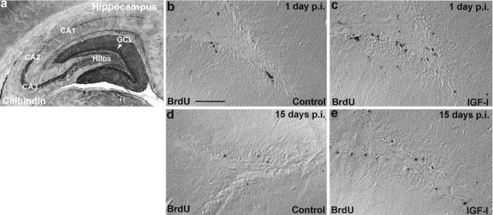
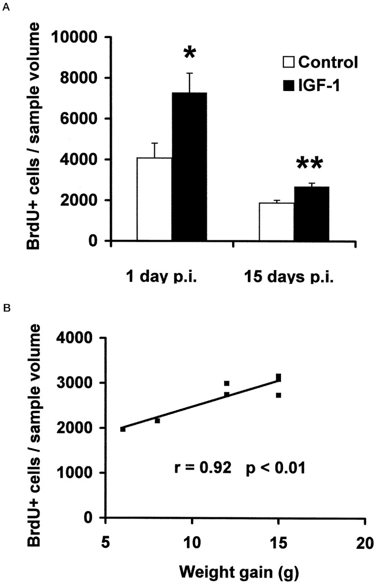
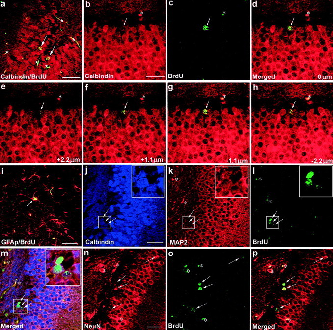
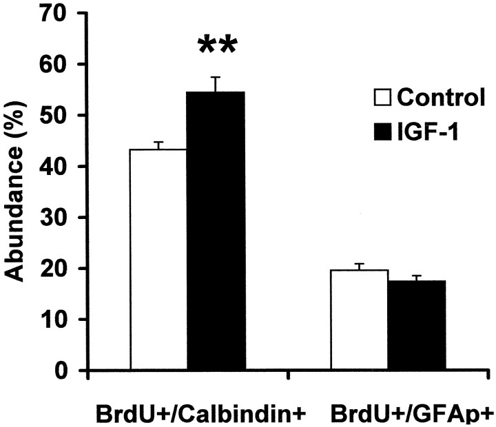
In several species, including humans, the dentate granule cell layer (GCL) of the hippocampus exhibits neurogenesis throughout adult life. The ability to regulate adult neurogenesis pharmacologically may be of therapeutic value as a mechanism for replacing lost neurons. Insulin-like growth factor-I (IGF-I) is a growth-promoting peptide hormone that has been shown to have neurotrophic properties. The relationship between IGF-I and adult hippocampal neurogenesis is to date unknown. The aim of this study was to investigate the effect of the peripheral administration of IGF-I on cellular proliferation in the dentate subgranular proliferative zone, which contains neuronal progenitor cells, and on the subsequent migration and differentiation of progenitor cells within the GCL. Using bromodeoxyuridine (BrdU) labeling, we found a significant increase of BrdU-immunoreactive progenitors in the GCL after 6 d of peripheral IGF-I administration. To determine the cell fate in progenitor progeny, we characterized the colocalization of BrdU-immunolabeled cells with cell-specific markers. In animals treated with IGF-I for 20 d, BrdU-positive cells increased significantly. Furthermore, the fraction of newly generated neurons in the GCL increased, as evaluated by the neuronal markers Calbindin D(28K), microtubule-associated protein-2, and NeuN. There was no difference in the fraction of newly generated astrocytes. Thus, our results show that peripheral infusion of IGF-I increases progenitor cell proliferation and selectively induces neurogenesis in the progeny of adult neural progenitor cells. This corresponds to a 78 +/- 17% (p < 0.001) increase in the number of new neurons in IGF-I-treated animals compared with controls.


Figures

Fig. 1.
Fig. 1.
BrdU immunohistochemistry in two experiments in which IGF-I was given for 6 d (corresponding to 1 d p.i. of BrdU) or 20 d (corresponding to 15 d p.i. of BrdU). a, The hippocampal region of the adult rat brain immunoperoxidase-stained for the neuronal marker Calbindin D28K. b–e, Differential interference contrast photomicrographs of BrdU-immunopositive cells in the hippocampus. A comparison of BrdU labeling in controls (b, d) and IGF-I-treated animals (c,e) at 1 and 15 d p.i. of BrdU is shown. (For quantification, see Fig. 2.) Scale bar, 100 μm.
Fig. 2.
Fig. 2.
Quantification of BrdU-positive cells in the adult rat hippocampus. A, The density of BrdU-positive cells (cells per cubic millimeter of sample volume) in the GCL 1 and 15 d p.i. was determined stereologically. IGF-I-treated animals (n = 4 at 1 d p.i.; n = 7 at 15 d p.i.) are hx rats given l-thyroxine, cortisol, and IGF-I, as described in Materials and Methods. Controls (n = 5 at 1 d p.i.; n = 8 at 15 d p.i.) are hx rats given l-thyroxine and cortisol only. Means ± SEM are given. *p < 0.05; **p < 0.01. B, Weight gain and the number of BrdU-labeled cells in the GCL at the individual level are compared. There is a correlation between S-IGF-I and BrdU-positive cells after 20 d of IGF-I treatment (15 d p.i.); animals with a relatively low serum IGF-I have relatively low numbers of BrdU-positive cells, and vice versa.
Fig. 3.
Fig. 3.
Colocalization of BrdU immunoreactivity (green–yellow in a, c–i, l, m, o, p) with immunoreactivity of the granule cell marker Calbindin D28K (red in a–h;blue in j,m), the astrocyte marker GFAP (red in i), the neuronal marker MAP2 (red in k,m), or the neuronal marker NeuN (red inn,p) (arrows indicate colocalization in a–p). Red blood cells and endothelial cells in several small blood vessels also emit nonspecificgreen and red fluorescence (asterisks in a–p). The specificity of BrdU and Calbindin D28K coexpression in three dimensions is demonstrated by a Z-series of focal planes above (e,f) and below (g,h) the focal plane shown in d(arrows in e–h indicate the same cell as in d). The merged image of b andc is shown in d, the merged image ofj, k, and l is shown inm, and the merged image of n and ois shown in p. Insets of eachboxed area in j–m are magnified 2.5×. Scale bars: a, i, 50 μm; b—h,j—m, n—p, 25 μm.
Fig. 4.
Fig. 4.
Percentage of the surviving BrdU-positive cells after 20 d of IGF-I treatment that differentiated into either neurons or astrocytes. Colocalization of BrdU immunoreactivity with cell-specific markers, either the granule cell marker Calbindin D28K or the astrocyte marker GFAP, was monitored to determine the phenotype of newborn cells after treatment with IGF-I, when compared with controls in the GCL (n = 5 for controls; n = 7 for IGF-I-treated animals). Means ± SEM are given. **p < 0.01.

References

    1. Altman J, Das GD. Autoradiographic and histological evidence of postnatal hippocampal neurogenesis in rats. J Comp Neurol. 1965;124:319–336. - PubMed
    1. Arsenijevic Y, Weiss S. Insulin-like growth factor-I is a differentiation factor for postmitotic CNS stem cell-derived neuronal precursors: distinct actions from those of brain-derived neurotrophic factor. J Neurosci. 1998;18:2118–2128. - PMC - PubMed
    1. Beach TG, Woodhurst WB, MacDonald DB, Jones MW. Reactive microglia in hippocampal sclerosis associated with human temporal lobe epilepsy. Neurosci Lett. 1995;191:27–30. - PubMed
    1. Beck KD, Powell-Braxton L, Widmer HR, Valverde J, Hefti F. IGF-I gene disruption results in reduced brain size, CNS hypomyelination, and loss of hippocampal granule and striatal parvalbumin-containing neurons. Neuron. 1995;14:717–730. - PubMed
    1. Bengzon J, Kokaia Z, Elmer E, Nanobashvili A, Kokaia M, Lindvall O. Apoptosis and proliferation of dentate gyrus neurons after single and intermittent limbic seizures. Proc Natl Acad Sci USA. 1997;94:10432–10437. - PMC - PubMed

Publication types

Substances