. 2000 Mar-Apr;7(2):97-103.
doi: 10.1101/lm.7.2.97.

Differential fear conditioning induces reciprocal changes in the sensory responses of lateral amygdala neurons to the CS(+) and CS(-)

Affiliations

Differential fear conditioning induces reciprocal changes in the sensory responses of lateral amygdala neurons to the CS(+) and CS(-)

D R Collins et al. Learn Mem. 2000 Mar-Apr.

Abstract

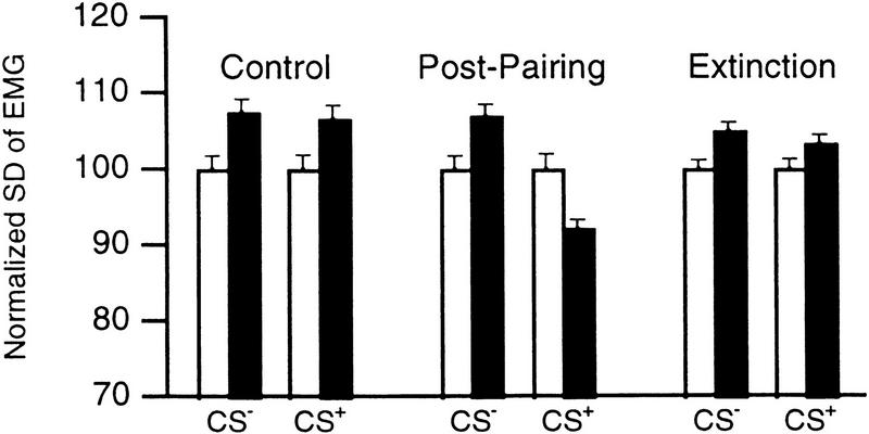
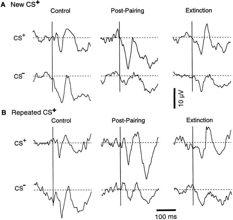
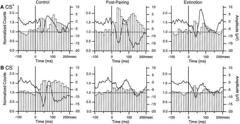
In classical fear conditioning, a neutral sensory stimulus (CS) acquires the ability to elicit fear responses after pairing to a noxious unconditioned stimulus (US). As amygdala lesions prevent the acquisition of fear responses and the lateral amygdaloid (LA) nucleus is the main input station of the amygdala for auditory afferents, the effect of auditory fear conditioning on the sensory responsiveness of LA neurons has been examined. Although conditioning was shown to increase CS-evoked LA responses, the specificity of the changes in responsiveness was not tested. Because conditioning might induce nonspecific increases in LA responses to auditory afferents, we re-examined this issue in conscious, head-restrained cats using a differential conditioning paradigm where only one of two tones (CS(+) but not CS(-)) was paired to the US. Differential conditioning increased unit and field responses to the CS(+), whereas responses to the CS(-) decreased. Such changes have never been observed in the amygdala except in cases where the CS(-) had been paired to the US before and fear responses not extinguished. This suggests that fear conditioning is not only accompanied by potentiation of amygdalopetal pathways conveying the CS(+) but also by the depression of sensory inputs unpaired to noxious stimuli.


Figures

Figure 1
Figure 1
Conditioning produces reversible CS+-evoked decreases in EMG activity. We computed the standard deviations (s.d.) of the EMG signal (filtered 100–1000 Hz; normalized to pretone values) during epochs of 500 msec obtained before (open bars) and 500 msec after (solid bars) tone onsets. Bars show Control (left), Post-pairing (middle), and Extinction (right) values.
Figure 2
Figure 2
Location of recorded cells in the LA nucleus. Scheme of three coronal sections arranged from rostral (left) to caudal (right). Dots indicate the location of cells recorded in conditioning experiments. (○) Cells recorded in experiments where tones were presented without US to determine whether their tone responses habituated. The location of recorded cells was determined from thionin-stained coronal sections, by combining micrometric readings with the placement of electrolytic lesions (see Materials and Methods for details). (BL) Basolateral nucleus of the amygdala; (BM) basomedial nucleus of the amygdala; (CE) central nucleus of the amygdala; (CL) claustrum; (GP) globus pallidus; (LA) lateral nucleus of the amygdala; (PU) putamen; (rh) rhinal sulcus. Bar, 2 mm.
Figure 3
Figure 3
Auditory-evoked field potentials and unit activity in the LA. Responses to 1-sec tones of 5 (A) and 10 (B) kHz. Vertical lines indicate tone onsets. Focal waves were digitally filtered (3–300 Hz, thick lines). Each tone was presented 20 times and the responses of 35 LA sites averaged. Superimposed on the focal waves are peristimulus histograms showing the summed activity of 72 LA neurons. Bins of 10 msec.
Figure 4
Figure 4
Conditioning causes opposite changes in focal LA responses to the CS+ and CS. Average auditory-evoked responses in sessions where a different (A) or the same (B) CS+ was used in the previous session. Broken lines indicate average voltage values before the tone onsets. The average amplitude of the field potentials (in μV) in A are, for the CS+: Control, 7.5 ± 2.9; Post-Pairing, 15.8 ± 3.79; Extinction, 8.6 ± 2.33. For the CS, Control, 13.3 ± 3.19; Post-Pairing, 4.8 ± 2.05; Extinction, 7.9 ± 3.09. In B, for the CS+, Control, 7.4 ± 2.11; Post-Pairing, 12.1 ± 3.4 ; Extinction, 8.5 ± 2.26. For the CS, they are Control, 13.9 ± 2.48; Post-Pairing, 5.4 ± 1.66; Extinction, 8.2 ± 2.17. Vertical bar, 10 μV; horizontal bar 100 msec.
Figure 5
Figure 5
Conditioning causes inverse changes in unit responses to the CS+ (A) and CS (B) in the LA. The responses of 72 neurons were summed and normalized to the average prestimulus bin height and superimposed on average focal waves (thick lines). Using the criteria of Fig. 3, short-latency increases in firing probability were significant (P < 0.05) in all conditions.
Figure 6
Figure 6
Effect of multiple presentations of the same tones on the responsiveness of LA cells. Averaged field (thick lines, right axis) and unit responses (histograms, left axis; n = 14) to presentations 1–10 (A) and 31–40 (B) of the same tones (3–10 kHz). Bins of ten msec. When the cells were recorded, the animals had never been presented these tones before and no foot shock had been administered in conjunction to auditory stimuli. Note that the spike counts were normalized to the average pretone activity.

References

    1. Amaral DG, Price JL, Pitkänen A, Carmichael ST. Anatomical organization of the primate amygdaloid complex. In: Aggleton JP, editor. The amygdala: Neurobiological aspects of emotion, memory, and mental dysfunction. New York, NY: Wiley-Liss; 1992. pp. 1–66.
    1. Bakin JS, Weinberger NM. Classical conditioning induces CS-specific receptive field plasticity in the auditory cortex of the guinea pig. Brain Res. 1990;536:271–286. - PubMed
    1. Bandler R. Induction of “rage” following microinjections of glutamate into midbrain but not hypothalamus of cats. Neurosci Lett. 1982;5:183–188. - PubMed
    1. Cahill L, Weinberger NM, Roozendaal B, McGaugh JL. Is the amygdala a locus of “conditioned fear”? Some questions and caveats. Neuron. 1999;23:227–228. - PubMed
    1. Collins DR, Paré D. Reciprocal changes in the firing probability of lateral and central medial amygdala neurons. J Neurosci. 1999;19:836–844. - PMC - PubMed

Publication types

LinkOut - more resources