Review
. 2000 Apr;13(2):236-301.
doi: 10.1128/CMR.13.2.236.

Zygomycetes in human disease

Affiliations
Review

Zygomycetes in human disease

J A Ribes et al. Clin Microbiol Rev. 2000 Apr.

Abstract

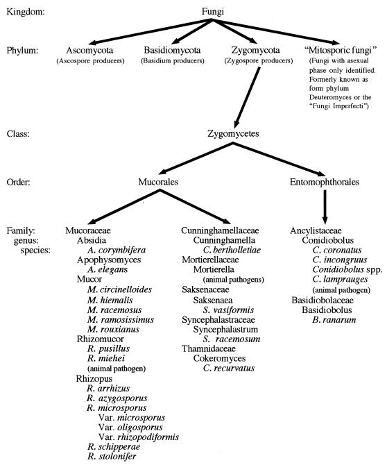
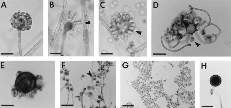
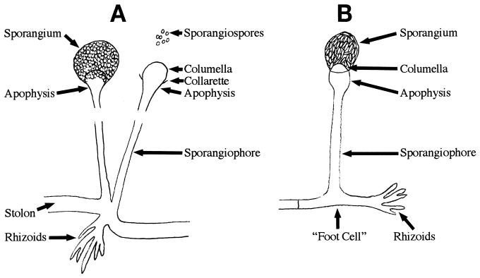
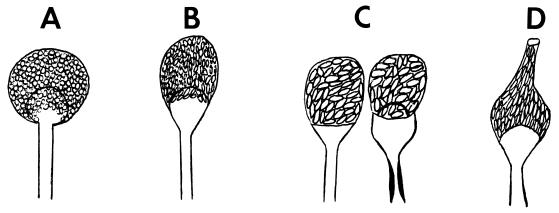
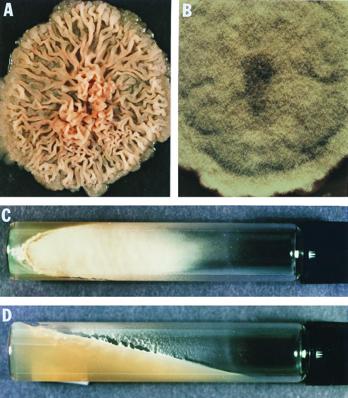
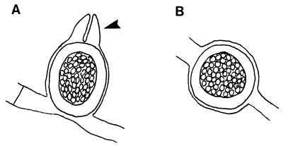
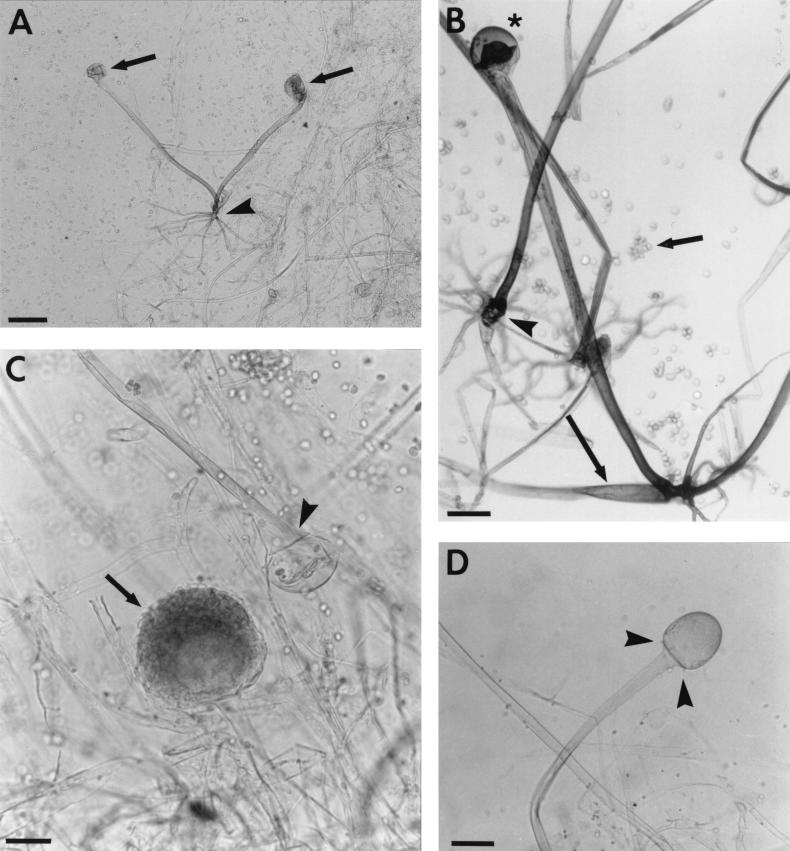
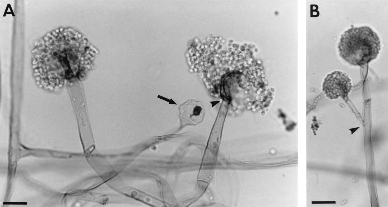
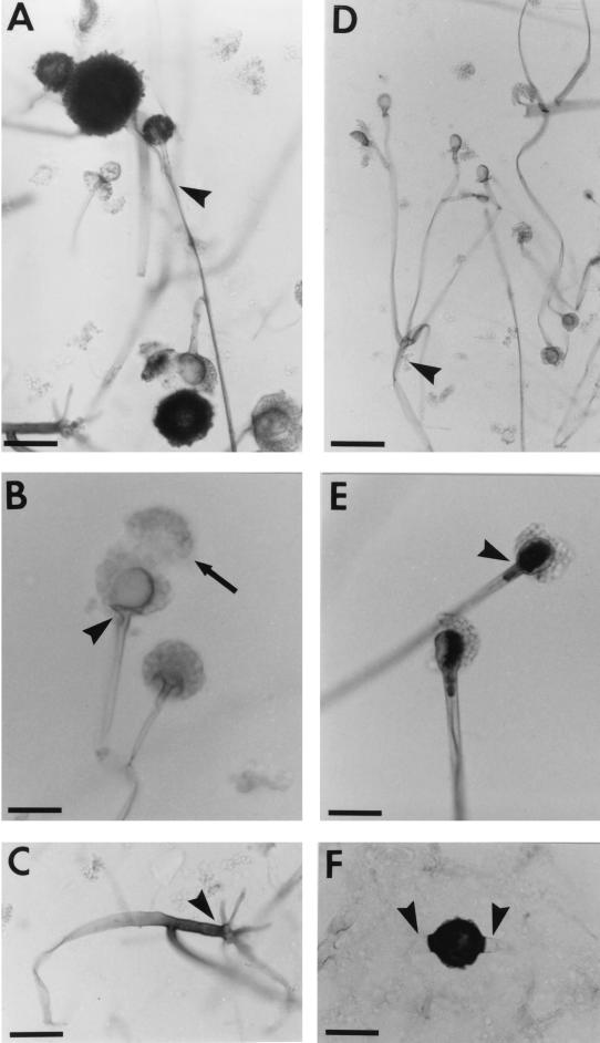
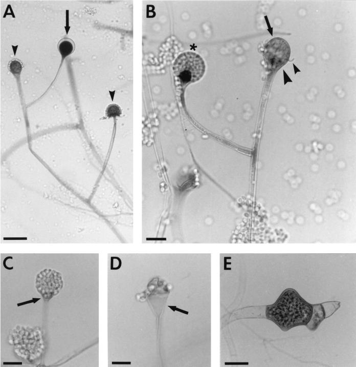
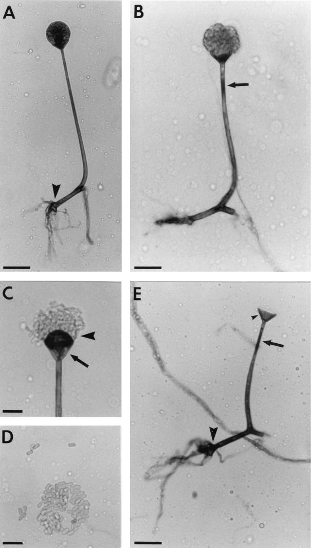
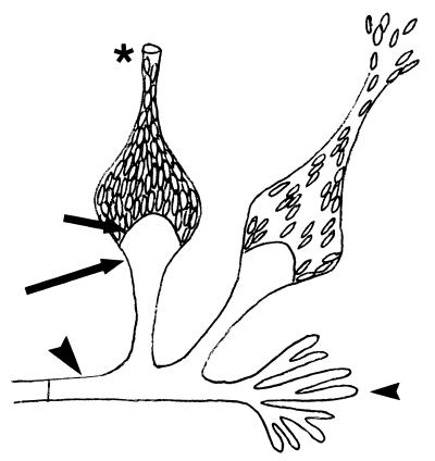
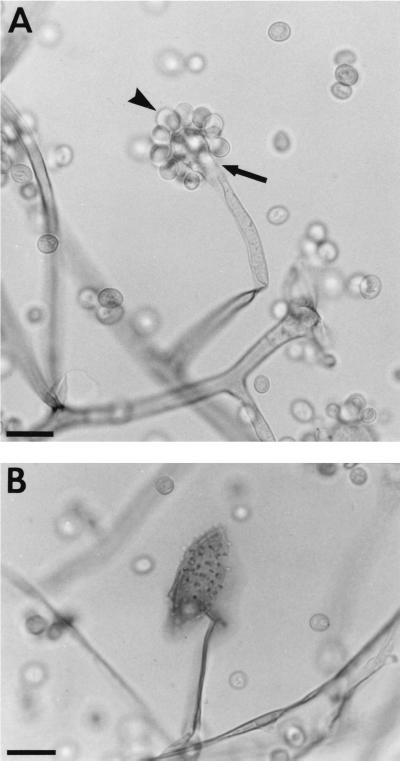
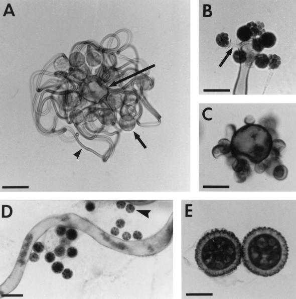
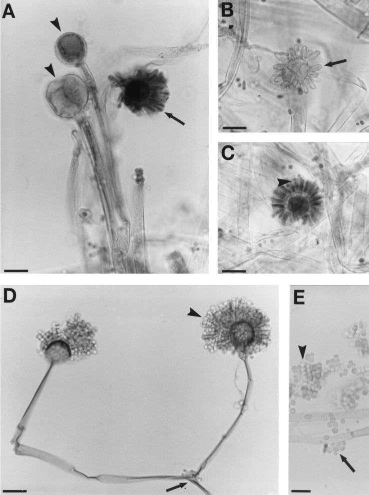
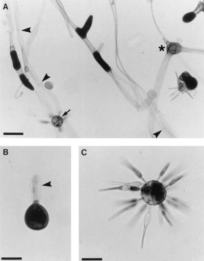
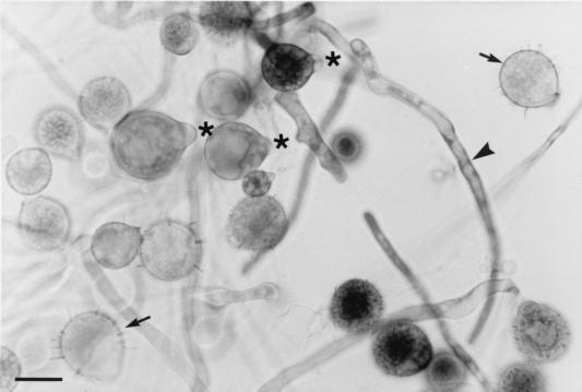
The Zygomycetes represent relatively uncommon isolates in the clinical laboratory, reflecting either environmental contaminants or, less commonly, a clinical disease called zygomycosis. There are two orders of Zygomycetes containing organisms that cause human disease, the Mucorales and the Entomophthorales. The majority of human illness is caused by the Mucorales. While disease is most commonly linked to Rhizopus spp., other organisms are also associated with human infection, including Mucor, Rhizomucor, Absidia, Apophysomyces, Saksenaea, Cunninghamella, Cokeromyces, and Syncephalastrum spp. Although Mortierella spp. do cause disease in animals, there is no longer sufficient evidence to suggest that they are true human pathogens. The spores from these molds are transmitted by inhalation, via a variety of percutaneous routes, or by ingestion of spores. Human zygomycosis caused by the Mucorales generally occurs in immunocompromised hosts as opportunistic infections. Host risk factors include diabetes mellitus, neutropenia, sustained immunosuppressive therapy, chronic prednisone use, iron chelation therapy, broad-spectrum antibiotic use, severe malnutrition, and primary breakdown in the integrity of the cutaneous barrier such as trauma, surgical wounds, needle sticks, or burns. Zygomycosis occurs only rarely in immunocompetent hosts. The disease manifestations reflect the mode of transmission, with rhinocerebral and pulmonary diseases being the most common manifestations. Cutaneous, gastrointestinal, and allergic diseases are also seen. The Mucorales are associated with angioinvasive disease, often leading to thrombosis, infarction of involved tissues, and tissue destruction mediated by a number of fungal proteases, lipases, and mycotoxins. If the diagnosis is not made early, dissemination often occurs. Therapy, if it is to be effective, must be started early and requires combinations of antifungal drugs, surgical intervention, and reversal of the underlying risk factors. The Entomophthorales are closely related to the Mucorales on the basis of sexual growth by production of zygospores and by the production of coenocytic hyphae. Despite these similarities, the Entomophthorales and Mucorales have dramatically different gross morphologies, asexual reproductive characteristics, and disease manifestations. In comparison to the floccose aerial mycelium of the Mucorales, the Entomophthorales produce a compact, glabrous mycelium. The asexually produced spores of the Entomophthorales may be passively released or actively expelled into the environment. Human disease with these organisms occurs predominantly in tropical regions, with transmission occurring by implantation of spores via minor trauma such as insect bites or by inhalation of spores into the sinuses. Conidiobolus typically infects mucocutaneous sites to produce sinusitis disease, while Basidiobolus infections occur as subcutaneous mycosis of the trunk and extremities. The Entomophthorales are true pathogens, infecting primarily immunocompetent hosts. They generally do not invade blood vessels and rarely disseminate. Occasional cases of disseminated and angioinvasive disease have recently been described, primarily in immunocompromised patients, suggesting a possible emerging role for this organism as an opportunist.


Figures

FIG. 1
FIG. 1
Taxonomic organization of the zygomycetes.
FIG. 2
FIG. 2
Microscopic morphology of Rhizopus spp., Aspergillus spp., and Candida spp. in tissue. (A) Rhizopus spp. in tissue section stained with GMS. The mucoraceous zygomycetes produce wide ribbon-like aseptate hyphae in tissues. There is a great deal of variation of hyphal width. Branching occurs at wide angles nearing 90° (arrowheads). A frothy or bubbly tissue appearance may be seen in areas of tissue where the hyphae are cross-sectioned (upper left hand corner of the frame). (B) Aspergillus spp. in tissue section stained with GMS. Aspergillus spp. produce thin hyphae with relatively consistent diameters. Hyphae are septate, with no constriction of the fungus seen at the point of septation (arrowheads). Blastoconidia are not produced, although areas where hyphae are cross-sectioned may be confused with yeast cells (asterisk). Hyphae branch at acute angles of about 45° (arrow). (C) Candida spp. in tissue section stained with GMS. Fungal elements in tissue appear as pseudohyphae with blastoconidia. Fungal elements constrict or “bud” at sites of septation (arrowheads). Branching occurs at acute angles (arrow). Pseudohyphae are thin, and their diameter is very similar to that seen for the true hyphae of the Aspergillus spp. All three panels are the same magnification. Bar, 10 μm.
FIG. 3
FIG. 3
Angioinvasion by mucoraceous zygomycetes. (A) H&E-stained section of a vessel filled with hyphae and inflammatory cells. Tissues infected with the mucoraceous zygomycetes often demonstrate extensive angioinvasion by fungal elements. Neutrophils are the predominant inflammatory cells responding to an infection with these agents. The inflammatory cells stain basophilic (dark), while the hyphae demonstrate an inconspicuous hyaline staining that can often be overlooked. Pale-staining hyphae (arrowheads) often look like hole or bubbles in the tissue. The hyphal elements are nearly obscured on this H&E section due to the inflammatory process seen. For these reasons, it is recommended that a fungus-specific stain such as GMS or PAS be performed to better characterize fungal elements in tissues having thrombosed vessels, extensive tissue necrosis, or hemorrhage. (B) H&E-stained section of a vessel with typical zygomycete hyphal elements in a patient with neutropenia. In contrast to panel A, the hyphal elements are much more prominent in this H&E preparation despite their hyaline staining, due largely to the absence of the inflammatory cells. (C) GMS-stained section of a vessel containing coenocytic fungal hyphae typical of the zygomycetes. Wide ribbon-like hyphae with broad-angle branching is seen (arrowhead). GMS provides excellent contrast to help distinguish hyphae, which stain black or dark gray, from the tissue elements or inflammatory cells, which stain with the pale counterstain. (D) GMS-stained section of a vessel with fungal hyphae penetrating the vessel wall. Infections originate in mucocutaneous sites, from which they may spread hematogenously. Fungal elements invade through the vessel walls from their tissue sites. Mycotic emboli disseminate and may thrombose small vessels in which they are lodged. Fungal elements may invade normal or devitalized tissues at these remote sites by directly penetrating the vessel wall (arrow). All four panels are the same magnification. Bar, 50 μm.
FIG. 4
FIG. 4
Gross morphology of Rhizopus, Mucor, and Absidia isolates in culture. (A) Rhizopus gross morphology on SABHI medium. Rhizopus spp. typically produce a very high, fibrous colony that rapidly fills the entire petri dish. This isolate has expanded to the lid (known as a “lid lifter”). It has produced abundant pigmented sporangia, which are seen as the dark areas peppering the otherwise pale mycelium. This morphology is characteristic of the Rhizopus spp. (B) Low-growing Mucor variant gross morphology on SABHI medium. Mucor spp. will show variation from culture to culture. This particular isolate has produced a low-growing, fibrous colony morphology that readily demonstrates the “woolly” or floccose growth characteristic of the Mucoraceae. Pigmentation is also variable, both within and among the Mucor spp. Increased pigmentation is generally reflective of areas of the mycelium that are rich in sporangia. Depending on the individual isolate, Mucor may be extremely floccose or low growing and may range from pure white to shades of gray or brown. (C) Absidia corymbifera gross colony morphology on SABHI medium. A. corymbifera produces a light-colored mycelium, generally cream or gray. The peppered appearance seen in the Rhizopus spp. is lacking despite the production of abundant sporangia. The mycelium of this isolate is firmly plastered to the lid of the petri dish, consistent with the lid-lifting properly of this fungus. (D) A. corymbifera, reverse side of the culture plate shown in panel C. The Mucoraceae are hyaline molds that produce a pale reverse in culture on standard media such as SABHI agar. Although some isolates such as Rhizopus spp. may have lightly pigmented hyphal elements, this is generally reflected as a pale yellow or brown reverse and not the darkly pigmented reverse of the dematiaceous fungi.
FIG. 5
FIG. 5
Reproductive structures of the Mucorales. (A) Sporangium. Development of asexual sporangiospore in sacks called sporangia is characteristic of the families Mucoraceae and Saksenaea. The aerial mycelium of these organisms terminate in swollen structures that develop into sporangial sacks and columellae. Sporangiospores develop asexually by free-cell cleavage within the sporangial membrane. At maturity, the sporangium of the Mucorales becomes deliquescent, releasing the sporangiospores. A related mechanism where sporangiospores are released from the sporangiospore by dissolution of a gelatinous plug is seen for Saksenaea vasiformis. The fungus pictured here demonstrates the sporangium produced by Mucor spp. Bar, 20 μm. (B) Merosporangia. Cylindrical or finger-like projections surround a swollen vesicle in this isolate of Syncephalastrum racemosum. A single row of sporangiospores form inside these tubular merosporangia (arrowhead). Spores may be released as entire merosporangial units or singly as the merosporangial membrane dissolves. This form of reproduction is characteristic of S. racemosum. Bar, 20 μm. (C and D) Sporangiola. Sporangiola or conidia develop singly around a swollen vesicle on stalks called sterigmata. Single celled sporangiola (arrowhead), typically produced by members of the genus Cunninghamella (C), are often echinate and form on short sterigmata. Those produced by Cokeromyces recurvatus (D) are multicelled (arrowhead) and occur on long recurving stalks. Bar, 20 μm. (E) Yeast. Several members of the order Mucorales are dimorphic. Yeast forms have been identified in vivo for some Mucor spp. as well as Cokeromyces recurvatus (pictured here). Yeast production in vitro often requires increased temperature of incubation, high carbon dioxide tension, or anaerobic culture conditions. Bar, 20 μm. (F and G) Chlamydospores and gemmae. These asexual reproductive structures are derived from the vegetative hyphae of certain species of the Mucorales. Chlamydospores may be formed intercalated with the mycelium (endogenous formation) (arrowhead in panel F), while gemmae are separated from the mycelium and often demonstrate yeast-like budding (exogenous formation) (G). Chlamydospores and gemmae are often considered together, since they may be difficult to differentiate and are similarly derived. These are produced by some but not all members of the Mucorales. Their morphology may vary substantially but is not particularly useful for species determination (355). Bar, 40 μm. (H) Zygospores. This is the only form of sexual reproduction employed by the zygomycetes. Zygospores may form within a single isolate without mating (homothallic reproduction) or may require mating with an appropriately oriented mating strain (heterothallic reproduction). Zygospore morphology is often characteristic for an organism when color, size, shape and surface decoration are taken into account. Mating of two isolates to produce mature zygospores provides definitive taxonomic identification of an unknown isolate. The zygospores pictured here are from Cokeromyces recurvatus. Bar, 40 μm.
FIG. 6
FIG. 6
Schematic diagram labeling the morphologic structures seen in the sporangium-producing Mucorales (not drawn to scale). (A) Rhizopus spp. (B) Apophysomyces elegans.
FIG. 7
FIG. 7
Morphologic variation seen in the sporangia produced by the Mucorales (not drawn to scale). (A) Globose sporangium. Nearly round or spherical sporangia represent the predominant morphology produced by the Mucorales. Globose sporangia are produced by Rhizopus, Mucor, Rhizomucor, and Mortierella spp. Although Absidia spp. generally produce pyriform-shaped sporangia, occasional isolates superficially appear to have round sporangia. Care must be taken to look for the presence of a prominent apophysis in these isolates. Rhizopus spp. may also have slight, inconspicuous apophysis, while Rhizomucor, Mucor, and Mortierella spp. do not. All but the Mortierella spp. will produce a columella. The size of the sporangial sack is also important in differentiating the various isolates from one another. (B and C) Pyriform sporangium. Pyriform or teardrop-shaped sporangia are typically produced by Absidia (B) and Apophysomyces elegans (C). The apophysis is very prominent in both of these genera. Absidia spp. produce a flask-shaped apophysis with a large columella producing the overall teardrop-shaped fruiting structures. A. elegans produce both a “martini glass” and “bell-shaped” apophyseal swelling. The large columella produced by this organism is often obscured by the spores in the sporangial sack. (D) Vasiform sporangium. Vase-like sporangia are produced by Saksenaea vasiformis. The apophysis is prominent and flask-shaped, ending in a large dome-shaped columella which protrudes into the sporangial sack. The sporangium swells and then tapers into a tubular neck, similar to what one sees with a bud vase, hence its name “vasiformis,” meaning vase like. At the apical end of the sporangial neck, a gelatinous plug is seen, which ultimately dissolves, releasing the sporangiospores.
FIG. 8
FIG. 8
Gross morphology of Basidiobolus, Conidiobolus, and Mucor colonies in culture. (A) Basidiobolus microsporus colony morphology on SABHI agar. Dense, waxy, folded, or furrowed colonies are produced on standard culture media. Colonies are low growing, lacking the floccose morphology seen in the typical isolate of the Mucorales (see Mucor morphology in panel B). This isolate produced a brownish orange colony that was pale on reverse. (B) Mucor colony morphology on SABHI agar. The floccose or “woolly” colony morphology produced by most of the Mucorales is well demonstrated in this Mucor isolate. Contrast the fibrous aerial mycelium with the glabrous morphology seen for both Basidiobolus (A) and Conidiobolus (C). This difference in colony morphology is one of the distinguishing features between the Mucorales and the Entomophthorales. (C) Conidiobolus coronatus colony morphology on Sabouraud dextrose agar slant. Similar to Basidiobolus spp., Conidiobolus spp. produce low-growing waxy (or sometimes powdery) folded and furrowed colonies that lack aerial mycelium. Satellite colonies arising from germination of ejected sporangiospores has lead to confluent growth in this tube containing a 6-week-old culture. (D) Conidiobolus coronatus tube culture, side view of the tube in panel C. Note the cloudy appearance of the glass surface of the tube. This is produced by the collection of a plethora of forcibly expelled sporangiospores which have become encrusted on the inside surface of the tube. This opacification of the culture tube (or petri dish lid) is characteristic of this organism.
FIG. 9
FIG. 9
Schematic diagram of characteristic zygospore morphology of Basidiobolus ranarum and Conidiobolus incongruus. (A) B. ranarum zygospores. Smooth, thick-walled zygospores are produced homothalically and demonstrate prominent conjugation beaks. These beaks represent the remnants of the conjugation tubes formed between the mating hyphal elements. (B) C. incongruus zygospore. Smooth, thick-walled zygospores are produced homothalically and may be distinguished from those produced by B. ranarum by the absence of the conjugation beaks.
FIG. 10
FIG. 10
Microscopic features of Rhizopus spp. in culture. (A) Low-power magnification (×4) of Rhizopus demonstrates the nodal occurrence of well-developed rhizoids in this genus (arrowhead). Sporangiophores are long and unbranched, usually terminating in large globose sporangia. In this figure, the sporangia have become deliquescent, releasing a plethora of sporangiospores seen dispersed in the background. The naked, somewhat collapsed columellae remain at the distal end of the sporangiophores (arrows). The sporangiophore themselves are pigmented light brown. Bar, 200 μm. (B) Rhizoids originate directly at the base of the sporangiophores, often from a gnarled, pigmented knot at the node (arrowhead). Stolons extend laterally from the node (long arrows). The naked columella has partially collapsed back on the sporangiophore (asterisk). At this higher power of magnification, the round to oval sporangiospore morphology is readily appreciated (short arrows). Bar, 25 μm. (C) An intact, mature globose sporangium is seen in this photomicrograph (arrow). The spherical columella is nearly obscured by the presence of the sporangiospores in the sporangial sack. A flattened, bare columella is also present, illustrating well the slight apophysis that is often seen in the Rhizopus spp. (arrowhead). Bar, 50 μm. (D) Upon deliquescence, small sporangial membrane remnants may adhere at the interface between the apophysis and the columella. These remnants are referred to as the collarette and are well illustrated in this photomicrograph (arrowheads). Note also the very slight apophysis at the top of the sporangiophore. This columella is intact and demonstrates the oval shape that may be seen in Rhizopus isolates. Bar, 25 μm.
FIG. 11
FIG. 11
Microscopic features of Mucor spp. in culture. (A) Stages of sporangial development. To the left, the mature, nearly globose sporangium is intact. The columella is partly obscured by the presence of sporangiospores within the sporangium. The sporangium to the far right is deliquescent. As the sporangial membrane dissolves, clumps of sporangiospores are released. The sporangiophore terminates without an apophyseal swelling (arrowhead). A bare, dome-shaped columella (arrow) is all that remains of the middle sporangium. Bar, 20 μm. (B) Mucor spp. produce branched sporangiophore (arrowhead). Also seen in Mucor spp. is a lack of both stolons and rhizoids. Bar, 30 μm.
FIG. 12
FIG. 12
Microscopic features of Rhizomucor pusillus and Rhizomucor miehei in culture. (A) R. pusillus produces branched sporangiophores (arrowhead) with globose sporangia often clustering at the aerial end. Bar, 50 μm. (B) Higher-power magnification of R. pusillus demonstrates a deliquescent sporangium (arrow) with an oval columella and no appreciable apophysis (arrowhead). The sporangiophore is again branched. Bar, 20 μm. (C) Rhizomucor spp. produce rhizoids that are primitive and inconspicuous (arrowhead). When they are present, they occur internodally, not at the base of the sporangiophore. Bar, 50 μm. (D) R. miehei produces sympodially branched sporangiophores that are very loose and open in morphology (arrowhead). The sporangia in this photomicrograph are all deliquescent, leaving little except the bare columellae behind. Bar, 100 μm. (E) Higher magnification of two mature sporangia of R. miehei demonstrates the oval columella, lack of apophysis (arrowhead), and small round sporangiospores. Bar, 25 μm. (F) Zygospores of R. miehei. Zygospores are produced homothalically in R. miehei but not R. pusillus. This is an important differentiating feature between these two isolates. These zygospores of R. miehei are spherical and thick walled and have a rough and/or “warty” surface decoration. The hyphal suspensors, the remnants of the conjugating mycelium, are still attached (arrowheads). When produced as a result of mating with an appropriate strain, the zygospores produced by R. pusillus are identical. Bar, 25 μm.
FIG. 13
FIG. 13
Microscopic features of Absidia corymbifera in culture. (A) Branched sporangiophores terminate in sporangia (arrow) or the columellar remnants after sporangiospore release (arrow heads). The intact sporangium is immature. The cytoplasm has not yet been divided to form the asexually produced sporangiospores. Bar, 60 μm. (B) This branched sporangiophore demonstrates an intact mature sporangium (asterisk) and the columellar remnants after deliquescence (arrow). Prominent collorettes (small arrowhead) are often seen after the dissolution of the sporangium. The columella (long arrow) is dome shaped with a flask-shaped apophysis (large arrowhead), both of which are clearly depicted in this photomicrograph. Bar, 20 μm. (C) This intact sporangium appears almost globose. Note, however, the prominent apophysis (arrow) that helps to differentiate Absidia from Rhizopus, Rhizomucor, and Mucor spp. Bar, 20 μm. (D) This deliquescent sporangium clearly demonstrates the flask-shaped apophysis (arrow). Bar, 20 μm. (E) Giant cells are produced by some but not all Absidia isolates. Giant cells are found intercalated within the vegetative mycelium and may be very pleomorphic. They may be round, oval, irregular or club shaped. They often appear in clusters. These are specialized forms of chlamydoconidia. Bar, 30 μm.
FIG. 14
FIG. 14
Microscopic features of Apophysomyces elegans in culture. (A) A. elegans sporulates only with a great deal of effort on the part of the microbiologist. This isolate grew as sterile mycelium on both SABHI and potato dextrose agars. Sporulation was stimulated on hay, straw, and grass infusion agars incubated at 37°C. Sporangiophores are unbranched ending in a “foot cell”-like structure with an adjacent tuft of rhizoids (arrowhead). The aerial portion of the sporangiophore widens into a very prominent apophysis and a pyriform sporangium. Bar, 20 μm. (B) Sporangiophores often have a hyperpigmented region of thickening just below the apophysis (arrow). This finding is peculiar to A. elegans. Bar, 20 μm. (C) This high-power (×60) photomicrograph demonstrates the prominent flask- or bell-shaped apophysis (arrow) and dome-shaped columella (arrowhead) produced by Apophysomyces. Similar to the other sporangium-forming zygomycetes, sporangiospores are released passively from the sporangium by dissolution of the sporangial membrane, as demonstrated in this figure. Bar, 20 μm. (D) Sporangiospores are large (average 5 by 5 to 8 μm) and are rectangular to oval. Bar, 20 μm. (E) After deliquescence, the typical “martini glass” apophysis is left (small arrowhead), with the columella having collapsed. The thickened pigmented portion of the sporangiophore is again well demonstrated (arrow). A prominent tuft of rhizoids is produced from the foot cell, adjacent to the sporangiophore (large arrowhead). Bar, 20 μm.
FIG. 15
FIG. 15
Schematic diagram of Saksenaea vasiformis culture characteristics. This schematic diagram demonstrates the morphologic features produced by S. vasiform in culture. Similar to Apophysomyces elegans, unbranched sporangiophores arise from a “foot cell”-like hyphal element (large arrowhead). Rhizoidal tufts arise lateral to the sporangiophores (small arrowhead). At the aerial end of the sporangiophore, a prominent flask-shaped apophysis forms (long arrow), giving rise to a dome-shaped columella (short arrow). The sporangium first swells and then tapers to a thin neck. The distal end of the sporangium is occluded by a gelatinous plug (asterisk), which dissolves with maturity, allowing the sporangiospores to be passively liberated.
FIG. 16
FIG. 16
Microscopic features of Cunninghamella bertholletiae in culture. (A) The branched sporangiophore of C. bertholletiae end in terminal swellings called vesicles (arrow). Single-celled sporangioles (arrowhead) are borne singly on short stalks called sterigmata, which entirely cover the vesicle. Bar, 20 μm. (B) This collapsed and disrupted vesicle bears the remnants of the sterigmata as short spines. Bar, 20 μm.
FIG. 17
FIG. 17
Microscopic features of Cokeromyces recurvatus in culture. (A) Sporangiolating vesicle (mature). Swollen vesicles (long arrow) form at the end of unbranched sporangiophores. Multicelled sporangioles (short arrow) develop at the end of recurving stalks called sterigmata (arrowheads). Bar, 20 μm. (B) Sporangiolating vesicle (immature). Early in growth, the vesicle is marginally swollen. The sterigmata are short and straight (arrow). At this stage of maturation, the morphology is similar to that seen in Cunninghamella spp. These two genera may be differentiated at this stage by the presence of multicelled sporangioles. Bar, 20 μm. (C) Yeast phase. C. recurvatus is dimorphic. The yeast phase may be induced by anaerobiosis and increased temperature of incubation (37°C in this isolate) or by incubation in a high-carbon-dioxide atmosphere. The yeast are typically produced as a large central cell surrounded by a crown of smaller yeast cells. This morphology has been compared to that of the yeast produced by Paracoccidioides braziliensis. The yeast phase is the form that has been identified in vivo. Bar, 20 μm. (D) Hyphae and sporangioles. The hyphal morphology is typical for the Mucorales, consisting of wide ribbon-like aseptate elements. Sporangioles are spherical and multicelled. Bar, 20 μm. (E) Zygospores. Zygospores are produced homothalically in isolates of C. recurvatus and may be produced focally in great abundance in a given culture. Zygospores are round and thick walled with a rough or echinate surface decoration. Bar, 20 μm.
FIG. 18
FIG. 18
Microscopic features of Syncephalastrum racemosum in culture. (A) Sporangial vesicles. Syncephalastrum produces an abundant aerial mycelium. Sporangiophores terminate in swollen vesicles (arrowheads). Tubular sacks called merosporangia then develop around the vesicle (arrow). Bar, 20 μm. (B and C) Merosporangia (immature). Merosporangial sacks develop as short finger-like projections from the vesicle surface (arrow in panel B). Initially, there is no internal detail (B), but with age, the protoplasm undergoes free cell cleavage to produce sporangiospores (arrowhead in panel C). At this stage of development, the merosporangial membrane is quite refractile. Note the abundant production of coenocytic vegetative mycelium. Bars, 20 μm. (D) Mature merosporangia. Sporangiospores form as a single column within the sporangial sack. With maturity, the merosporangial membrane becomes deliquescent. At this stage, the sporangiospores may appear like strings of conidia (arrowhead). Care must be taken to avoid misidentifying this organism as Aspergillus spp. The presence of branched sporangiophores (arrow) and the production of coenocytic hyphae together with the lack of metulae and phialides will aid in differentiating these two organisms. Bar, 20 μm. (E) Sporangiospores. Sporangiospores may be released from vesicles as entire merosporangial units (arrowhead) or as individual spores (arrow). Bar, 20 μm.
FIG. 19
FIG. 19
Basidiobolus microscopic morphology on slide culture. (A) Low-power magnification of B. microsporus. Basidiobolus produces wide ribbon-like hyphae that are occasionally septate (arrowhead). An empty sporangiophore that has expelled its sporangiospore is seen (asterisk). Note the sporangiospore that has undergone cleavage to produce multiple meristospores (arrow). Bar, 50 μm. (B) Sporangiospore (ballitospore or conidiospore). Actively expelled, single-celled sporangiospores are characterized by a hyphal tag (arrowhead) that represents the remnent of their sporangiophore wall. These spores are generally round or may have flattened apices. Bar, 20 μm. (C) Sporangiolating sporangiospore. Sporangiospores may undergo cleavage to produce multicelled meristospores. Secondary sporangioles form a crown around the original sporangiospore. These represent the passively released population of sporangioles. Bar, 20 μm.
FIG. 20
FIG. 20
Conidiobolus coronatus microscopic morphology on slide culture. Thin-walled hyaline hyphae are coenocytic in morphology. Occasional septations (arrowhead) are seen, increasing in number with culture age. Round and pyriform sporangiospores (conidiospores) are produced in abundance. Many have a prominent papilla at one side (asterisks). Some conidiospores are encircled by many hair-like processes called villae. These are referred to as villous conidiospores and are the structures from which this organism derives its name “coronatus,” meaning crown (arrow). Bar, 25 μm.

References

    1. Abdel-Hafez A I I, El-Sharouny H M M. Keratinophilic and saprophytic fungi isolated from student's nails in Egypt. J Basic Microbiol. 1990;30:3–11. - PubMed
    1. Abdel-Hafez A I I, El-Sharouny H M M. The occurrence of keratinophilic fungi in sewage sludge from Egypt. J Basic Microbiol. 1990;30:73–79. - PubMed
    1. Abdel-Hafez S I I. Composition of fungal flora of four cereal grains in Saudi Arabia. Mycopathologia. 1984;85:53–57. - PubMed
    1. Abdou R F, Magalla S E, Moharram A M, Abdel-Gawad K M, Sherif T H I, El-Syed Mahmood A L, Lottfy A E. Cytologic effects of fungal metabolites produced by fungi isolated from Egyptian poultry foodstuffs. J Basic Microbiol. 1989;29:131–139. - PubMed
    1. Abramowitz I. Fatal perforations of the stomach due to mucormycosis of the gastrointestinal tract. S Afr Med J. 1964;38:93–94. - PubMed