. 2000 May;74(9):4004-16.
doi: 10.1128/jvi.74.9.4004-4016.2000.

Role of the cytoplasmic tail of pseudorabies virus glycoprotein E in virion formation

Affiliations

Role of the cytoplasmic tail of pseudorabies virus glycoprotein E in virion formation

A R Brack et al. J Virol. 2000 May.

Abstract

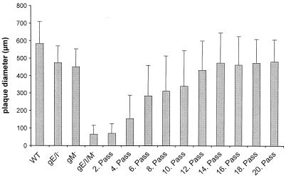
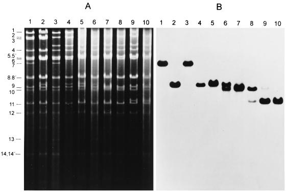
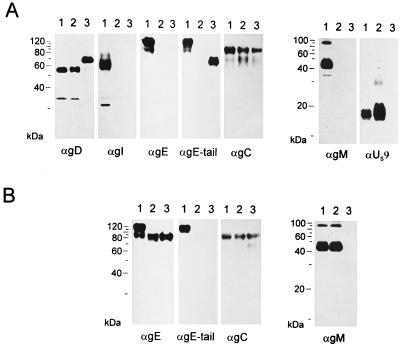
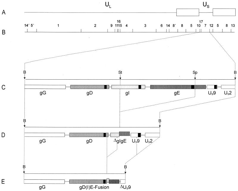
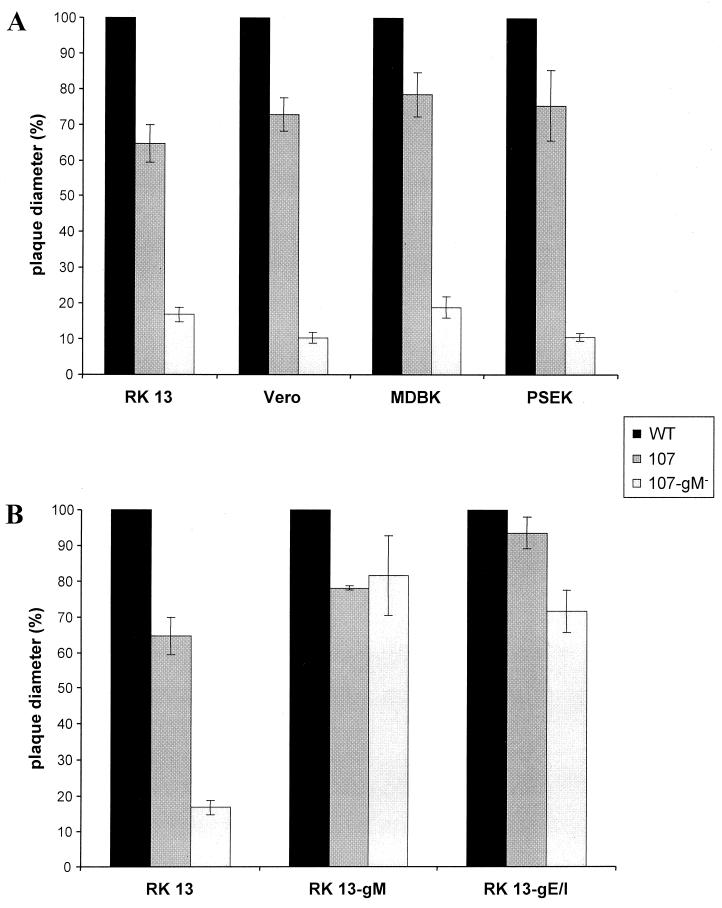
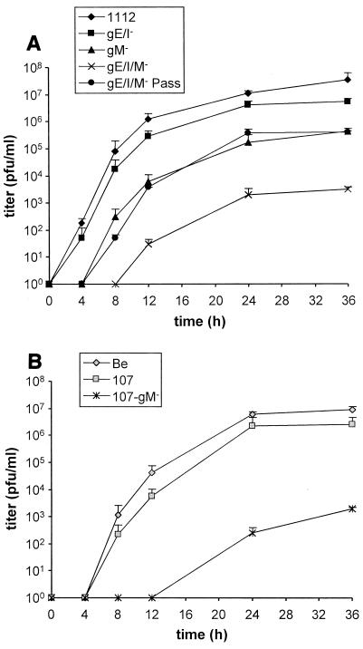
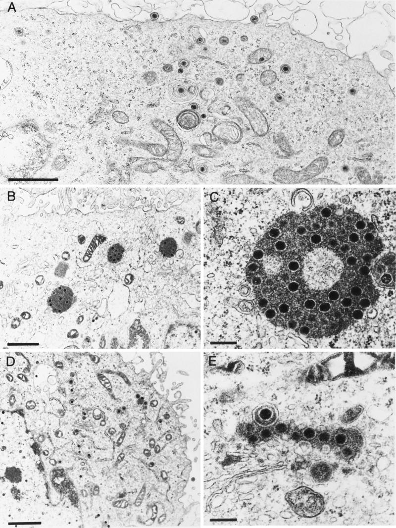
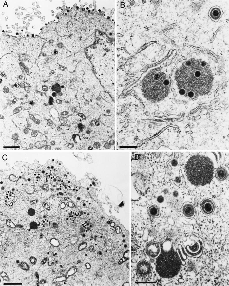
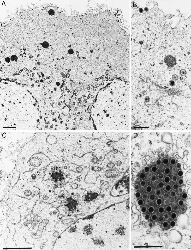
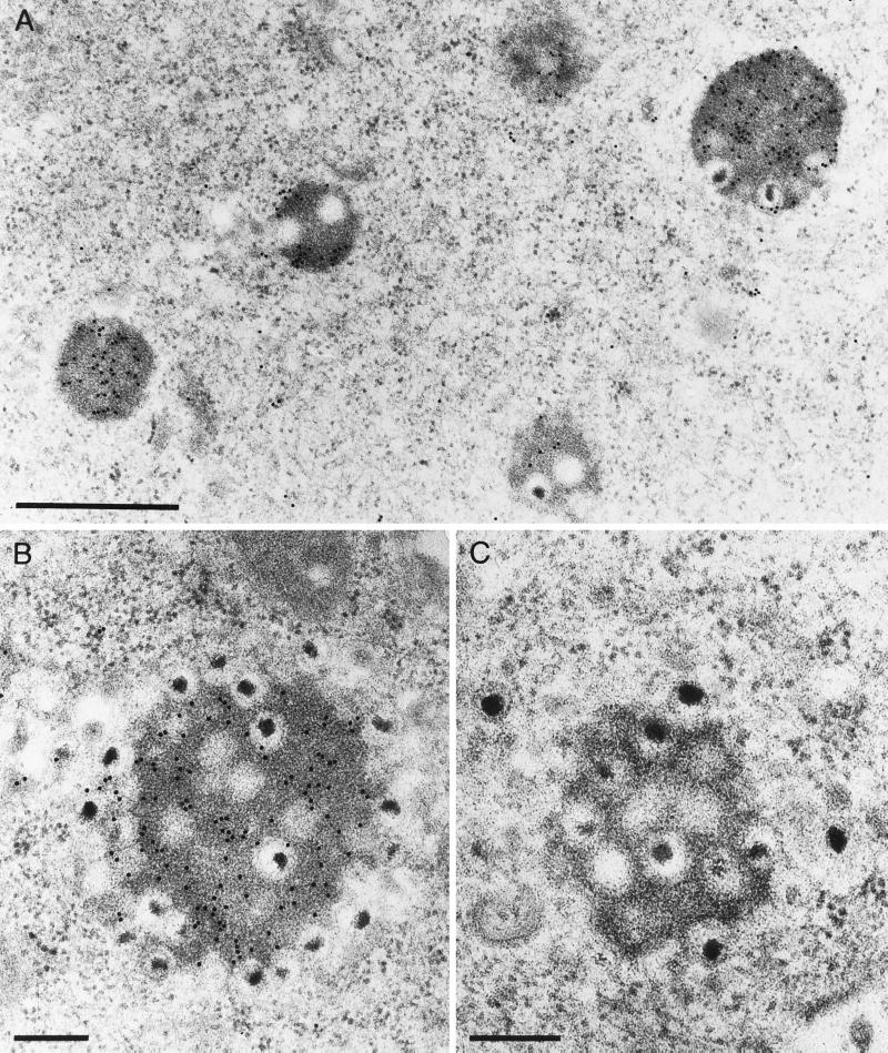
Glycoproteins M (gM), E (gE), and I (gI) of pseudorabies virus (PrV) are required for efficient formation of mature virions. The simultaneous absence of gM and the gE/gI complex results in severe deficiencies in virion morphogenesis and cell-to-cell spread, leading to drastically decreased virus titers and a small-plaque phenotype (A. Brack, J. Dijkstra, H. Granzow, B. G. Klupp, and T. C. Mettenleiter, J. Virol. 73:5364-5372, 1999). Serial passaging in noncomplementing cells of a virus mutant unable to express gM, gE, and gI resulted in a reversion of the small-plaque phenotype and restoration of infectious virus formation to the level of a gM(-) mutant. Genetic analyses showed that reversion of the phenotype was accompanied by a genomic rearrangement which led to the fusion of a portion of the gE gene encoding the cytoplasmic domain to the 3' end of the glycoprotein D gene, resulting in expression of a chimeric gD-gE protein. Since this indicated that the intracytoplasmic domain of gE was responsible for the observed phenotypic alterations, the UL10 (gM) gene was deleted in a PrV mutant, PrV-107, which specifically lacked the cytoplasmic tail of gE. Regarding one-step growth, plaque size, and virion formation as observed under the electron microscope, the mutant lacking gM and the gE cytoplasmic tail proved to be very similar to the gE/I/M triple mutant. Thus, our data indicate that it is the cytoplasmic tail of gE which is responsible for the observed phenotypic effects in conjunction with deletion of gM. We hypothesize that the cytoplasmic domain of gE specifically interacts with components of the capsid and/or tegument, leading to efficient secondary envelopment of intracytoplasmic capsids.


Figures

FIG. 1
FIG. 1
Reversion of small-plaque phenotype by passaging of PrV-gE/I/M in Vero cells. Vero cells were infected with PrV-gE/I/M. After development of CPE, cells were trypsinized and reseeded with uninfected cells. For each second passage, a parallel plate was fixed, stained with X-Gal, and used for determination of plaque size. The average diameters of plaques are shown. Bars indicate standard deviation. A total of 30 plaques were measured for each assay.
FIG. 2
FIG. 2
Southern blot analysis of PrV-gE/I/MPass. Southern blot analysis was performed with DNAs from wild-type PrV-Ka (lanes 1), PrV-gE/I (lanes 2), PrV-gM (lanes 3), PrV-gE/I/M (lanes 4), and PrV-gE/I/M passaged 2 (lanes 5), 4 (lanes 6), 6 (lanes 7), 10 (lanes 8), and 20 times (lanes 9) as well the single-plaque isolate PrV-gE/I/MPass (lanes 10) after cleavage with BamHI. (A) Ethidium bromide-stained gel. (B) Hybridization with genomic BamHI fragment 7 derived from the US region (see Fig. 4 for location). The locations of the BamHI fragments of PrV wild-type DNA are on the left.
FIG. 3
FIG. 3
Western blot analysis. Purified virions of PrV-Ka (A, lanes 1), PrV-gE/I/M (A, lanes 2), PrV-gE/I/MPass (A, lanes 3), PrV-Be (B, lanes 1), PrV-107 (B, lanes 2), and PrV-107-gM (B, lanes 3) were lysed, and proteins were separated in an SDS–10 or 13% polyacrylamide gel. After electrophoretic transfer, nitrocellulose membranes were probed with antibodies against gD, gI, gE, the gE cytoplasmic tail, gC, gM, or the US9 protein. After incubation with peroxidase-conjugated secondary antibody, bound antibody was visualized by chemiluminescence recorded on X-ray films.
FIG. 4
FIG. 4
Genomic arrangement in the US region of PrV-gE/I/MPass. (A) Schematic diagram of the PrV genome. (B) BamHI restriction fragment map. UL, unique long region; US, unique short region. Open boxes, inverted repeat sequences which bracket the US region. (C) Enlargement of BamHI fragment 7 located in the US region, with relevant cleavage sites (B, BamHI; St, StuI; Sp, SphI). (D) Genomic arrangement in the US regions of PrV-gE/I and PrV-gE/I/M. It is evident that by deletion of gI sequences from the StuI site and gE sequences up to the SphI site (36), the 5′ terminus of the gI gene had been fused to the 3′ terminus of the gE gene. Note that the gE sequences in the ΔgI/gE fusion gene are out of frame with the gI sequences. (E) Deletion events that occurred during passaging were mapped by sequencing. One deletion resulted in the formation of a chimeric gD(I)E hybrid gene, in which the gE sequences are in frame with the gD sequences and the gI remnant is out of frame. Another rearrangement led to truncation of the US9 gene and at least partial deletion of the US2 gene. The locations of the gene sequences encoding the transmembrane portions of the gD, gI, gE, and US9 proteins are indicated by black boxes.
FIG. 5
FIG. 5
Plaque sizes of PrV-107 and PrV-107-gM. RK13, Vero, MDBK, and PSEK cells (A), and RK13, RK13-gM, and RK13-gE/I cells (B) were infected under plaque assay conditions with PrV-107 and PrV-107-gM. Two days after infection, plaque diameters were measured microscopically and compared with the average diameter of plaques induced by parental PrV-Be (WT, solid bars), which was set at 100%. Average values and standard deviations after measurement of at least 30 plaques in three independent experiments each are indicated.
FIG. 6
FIG. 6
One-step growth analysis. RK13 cells were infected at an MOI of 5 with PrV-1112, PrV-gE/I, PrV-gM, PrV-gE/I/M, and PrV-gE/I/MPass (A) or PrV-Be, PrV-107, and PrV-107-gM (B). At the indicated times after infection, supernatant and cells were harvested, titers were determined on RK13 cells, and the titers were added. Average values and standard deviations of two independent experiments are shown.
FIG. 7
FIG. 7
Electron microscopy of RK13 cells infected with (A) PrV-Ka, (B and C) PrV-gE/I/M, and (D and E) PrV-gE/I/MPass, analyzed 16 h after infection. Bars: (A, B, and D) 1 μm; (C and E) 250 nm.
FIG. 8
FIG. 8
Electron microscopy of RK13 cells infected with (A and B) PrV-gM and (C and D) PrV-gE/I, analyzed 16 h after infection. Bars: (A and C) 1 μm; (B and D) 250 nm.
FIG. 9
FIG. 9
Electron microscopy of RK13 cells infected with (A and B) PrV-107 and (C and D) PrV-107-gM, analyzed 16 h after infection. Bars: (A and D) 2 μm; (B and C) 500 nm.
FIG. 10
FIG. 10
Immunoelectron microscopy of PrV-gE/I/M-infected RK13 cells analyzed 16 h after infection. Thin sections were incubated with either a monospecific anti-PrV UL49 antiserum (A and B) or a control antiserum directed against Newcastle disease virus (C). Bars: (A) 750 nm; (B and C) 250 nm.

References

    1. Alconada A, Bauer U, Sodeik B, Hoflack B. Intracellular traffic of herpes simplex virus glycoprotein gE: characterization of the sorting signals required for its trans-Golgi network localization. J Virol. 1999;73:377–387. - PMC - PubMed
    1. Babic N, Klupp B G, Brack A, Mettenleiter T C, Ugolini G, Flamand A. Deletion of glycoprotein gE reduces the propagation of pseudorabies virus in the nervous system of the mouse after intranasal inoculation. Virology. 1996;219:279–284. - PubMed
    1. Becker C H. Zur primären Schädigung vegetativer Ganglien nach Infektion mit dem Herpes suis Virus bei verschiedenen Tierarten. Experientia. 1967;23:209–217. - PubMed
    1. Brack A R, Dijkstra J, Granzow H, Klupp B G, Mettenleiter T C. Inhibition of virion maturation by simultaneous deletion of glycoproteins E, I, and M of pseudorabies virus. J Virol. 1999;73:5364–5372. - PMC - PubMed
    1. Brideau A D, Banfield B W, Enquist L W. The Us9 gene product of pseudorabies virus, an alphaherpesvirus, is a phosphorylated, tail-anchored type II membrane protein. J Virol. 1998;72:4560–4570. - PMC - PubMed

Publication types

MeSH terms

Substances

LinkOut - more resources