. 2000 May;74(9):4028-38.
doi: 10.1128/jvi.74.9.4028-4038.2000.

Establishment and characterization of molecular clones of porcine endogenous retroviruses replicating on human cells

Affiliations

Establishment and characterization of molecular clones of porcine endogenous retroviruses replicating on human cells

F Czauderna et al. J Virol. 2000 May.

Abstract

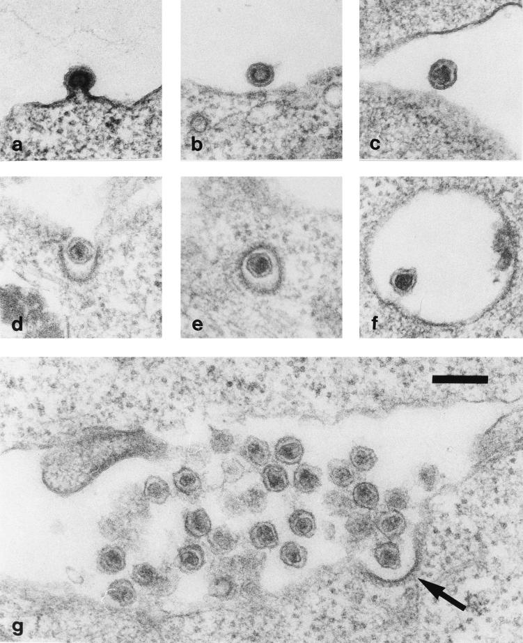
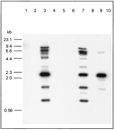
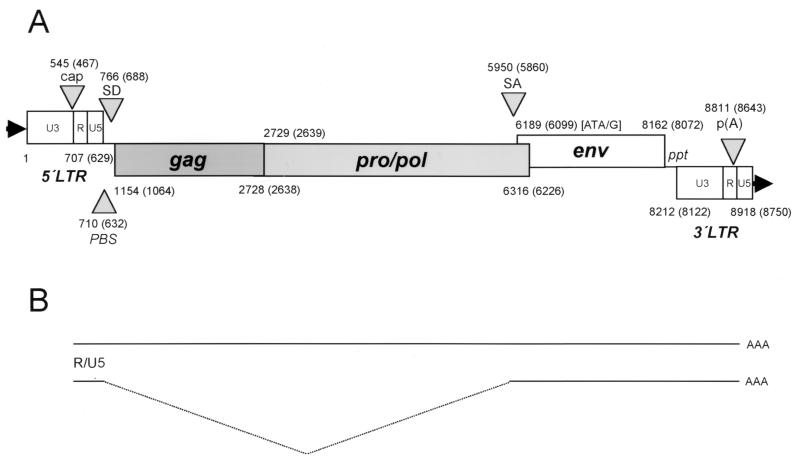
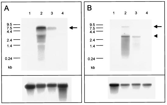
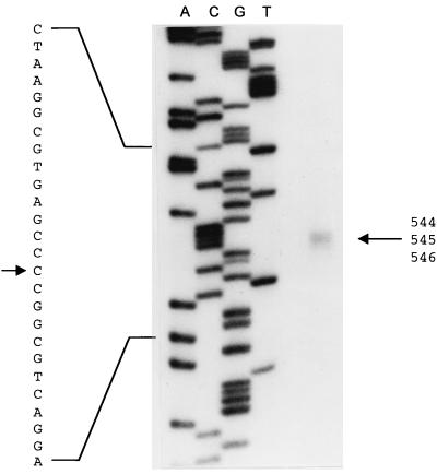
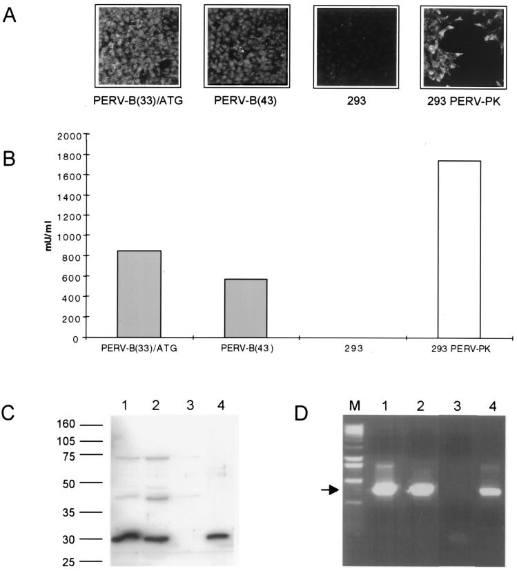
The use of pig xenografts is being considered to alleviate the shortage of allogeneic organs for transplantation. In addition to the problems overcoming immunological and physiological barriers, the existence of numerous porcine microorganisms poses the risk of initiating a xenozoonosis. Recently, different classes of type C porcine endogenous retoviruses (PERV) which are infectious for human cells in vitro have been partially described. We therefore examined whether completely intact proviruses exist that produce infectious and replication-competent virions. Several proviral PERV sequences were cloned and characterized. One molecular PERV class B clone, PERV-B(43), generated infectious particles after transfection into human 293 cells. A second clone, PERV-B(33), which was highly homologous to PERV-B(43), showed a G-to-A mutation in the first start codon (Met to Ile) of the env gene, preventing this provirus from replicating. However, a genetic recombinant, PERV-B(33)/ATG, carrying a restored env start codon, became infectious and could be serially passaged on 293 cells similar to virus clone PERV-B(43). PERV protein expression was detected 24 to 48 h posttransfection (p. t.) using cross-reacting antiserum, and reverse transcriptase activity was found at 12 to 14 days p.t. The transcriptional start and stop sites as well as the splice donor and splice acceptor sites of PERV mRNA were mapped, yielding a subgenomic env transcript of 3. 1 kb. PERV-B(33) and PERV-B(43) differ in the number of copies of a 39-bp segment in the U3 region of the long terminal repeat. Strategies to identify and to specifically suppress or eliminate those proviruses from the pig genome might help in the production of PERV-free animals.


Figures

FIG. 1
FIG. 1
Morphology of PERV. Ultrathin sections showing PERV particles budding from (a to c) and infecting (d to f) human kidney cells (293 line). Budding proceeds in a typical C-type manner, while virus entry follows the pathway of receptor-mediated endocytosis via coated pits (d and arrow in g) and coated vesicles (e) towards endosomes (f). The diameter of the particles is approximately 110 nm. The arrow points to a virus particle during cell entry. The bar in panel g represents 200 nm. 293 cells were infected with originally PK15-derived PERV particles, which can be passaged on 293 cells.
FIG. 2
FIG. 2
Presence of PERV sequences in different genomes and restriction enzyme analysis of PERV-infected 293 cell line. Southern blot analyses of EcoRI-digested genomic DNA samples from different species using a PERV pro-pol probe. Lane 1, mouse; lane 2, dog; lane 3, pig PBMC; lane 4, sheep; lane 5, goat; lane 6, bovine; lane 7, pig (PK15 cell line); lane 8, human 293 cell line; lane 9, human cells infected with PERV (293 cell line infected with PK15-derived PERV); lane 10, human teratocarcinoma cell line GH. A 753-bp pro-pol fragment served as the hybridization probe. The sizes of HindIII-digested λ are given.
FIG. 3
FIG. 3
Proviral organization of clones PERV-B(33) and PERV-B(43). (A) Proviral sequences are 8,918 and 8,760 bp long for PERV-B(33) and PERV-B(43), respectively. Genes and ORFs are shown as open boxes; the numbers indicate the first and last nucleotide in the PERV-B(33) and PERV-B(43) (in parentheses) sequences. A 39-bp repeat is found four times in the U3 element of the PERV-B(33) LTRs, whereas the PERV-B(43) LTRs harbor two 39-bp repeat elements. Cap, transcriptional start site; PBS, primer binding site (tRNAGly4); SD, splice donor; SA, splice acceptor; ppt, polypurine tract; p(A), poly(A) addition site. (B) Two mRNA species are expressed from proviral PERV. A full-length (8.3 kb) and a subgenomic (3.1 kb) env mRNA are indicated.
FIG. 4
FIG. 4
PERV RNA expression pattern. Northern blot analysis of total RNA (A) and oligo(dT)-selected RNA (B) from different cell lines. Equal amounts of RNA were separated on sodium phosphate-agarose gels, transferred to nylon membranes, and hybridized with a radiolabeled pro-pol probe (A) and with a U5 probe (B). A β-actin probe served as a control (lower panels). Lane 1, human kidney cell line 293; lane 2, 293 cells infected with PERV; lane 3, porcine kidney cell line PK15; lane 4, human teratocarcinoma cell line GH. A full-length transcript of about 8.3 kb is indicated (arrows). A subgenomic env transcript of ∼3.1 kb hybridizes with the U5 probe (arrowhead in panel B). RNA prepared from uninfected 293 cells and the human teratocarcinoma cell line GH served as negative controls.
FIG. 5
FIG. 5
Identification of the transcription initiation site of PERV mRNA. Polyadenylated RNA prepared from the cell line 293 PERV-PK was reverse transcribed using the 32P-end-labeled oligonucleotide PERV-PK-REV. The cDNA was separated by SDS–6% PAGE together with a four-lane Sanger sequencing reaction performed with the same unlabeled primer and the cloned 5′ LTR (pPERV-PK26/34) as the template. Two to three end-labeled transcripts were detected on the autroradiograph. The cap site was assigned to nt 545 (C) in the PERV-B(33) LTR (arrows). The lower-strand sequence is given on the left side.
FIG. 6
FIG. 6
Analysis of retrovirus expression after transfection of molecular clones PERV-B(33) and PERV-B(43). (A) Indirect immunofluorescence analysis of PERV Gag expression 12 weeks p.t. using cross-reacting FeLV CA antiserum. Molecular clones PERV-B(33)/ATG and PERV-B(43) in 293 cells were used at 12 weeks p.t.; uninfected 293 cells and 293 cells productively infected with PERV derived from PK15 (293 PERV-PK) served as controls. Expression of Gag from PERV-B(33)/ATG and PERV-B(43) was detected as early as 24 h p.t. in 20 to 50% of cells (not shown). (B) RT activities in cell-free supernatants 12 weeks p.t. RT produced by PERV-B(33)/ATG and PERV-B(43) was detected as early as 12 to 14 days p.t. (not shown). The controls were the same as in A. (C) Immunoblot analysis of PERV Gag proteins. Sucrose gradient-purified particles from PERV-producing cells [PERV-B(33)/ATG and PERV-B(43) at 12 weeks p.t.] were separated by SDS-PAGE. The p30 capsid protein (arrow) cross-reacts with FeLV CA antiserum. Lane 1, PERV-B(33)/ATG; lane 2, PERV-B(43); lane 3, 293 cell line; lane 4, 293 PERV-PK cell line. The positions of molecular mass markers are indicated (in kilodaltons). (D) DNA PCR of PERV pro-pol sequences demonstrating integration of proviral PERV sequences. Lane 1, PERV-B(33)/ATG; lane 2, PERV-B(43); lane 3, 293 cell line; lane 4, 293 PERV-PK cell line. The arrow marks an 812-bp amplification product obtained with primers PK45 and PK47. Lane M, molecular size markers (1-kb ladder; Life Technologies).

References

    1. Akiyoshi D E, Denaro M, Zhu H, Greenstein J L, Banerjee P T, Fishman J A. Identification of a full-length cDNA for an endogenous retrovirus of minature swine. J Virol. 1998;72:4503–4507. - PMC - PubMed
    1. Allan J S. Xenotransplantation at a crossroads: prevention versus progress. Nat Med. 1996;2:18–21. - PubMed
    1. Armstrong J A, Porterfield J S, Madrid A T D. C-type virus particles in pig kidney cell lines. J Gen Virol. 1971;10:195–198. - PubMed
    1. Bach F H, Robson S C, Winkler H, Ferran C, Stuhlmeier K M, Wrighton C J, Hancock W W. Barriers to xenotransplantation. Nat Med. 1995;1:869–873. - PubMed
    1. Battini J L, Danos O, Heard J M. Receptor-binding domain of murine leukemia virus envelope glycoproteins. J Virol. 1995;69:713–719. - PMC - PubMed

Publication types

LinkOut - more resources