. 2000 May;74(9):4085-92.
doi: 10.1128/jvi.74.9.4085-4092.2000.

The vaccinia virus A14.5L gene encodes a hydrophobic 53-amino-acid virion membrane protein that enhances virulence in mice and is conserved among vertebrate poxviruses

Affiliations

The vaccinia virus A14.5L gene encodes a hydrophobic 53-amino-acid virion membrane protein that enhances virulence in mice and is conserved among vertebrate poxviruses

T Betakova et al. J Virol. 2000 May.

Abstract

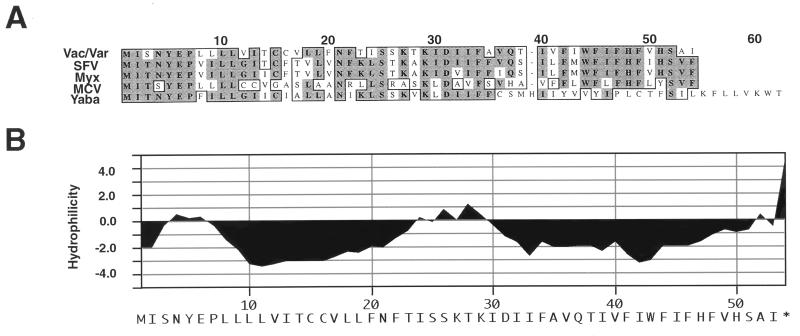
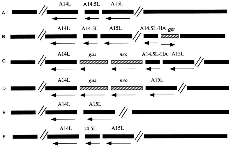
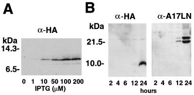
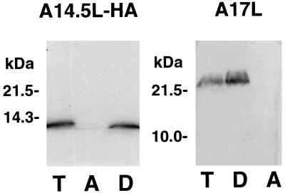
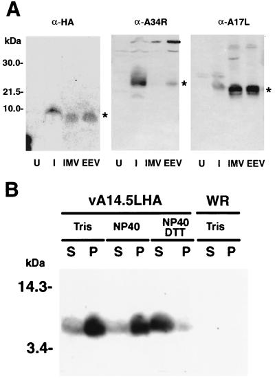
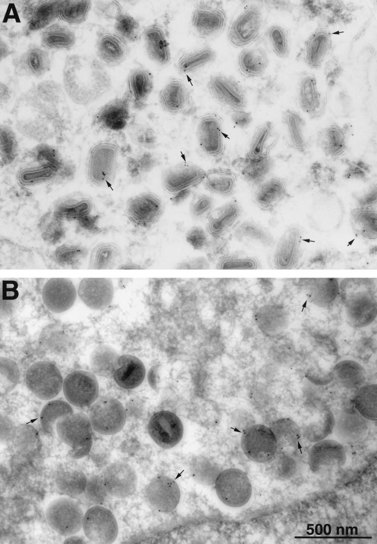
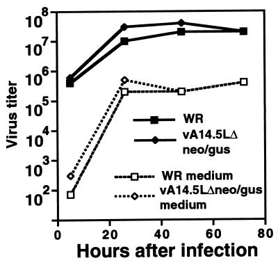
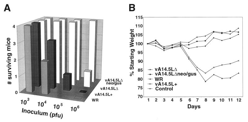
A short sequence, located between the A14L and A15L open reading frames (ORFs) of vaccinia virus, was predicted to encode a hydrophobic protein of 53 amino acids that is conserved in orthopoxviruses, leporipoxviruses, yatapoxiruses, and molluscipoxviruses. We constructed a recombinant vaccinia virus with a 10-codon epitope tag appended to the C terminus of the A14.5L ORF. Synthesis of the tagged protein occurred at late times and was blocked by an inhibitor of DNA replication, consistent with regulation by a predicted late promoter just upstream of the A14.5L ORF. Hydrophobicity of the protein was demonstrated by extraction into the detergent phase of Triton X-114. The protein was associated with purified vaccinia virus particles and with membranes of immature and mature virions that were visualized by electron microscopy of infected cells. Efficient release of the protein from purified virions occurred after treatment with a nonionic detergent and reducing agent. A mutant virus, in which the A14.5L ORF was largely deleted, produced normal-size plaques in several cell lines, and the yields of infectious intra- and extracellular viruses were similar to those of the parent. In contrast, with a mouse model, mutant viruses with the A14.5L ORF largely deleted were attenuated relative to that of the parental virus or a mutant virus with a restored A14.5L gene.


Figures

FIG. 1
FIG. 1
Sequence homology and predicted hydrophobicity of the A14.5L ORF. (A) The sequence of the vaccinia virus (Vac) A14.5L ORF and the identical variola virus (Var) ORF is shown aligned with the orthologous ORFs of Shope fibroma virus (SFV), myxoma virus (Myx), molluscum contagiosum virus (MCV), and Yaba monkey tumor virus (Yaba). The numbers correspond to the codons of the longest ORF. (B) Hydrophilicity plot of the vaccinia virus A14.5L ORF.
FIG. 2
FIG. 2
Representations of parental and recombinant viral genomes. (A) Parental vaccinia virus strain WR. (B) vT7lacOI-A14.5LHA. (C) vA14.5LHA. (D) vA14.5LΔneo/gus. (E) vA14.5LΔ. (F) vA14.5L+. The vaccinia virus ORFs A14L, A14.5L, and A15L; the antibiotic selection markers neo and gpt; and the reporter gene gus are indicated above the bars. An arrow below the bar indicates the direction of transcription of each ORF. The symbol // signifies additional portions of the vaccinia virus genome. vT7lacOI-A14.5LHA contains a T7 RNA polymerase gene, an E. coli lac repressor gene, and a T7 promoter and lac operator regulating expression of A14.5L-HA, which are not shown. VA14.5L+ is a revertant of vA14.5LΔneo/gus and has the same gene arrangement as the parental vaccinia virus.
FIG. 3
FIG. 3
Expression of the HA-tagged A14.5L ORF. (A) Expression of the epitope-tagged A14.5L protein from an IPTG-inducible bacteriophage T7 promoter. BS-C-1 cells, in a six-well plate, were infected with 10 PFU of vT7lacOI-A14.5LHA per cell. After 1 h at 37°C, fresh medium containing 0 to 200 μM IPTG was added to each monolayer. The cells were harvested at 18 h and lysed in 50 μl of extraction buffer (4), and 20 μl of cleared supernatant was analyzed by SDS-PAGE and Western blotting with MAb 12CA5 to the HA epitope. (B) Expression of the epitope-tagged A14.5L protein from the natural A14.5L promoter. A Western blot of lysates from BS-C-1 cells harvested at 2 to 24 h after infection with vA14.5LHA is shown. BS-C-1 cells were infected with vA14.5LHA, as in panel A, except that no IPTG was added. At intervals from 2 to 24 h, cells from duplicate wells were harvested and analyzed by Western blotting with MAb 12CA5 or polyclonal antibody to the A17L protein. The positions and masses of marker proteins are indicated to the left of each panel.
FIG. 4
FIG. 4
Partition of the A14.5L-HA protein in the Triton X-114 detergent phase. BS-C-1 cells were infected with vA14.5LHA (10 PFU/cell). After 18 h, the cells were washed with PBS and resuspended in 0.2 ml of 10 mM Tris-HCl, 0.15 M NaCl, and 1% (vol/vol) Triton X-114 (pH 7.4). The phase separation was carried out as described previously (14). Equal volumes of total extract (T), the aqueous phase (A), and the detergent phase (D) were analyzed by SDS-PAGE and Western blotting with MAb 12CA5 or polyclonal antibody to the A17L protein. The positions and masses of marker proteins are indicated to the left.
FIG. 5
FIG. 5
Association of the A14L-HA protein with the membrane fraction of virus particles. (A) BS-C-1 cells were mock infected or infected with 10 PFU of vA14.5LHA per cell. After 18 h, the medium was harvested, and the cells were disrupted with a Dounce homogenizer. Virus particles were purified by sedimentation through a sucrose cushion and CsCl centrifugation (23). The virus band was diluted with Tris-HCl (pH 9), collected by centrifugation, and resuspended in Tris buffer. The lysates of mock-infected cells (U) or infected cells (I) were analyzed in parallel with CsCl gradient fractions by SDS-PAGE and Western blotting with MAb 12CA5 (α-HA) or polyclonal antibody to the A34R protein (α-A34R) or A17L protein (α-A17R). Fractions containing IMV and IEV or EEV are indicated. Small differences in the electrophoretic mobilities of proteins from lysates and CsCl gradients could be due to the ionic strengths. The bands corresponding to A14.5L-HA, A34R, and A17L are indicated by asterisks. The positions and masses of marker proteins are indicated to the left. (B) Vaccinia virions, purified by sucrose gradient centrifugation (9) from cells infected with vaccinia virus WR or vA14.5LHA, were incubated with 10 mM Tris (pH 7.4) or 1% NP-40 in 10 mM Tris (pH 7.4) in the presence or absence of 10 mM dithiothreitol. Soluble (S) and insoluble (P) fractions were separated by centrifugation and resuspended to equal volumes in SDS-containing sample buffer. An equivalent amount of each fraction was subjected to electrophoresis, and the separated proteins were transferred to a membrane and analyzed by Western blotting with anti-HA MAb 12CA5.
FIG. 6
FIG. 6
Immunogold labeling of viral membranes with antibody to the HA tag. BSC-1 cells that had been infected with vA14.5L-HA for 24 h were fixed in paraformaldehyde, cryosectioned, and incubated with a mouse MAb to the HA tag, rabbit IgG to mouse IgG, and then with 10-nm-diameter gold particles conjugated to protein A. Electron microscopic images are shown with a 500-nm marker. (A) Field containing large numbers of IEV. (B) Field containing large numbers of immature virons. Arrows point to examples of gold particles.
FIG. 7
FIG. 7
Replication of an A14.5L deletion mutant. B-SC-1 cells in a six-well plate were inoculated with 10 PFU of vaccinia virus strain WR or vA14.5LΔneo/gus per cell for 1 h at 37°C. The cells were then washed twice with medium. At appropriate times, the medium was removed and saved, and the cells were harvested by scraping. The media and cells were centrifuged, and the virus titers in the supernatants of the former (dashed lines) and the pellets of the latter (solid lines) were determined by plaque assay on BS-C-1 cells.
FIG. 8
FIG. 8
Virulence of parental and mutant vaccinia viruses in mice. Groups of four mice were inoculated intranasally with 103 to 106 PFU of purified vaccinia virus strain WR, vA14.5LΔneo/gus, vA14.5LΔ, and vA14.5L+. (A) Death of animals. (B) Percentage of original weight of mice in each group infected with 103 PFU.

References

    1. Alexander W A, Moss B, Fuerst T R. Regulated expression of foreign genes in vaccinia virus under the control of bacteriophage T7 RNA polymerase and the Escherichia coli lac repressor. J Virol. 1992;66:2934–2942. - PMC - PubMed
    1. Amegadzie B Y, Ahn B-Y, Moss B. Characterization of a 7-kilodalton subunit of vaccinia virus DNA-dependent RNA polymerase with structural similarities to the smallest subunit of eukaryotic RNA polymerase II. J Virol. 1992;66:3003–3010. - PMC - PubMed
    1. Antoine G, Scheiflinger F, Dorner F, Falkner F G. The complete genomic sequence of the modified vaccinia Ankara strain: comparison with other orthopoxviruses. Virology. 1998;244:365–396. - PubMed
    1. Betakova T, Wolffe E J, Moss B. Regulation of vaccinia virus morphogenesis: phosphorylation of the A14L and A17L membrane proteins and C-terminal truncation of the A17L protein are dependent on the F10L protein kinase. J Virol. 1999;73:3534–3543. - PMC - PubMed
    1. Bordier C. Phase separation of integral membrane proteins in Triton X-114 solution. J Biol Chem. 1990;256:1604–1607. - PubMed

MeSH terms

LinkOut - more resources