. 2000 May;74(9):4102-9.
doi: 10.1128/jvi.74.9.4102-4109.2000.

Protection of the villus epithelial cells of the small intestine from rotavirus infection does not require immunoglobulin A

Affiliations

Protection of the villus epithelial cells of the small intestine from rotavirus infection does not require immunoglobulin A

C M O'Neal et al. J Virol. 2000 May.

Abstract

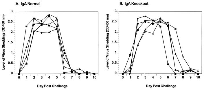
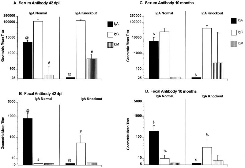
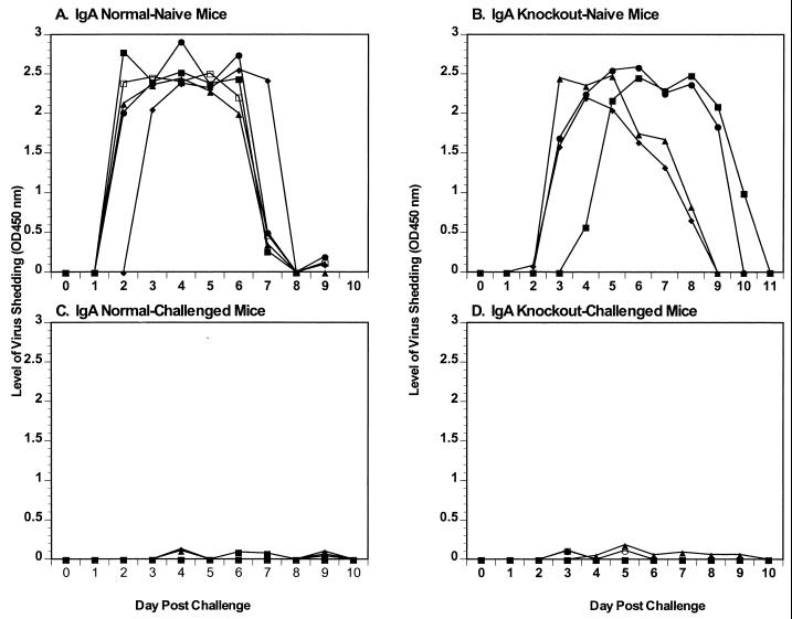
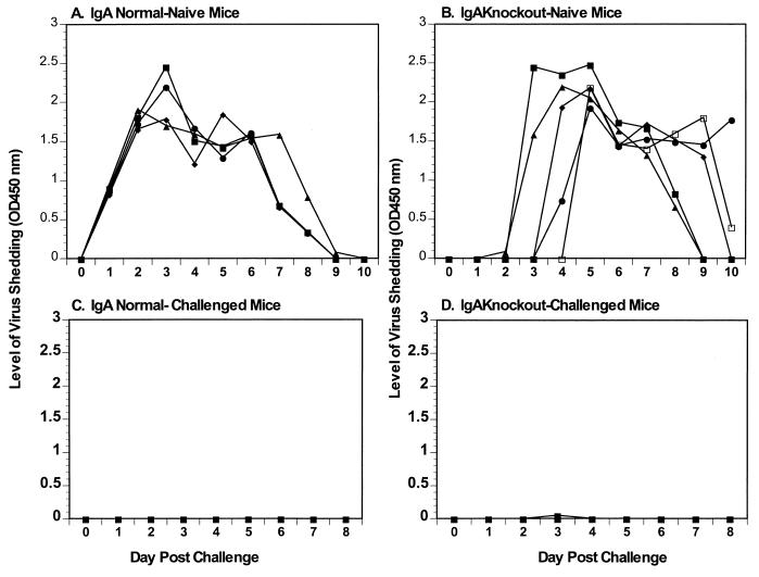
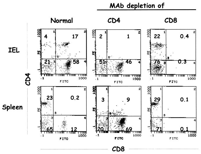
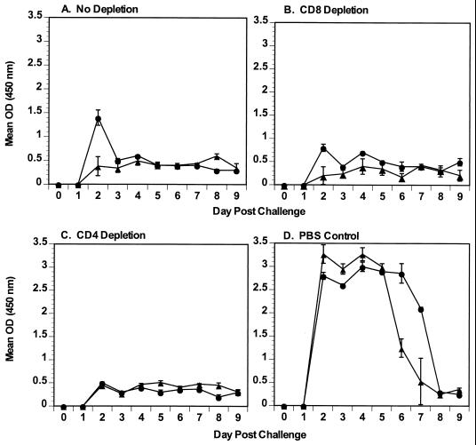
Immunoglobulin A (IgA) is the primary immune response induced in the intestine by rotavirus infection, but vaccination with virus-like particles induces predominantly IgG, not IgA. To definitively assess the role of IgA in protection from rotavirus infection, IgA knockout mice, which are devoid of serum and secretory IgA, were infected and then rechallenged with murine rotavirus at either 6 weeks or 10 months. Following primary rotavirus infection, IgA knockout mice cleared virus as effectively as IgA normal control mice. Rotavirus-infected IgA knockout mice produced no serum or fecal IgA but did have high levels of antirotavirus serum IgG and IgM and fecal IgG, whereas IgA normal control mice made both serum IgA and IgG and fecal IgA. Both IgA normal and IgA knockout mice were totally protected from rotavirus challenge at 42 days. Ten months following a primary infection, both IgA normal and knockout mice still had high levels of serum and fecal antirotavirus antibody and were totally protected from rotavirus challenge. To determine if compensatory mechanisms other than IgG were responsible for protection from rotavirus infection in IgA knockout mice, mice were depleted of CD4(+) T cells or CD8(+) T cells. No changes in the level of protection were seen in depleted mice. These data show that fecal or systemic IgA is not essential for protection from rotavirus infection and suggest that in the absence of IgA, IgG may play a significant role in protection from mucosal pathogens.


Figures

FIG. 1
FIG. 1
Rotavirus shedding curves following primary infection with ECwt. IgA normal (A) and knockout (B) mice were orally infected with 105 SD50 of ECwt. Fecal samples were collected for 11 days, and levels of virus antigen shedding were determined by ELISA. The y axis represents the level of virus shed as determined by ELISA.
FIG. 2
FIG. 2
Serum and fecal rotavirus-specific antibody responses in IgA normal and knockout mice at 42 dpi and 10 months following primary infection with 105 SD50 of ECwt. Serum (A and C) and fecal (B and D) antirotavirus IgA, IgG, and IgM antibodies (as indicated) were measured 42 days (A and B) or 10 months (C and D) following infection with ECwt. Antibody titers were measured for individual mice, and results are plotted as GMTs of the groups (n = 10 per group). Error bars represent 1 standard error of the mean. Significant differences in GMT are indicated (@, #, $, and %, P = 0.001); Mann-Whitney U test).
FIG. 3
FIG. 3
Rotavirus antigen shedding curves following challenge with ECwt. IgA normal and knockout mice were orally infected with 105 SD50 of ECwt. Rotavirus antigen shedding curves for IgA normal (A) and knockout (B) control mice receiving a primary infection on 42 dpi and IgA normal (C) and knockout (D) mice challenged on 42 dpi are shown. Fecal samples were collected for 11 days, and levels of virus antigen shedding were determined by ELISA. The y axis represents the level of virus shed as determined by ELISA.
FIG. 4
FIG. 4
Rotavirus antigen shedding curves following challenge infection with ECwt. (A and B) Rotavirus antigen shedding curves for primary infection of 3-month-old IgA normal (A) and knockout (B) control mice. IgA normal (C) and IgA knockout (D) mice were orally challenged with 105 SD50 of ECwt 10 months following the primary infection. Fecal samples were collected for 11 days, and levels of virus antigen shedding were determined by ELISA. The y axis represents the level of virus antigen shed as determined by ELISA.
FIG. 5
FIG. 5
Flow cytometric analyses of IgA knockout mice depleted of CD4+ or CD8+ T cells. In IgA knockout mice depleted of CD4+ or CD8+ T cells, spleen and IELs were isolated, and flow cytometry was performed to confirm depletion. PE-stained CD4+ T cells are shown on the vertical axis; FITC-stained CD8+ T cells are shown on the horizontal axis. Numbers in quadrants indicate percentages of stained cells in the compartments.
FIG. 6
FIG. 6
Protection from rotavirus challenge in IgA normal and knockout mice depleted of CD4+ or CD8+ T cells. Mice were infected with 105 SD50 of ECwt on day 0 and then challenged at 42 dpi with the same dose of ECwt rotavirus. Individual stool samples were collected daily and quantitated for levels of rotavirus antigen by ELISA. Results are plotted as the mean OD reading for each group of mice (four or five mice per group). The y axis represents the mean level of virus antigen shed by IgA normal (●) and knockout (▴) mice as determined by ELISA. (A) Mice infected with rotavirus, not depleted of any cell type, and then challenged with rotavirus; (B) mice infected with rotavirus, depleted of CD8 cells, and then challenged with rotavirus; (C) mice infected with rotavirus, depleted of CD4 cells, and then challenged with rotavirus; (D) nondepleted mice that received their primary infection on the same day that the other groups were challenged with rotavirus. Error bars represent the standard error of the mean.

References

    1. Blanchard T G, Czinn S J, Redline R W, Sigmund N, Harriman G R, Nedrud J G. Antibody-independent protective mucosal immunity to gastric helicobacter infection in mice. Cell Immunol. 1999;191:74–80. - PubMed
    1. Choi A H-C, Basu M, McNeal M M, Clements J D, Ward R L. Antibody-independent protection against rotavirus infection of mice stimulated by intranasal immunization with chimeric VP4 or VP6 protein. J Virol. 1999;73:7574–7581. - PMC - PubMed
    1. Ciarlet M, Crawford S E, Barone C, Bertolotti-Ciarlet A, Ramig R F, Estes M K, Conner M E. Subunit rotavirus vaccine administered parenterally to rabbits induces active protective immunity. J Virol. 1998;72:9233–9246. - PMC - PubMed
    1. Coffin S E, Klinek M, Offit P A. Induction of virus-specific antibody production by lamina propria lymphocytes following intramuscular inoculation with rotavirus. J Infect Dis. 1995;172:874–878. - PubMed
    1. Conner M E, Crawford S E, Barone C, Estes M K. Rotavirus vaccine administered parenterally induces protective immunity. J Virol. 1993;67:6633–6641. - PMC - PubMed

MeSH terms